0540
Three-dimensional holographic visualization of high-resolution myocardial scar on HoloLens1Department of Medicine, Beth Israel Deaconess Medical Center and Harvard Medical School, Boston, MA, United States, 2Department of Computer Science, Technical University of Munich, Munich, Germany, 3Department of Radiology, Beth Israel Deaconess Medical Center and Harvard Medical School, Boston, MA, United States
Synopsis
We present a framework for 3D holographic visualization of high-resolution 3D late gadolinium enhancement (LGE) of myocardial scar on augmented-reality glass HoloLens via two approaches; 1) voxel-wise 3D scar rendering and 2) surface projection of the scar. Holographic visualization of high-resolution 3D LGE data will provide a true 3D perception of the complex scar architecture with an immersive experience to explore the clinical standard LGE images in a more interactive and interpretable way, which may facilitate MR-guided scar-related ventricular tachycardia ablation.
Purpose
Late gadolinium enhancement (LGE) imaging is a clinical gold standard for the imaging of myocardial infarction (MI).1 In particular, the detailed scar depiction of LGE has been suggested to be useful for planning and guiding scar-related ventricular tachycardia (VT) ablation procedure by characterizing the location and extent of the scar.2,3 However, the current practice for interpreting LGE scans is based on the stack of multiple 2D slices, which makes it difficult to understand the complex three-dimensional (3D) architecture of the scar. Also, the lack of interactivity in merging and visualizing LGE data during electrophysiology makes MR-guided VT ablation challenging. Augmented reality (AR) has emerged as a potential tool for visualizing complex medical data, particularly in planning and during procedures.4 In particular, HoloLens5 enables 3D holographic visualization, which can provide an immersive experience to interactively explore data in 3D space. Recently, 3D LGE imaging with isotropic spatial resolution has been enabled by advanced acquisition and reconstruction techniques.6 Based on these advancements, we sought to present a framework for 3D holographic visualization of LGE data on AR HoloLens to provide a true 3D perception of the scar architecture with interactive data exploration, which may be useful for guiding VT ablation based on LGE images.Methods
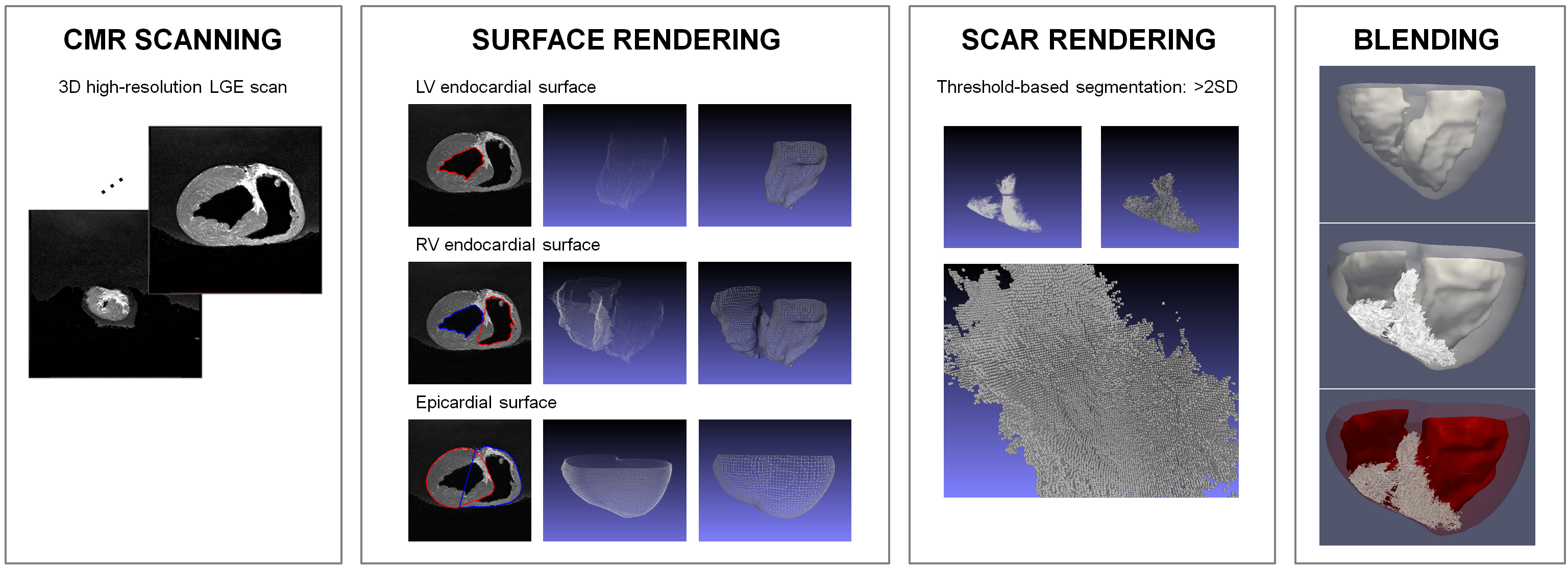
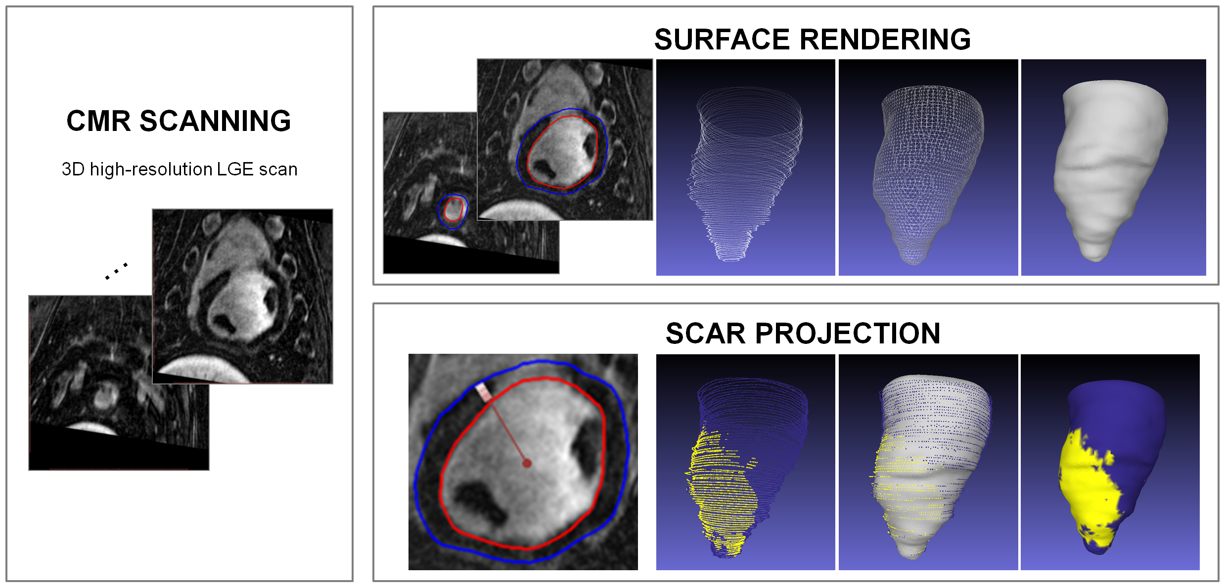
We propose a framework for 3D holographic visualization based on high-resolution 3D LGE images via two approaches; 1) voxel-wise 3D scar rendering and 2) surface projection of the scar. The initial step of the framework for both approaches was reconstructing endo-/ epicardial surface meshes. Endo-/ epicardial contours were first manually delineated from LGE images in all slices by an experienced reader on the short-axis view. Contours in all slices were merged into a set of point clouds for each surface. Clustering decimation was performed to organize point clouds based on the cell of 3D grid, followed by computation of the normals of all vertices based on the 10 neighboring points. Endo-/ epicardial triangular surface meshes were then generated using a Poisson surface reconstruction.
Voxel-wise 3D scar rendering
All voxels in the myocardium were bimodal fitted and scar tissues were defined as voxels with signal intensity higher than 2SDs of the healthy region.7 To preserve high-resolution submillimeter information of LGE voxels, each voxel of the scar was rendered into a cube, which carries the actual resolution of the LGE scan. All duplicated vertices and faces were removed to reduce the size of the rendered scar mesh. All layers of rendered surface and scar mesh were then blended with different transparencies and colors to enhance visibility and perception. The proposed framework for voxel-wise 3D scar rendered holographic visualization of the high-resolution LGE data is described in Fig. 1.
Surface projection of the scar
LGE voxels in the subendocardial region (0-33% from endocardial to epicardial), was projected onto the endocardial points. Scar was defined using a full-width at half-maximum (FWHM) technique.7 Endocardial points with projected scar information were then transferred to a texture, which was followed by a texture mapping performed on the parametrized Poisson surface of the endocardial mesh. The proposed framework for 2D surface scar projection holographic visualization of high-resolution LGE data is described in Fig. 2. This proposed framework was performed using Matlab (MathWorks) and MeshLab,8 which was exported to FBX file format on Blender. Created 3D LGE models were visualized on HoloLens using a 3D Viewer Beta (Microsoft).
CMR imaging protocol
In-vivo and ex-vivo CMR imaging was performed using a 1.5T Philips scanner. High-resolution 3D LGE images with random under-sampled accelerated acquisition6 were acquired 15-25 minutes after injection of 0.2mmol/kg gadobenate dimeglumine. A respiratory navigator with adaptive acquisition window9 was used for prospective motion correction. Imaging parameters were as follows: In-vivo imaging, gradient echo imaging sequence; TR/TE=6.1/2.7ms; field of view=280×280×112mm3; flip angle=25°; isotropic spatial resolution=1.0×1.0×1.0mm3; Ex-vivo imaging, TR/TE=17/8ms; field of view=130×130×100mm3; flip angle=25°, isotropic spatial resolution=0.4×0.4×0.5mm3.
Results/ Discussion
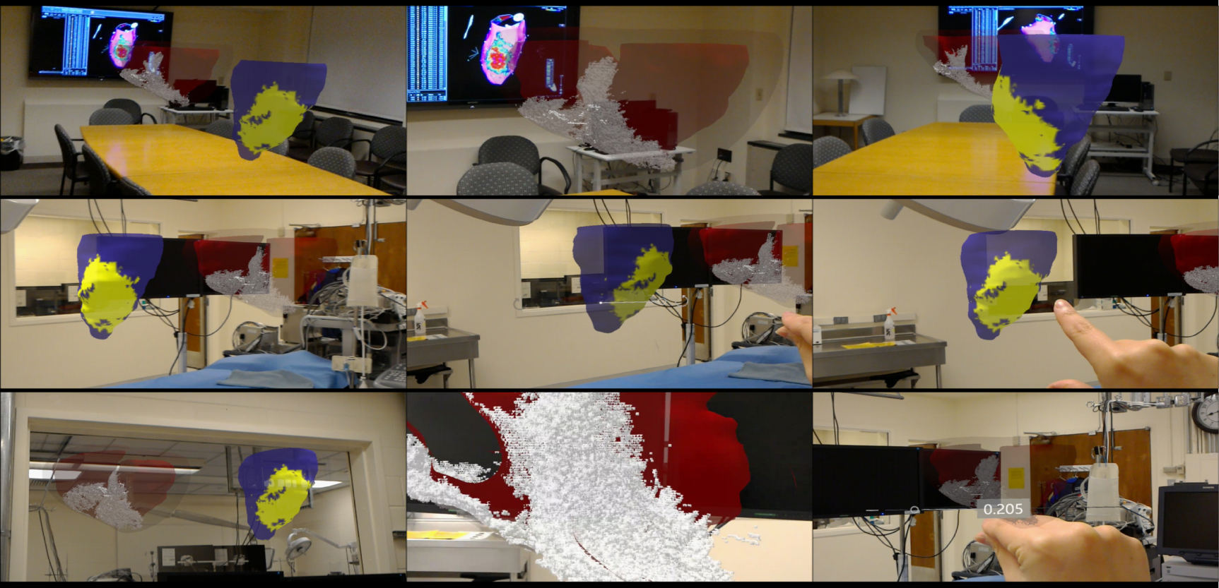
The demonstration of the holographic visualization of the 3D LGE data using the proposed frameworks was recorded on HoloLens and is presented in Fig 3. 3D holographic visualization of LGE data enables us to take full advantages of the 3D MR sequences, and to better understand the complex 3D scar architecture. Holographic visualization provides an embodied experience to explore the clinical standard LGE images in a more interactive and interpretable way. AR also allows for the combination of holographic 3D LGE data interacting with real-world environments, such as a patient’s body or a surgical suite. This will facilitate data fusion and exploration of LGE and electrophysiology data, which will support the planning and procedure of scar-related VT ablation.Acknowledgements
No acknowledgement found.References
1. Kim RJ, Fieno DS, Parrish TB, et al. Relationship of MRI delayed contrast enhancement to irreversible injury, infarct age, and contractile function. Circulation. 1999;100:1992-2002.
2. Dickfeld T, Tian J, Ahmad G, et al. MRI-guided ventricular tachycardia ablation integration of late gadolinium-enhanced 3D scar in patients with implantable cardioverter-defibrillators. Circulation: Arrhythmia and Electrophysiology. 2009;4:172-184.
3. Andreu D, Berruezo A, Ortiz-Pérez JT, et al. Integration of 3D electroanatomic maps and magnetic resonance scar characterization into the navigation system to guide ventricular tachycardia ablation. Circulation: Arrhythmia and Electrophysiology. 2009;4:674-683.
4. Riva G, Wiederhold BK. The New Dawn of Virtual Reality in Health Care: Medical Simulation and Experiential Interface. Stud Health Technol informatics. 2014;219:3-6.
5. Taylor AG. What Is the Microsoft HoloLens? In: Develop Microsoft HoloLens Apps Now. Apress; 2015:3-7.
6. Akçakaya M, Rayatzadeh H, Basha TA, et al. Accelerated late gadolinium enhancement cardiac MR imaging with isotropic spatial resolution using compressed sensing: initial experience. Radiology. 2012;264:691-699.
7. Flett AS, Hasleton J, Cook C, et al. Evaluation of techniques for the quantification of myocardial scar of differing etiology using cardiac magnetic resonance. JACC: Cardiovasc Imaging. 2008;4:150-156.
8. Cignoni P, Callieri M, Corsini M, Dellepiane M, Ganovelli F, Ranzuglia G. Meshlab: an open-source mesh processing tool. Eurographics Ital Chapter Conf. 2007;2008:129-136.
9. Moghari MH, Chan RH, Hong SN, et al. Free-breathing cardiac MR with a fixed navigator efficiency using adaptive gating window size. Magn Reson Med. 2012;68:1866-1875.
Figures