0521

Fast multi-slice B1 and B0 mapping (B01TIAMO) for 32-channel pTx body MRI at 7 Tesla
Sascha Brunheim1,2, Sören Johst1, Stephan Orzada1, Jose P Marques3, Marcel Gratz1,2, Mark E Ladd1,4, and Harald H Quick1,2

1Erwin L. Hahn Institute for Magnetic Resonance Imaging, University Duisburg-Essen, Essen, Germany, 2High Field and Hybrid MR Imaging, University Hospital Essen, Essen, Germany, 3Donders Institute for Brain, Cognition and Behaviour, Radboud University Nijmegen, Nijmegen, Netherlands, 4Medical Physics in Radiology, German Cancer Research Center (DKFZ), Heidelberg, Germany

Synopsis

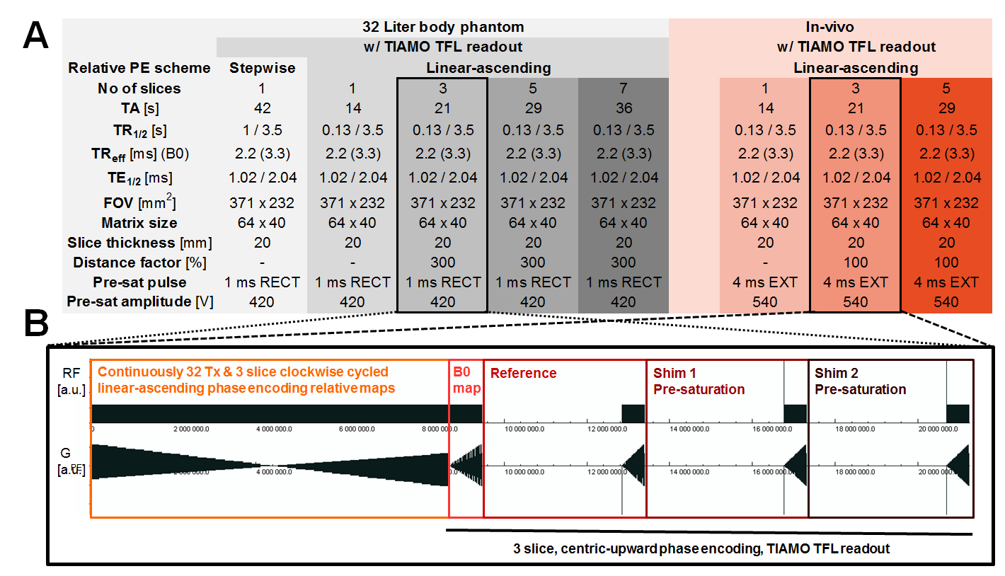
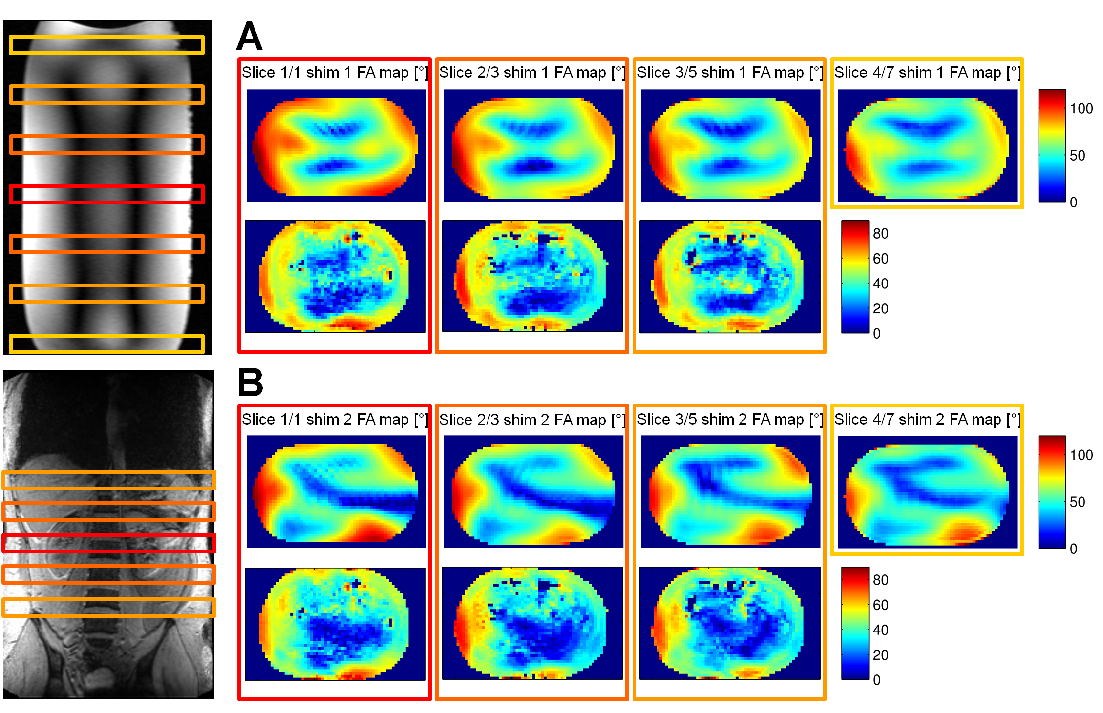
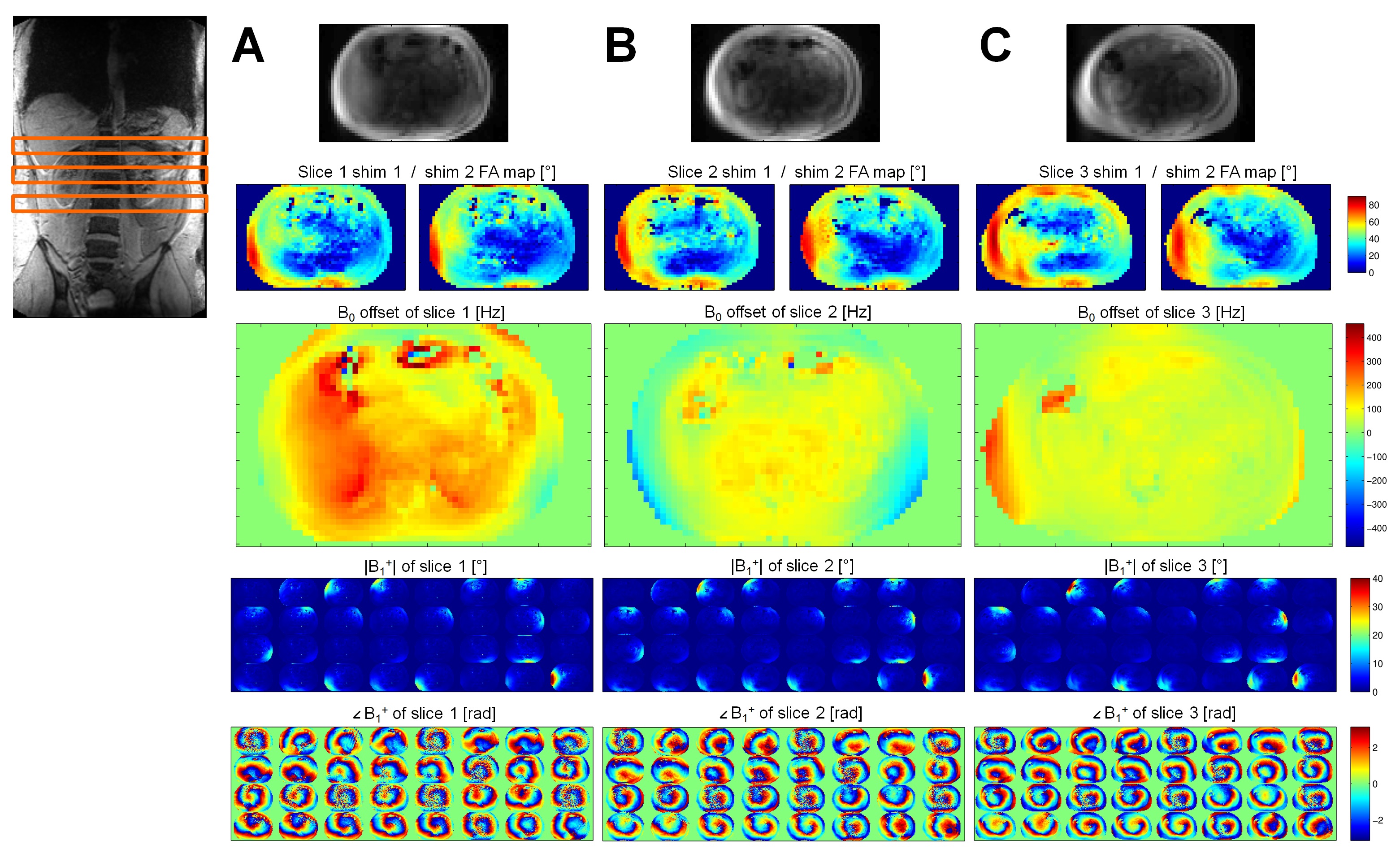
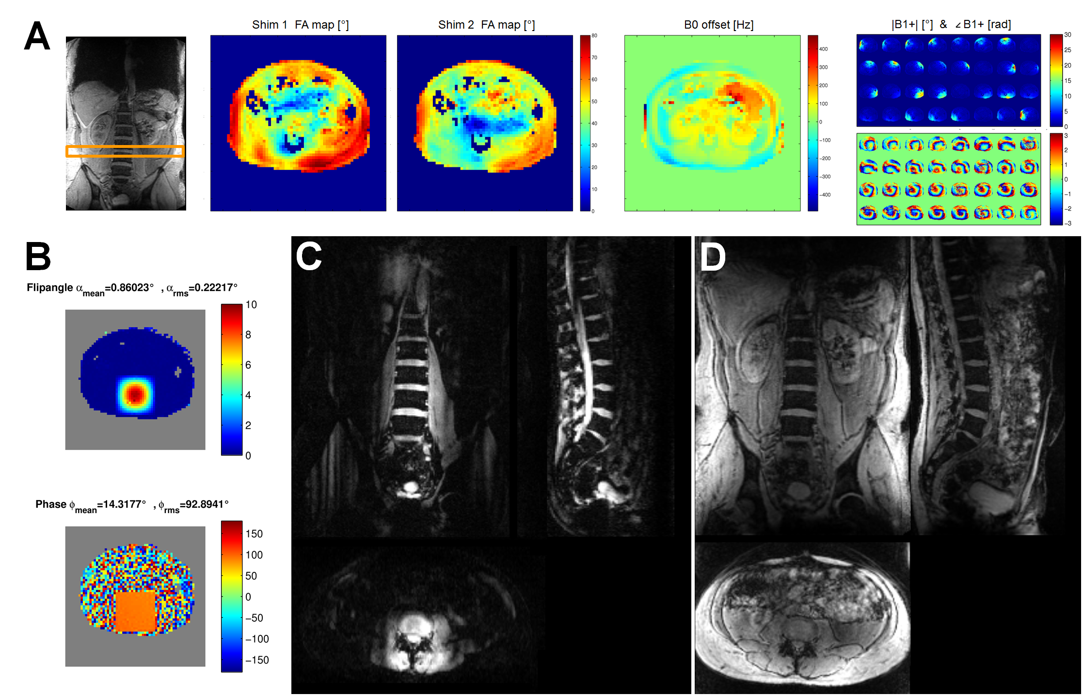
The aim of this work was to improve the recently developed B1TIAMO method at ultra-high magnetic field for a 32-channel body transceiver array by providing complete information about both the B0 and B1+ distribution within the human abdomen without movement artifacts by breathing. Therefore, a fast multi-slice version including two-echo B0 maps with time interleaved acquisition of modes (B01TIAMO) was introduced. Furthermore, B1+ phase calculation was improved by geometric-decomposition coil compression, resulting in accurate single-channel B1+ and B0 maps for three different slices within only 21s versus the 42s step-by-step measurement for a single slice with normal B1TIAMO.

Purpose

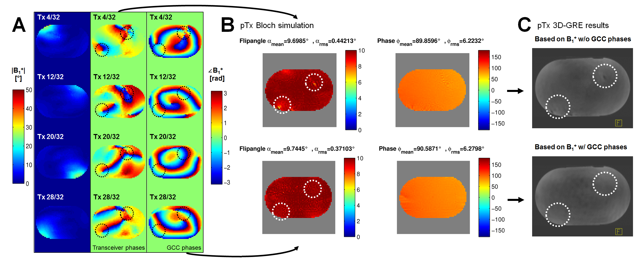
Parallel transmit (pTx) body imaging1,2 requires accurate knowledge of the individual transmit channel magnitude/phase distribution (B1+) over a large field-of-view supplemented by the local frequency offset of B0. For non-moving body regions, these datasets are often acquired over several minutes, which is not feasible for the human torso due to breathing and organ motion. Furthermore, B1+ mapping at ultra-high magnetic field is limited for such a large volume because a high dynamic flip angle (FA) range is necessary for absolute scaling. Hence, low local signal-to-noise ratios (SNR) additionally result in phase singularities due to normalization onto one transceiver (Tx/Rx) channel. Therefore, in this work the B1TIAMO technique3 for fast and accurate B1+ mapping of a 32-channel Tx/Rx array was expanded not only by the geometric-decomposition coil compression (GCC)4 post-processing, but a much faster joint multi-slice measurement together with time interleaved acquisition of modes5 B0 mapping was also included.

Methods

Data were acquired on a 7T ultrahigh field MRI system (Magnetom 7T, Siemens Healthcare, Erlangen, Germany) equipped with a custom-built 32-channel Tx/Rx remote body array6. The amplitudes and phases of the individual transmit channels were controlled by an add-on system including online SAR supervision7,8. Firstly, the B1TIAMO data for a single slice were calculated in a standard way by normalizing the phases onto one Tx/Rx channel9. For comparison, the relative data were separated by a twofold singular value decomposition including the creation of a virtual coil set by linear combinations of Rx over the phase-encoding direction while GCC and alignment was used to derive smooth single Tx phases. Both of these single-slice B1+ datasets allowed pTx pulse calculation for a homogeneous axial slice-excitation within phantom. Secondly, B1TIAMO was extended to acquire fast multi-slice B0/B1+ maps with parameters listed in Figure 1A, designated B01TIAMO. In general, for each of the turbo fast low angle shot (TFL) readouts, an additional cycling through all of the slices was introduced (see Figure 1B). The single-channel relative maps were not acquired step-by-step but in continuously alternated clockwise coil and slice cycling with an overall linear-ascending phase-encoding scheme. The B0 maps were calculated from a continuously alternated slice cycled centric-upward TIAMO TFL readout for a short asymmetric in-phase two-echo10 timing. The performance of the multi-slice sequence was validated for the two complementary shim setting FA maps against single-slice B1TIAMO in a 32-Liter human body phantom (T1=530ms; T2=300ms; εr=45; σ=0.55S/m). Finally, multi-slice axial FA, B0, and single-channel B1+ maps were acquired in breath hold (see Figure 1) and calculated for a healthy male volunteer (1.86m; 80kg) to compute an in-vivo pTx pulse excitation pattern.

Results

Figure 2 illustrates the effect of the GCC-calculated phase distribution on four exemplary single-channel B1+ maps. In contrast to the normal dataset with normalized transceiver phases, major deviations in the calculated magnitudes are absent for B1+ with GCC within the Bloch simulation. This prediction agrees well with the measurement results of the 2D spiral pTx excitation. As stated previously for the phantom, especially the effect of the prolonged centric-upward TFL readout train after one single FA encoding non-selective pre-saturation pulse for multiple slices was investigated for the complementary shims of B01TIAMO. The multi-slice results of each central slice show a general reduction of measured absolute FA compared to the values acquired from a single slice (see Figure 3). Within the phantom, the FAmean for 3/5/7 slices is 83.6%/82.3%/78.7% of FAmean for one slice. In-vivo the mean FA of 3/5 slices for the central slice of both shims amounts to 92.9%/92.4% of FAmean for one slice. In Figure 4 the complete B01TIAMO results for 3 axial slices of the kidney region acquired within 21 seconds are depicted, including B0 maps which show no artefacts due to insufficient SNR apart from air-filled cavities. Lastly, the feasibility of the improved B01TIAMO technique for an abdominal pTx application is demonstrated for the lower part of the spinal cord (see Figure 5).

Discussion and Conclusion

The overall B1+ data quality of the B1TIAMO technique could be improved by the GCC method. Moreover, for the purpose of pTx applications, the additional information from B0 mapping without significant total measurement time prolongation is recommended. For multi-slice B01TIAMO, the absolute FA map signal loss of 10-20%, depending on T1 time, has to be considered; hence, splitting up the acquisition into more pre-saturation blocks should be investigated to ameliorate the signal loss. However, this effect might be outweighed by the significant time gain for in-vivo B0/B1+ mapping. As an outlook, B0/B1+ information in three orthogonal directions over a large field-of-view would facilitate elaborate 3D abdominal pTx.

Acknowledgements

The research leading to these results has received funding from the European Research Council under the European Union's Seventh Framework Programme (FP/2007-2013) / ERC Grant Agreement n. 291903 MRexcite.

References

[1] Katscher U, Boernert P, Leussler C, et al. Transmit SENSE. (2003) MRM 49(1): 144-50.

[2] Grissom W, Yip CY, Zhang Z, et al. Spatial domain method for the design of RF pulses in multicoil parallel excitation. (2006) MRM 56(3): 620-9.

[3] Brunheim S, Orzada S, Gratz M et al. Combined B1 mapping with TIAMO for fast and accurate multi-channel RF shimming in 7 Tesla body MRI. (2016) Proc. Intl. Soc. Mag. Reson. Med. 16: p. 936.

[4] Zhang T, Pauly JM, Vasanawala SS, Lustig M. Coil compression for accelerated imaging with Cartesian sampling. (2013) MRM 69(2): 571-582.

[5] Orzada S, Maderwald S, Poser BA et al. RF excitation using time interleaved acquisition of modes (TIAMO) to address B1 inhomogeneity in high-field MRI. (2010) MRM 64(2): 327-333.

[6] Orzada S, Bitz AK, Kraff O et al. A 32-channel integrated body coil for 7 Tesla whole-body imaging. (2016) Proc. Intl. Soc. Mag. Reson. Med. 24: p. 167.

[7] Shooshtary S, Gratz M, Ladd ME, Solbach K. High-Speed RF Modulation System for 32 Parallel Transmission Channels at 7T. (2014) Proc. Intl. Soc. Mag. Reson. Med. 22: p. 544.

[8] Orzada S, Bitz AK, Solbach K, Ladd ME. A receive chain add-on for implementation of a 32-channel integrated Tx/Rx body coil and use of local receive arrays at 7 Tesla. (2015) Proc. Intl. Soc. Mag. Reson. Med. 23: p. 3134.

[9] Van de Moortele PF, Snyder C, DelaBarre et al. Calibration tools for RF shim at very high field with multiple element RF coils: From ultra fast local relative phase to absolute magnitude B1+ mapping. (2007) Proc. Intl. Soc. Mag. Reson. Med. 15: p. 1676.

[10] Robinson S, Grabner G, Witoszynskyj S, Trattnig S. Combining phase images from multi-channel RF coils using 3D phase offset maps derived from a dual-echo scan. (2011) MRM 65(6): 1638–1648.

Figures

Fig 1. A: B01TIAMO sequence parameters used for the large body phantom and for in-vivo measurements. For comparison, in the first column parameters for the time-consuming stepwise relative mapping together with B0 and two-mode FA maps with TIAMO TFL readout for one slice are also listed, resulting in a total acquisition time (TA) of 42 seconds. B: Each measurement includes an additional B0 mapping block with TIAMO TFL readout that was embedded into the B1TIAMO scheme. As an example the complete B01TIAMO sequence for three slices is depicted.

Fig 2. A: Four exemplary axial B1+ results for the body phantom. From left to right: Magnitude, corresponding transceiver phases normalized onto channel 32 (Tx 32) and GCC-derived transmit phases. Phase singularities originating from Tx 32 are marked with circles. B: The complete 32-channel information with transceiver phases (upper row) or GCC transmit phases (lower row) were employed for a Bloch simulation of a pTx pulse. Signal dropouts are visible when using transceiver phases. C: Experimental validation by a homogeneous non-slice-selective pTx excitation. Signal dropouts are visible when using transceiver phases (upper row), but not when using GCC (lower row).

Fig 3. A+B: B01TIAMO FA map comparisons in a central slice of the body phantom (upper rows) and in-vivo for the kidney region (lower rows) for two complementary RF shim modes. A: First RF shim mode FA distribution from left to right: for a single-slice acquisition, a combined measurement of 3 slices, 5 slices, and 7 slices (body phantom only). B: Second RF shim mode FA maps. The single-/multi-slice acquisition assignment of each central axial slice is color coded and shown on a sagittal/coronal image for the phantom/abdomen.

Fig 4. In-vivo multi-slice FA maps for two shim modes, B0 frequency offsets, and 32 single-channel B1+ maps acquired by B01TIAMO for the kidney region. The individual B1+ phase distributions were derived by the GCC method. A: Axial slice 1 for the upper part of the kidneys including the liver. B: Central slice 2 data corresponding to Figure 3. C: Slice 3 for the lower part of the kidneys.

Fig 5. A+B: The results of B01TIAMO for a single axial slice at intervertebral disc L3/4 were employed to calculate a non-slice-selective rectangular 2D spiral pTx excitation with a RF pulse length of 3.4ms for the spine. C: 3D reconstructed axial, sagittal and coronal slice of the spinal cord’s lower part during free breathing measured with the pTx pulse computed in (B) and gradient-echo readout with a preceding fat saturation pulse. D: Gradient-echo TIAMO images within breath hold for anatomical orientation which were reconstructed from coronal acquired slices.

Proc. Intl. Soc. Mag. Reson. Med. 25 (2017)
0521