0504
Combined tract-based analysis of diffusion fractional anisotropy and quantitative susceptibility mapping: a joint assessment of axonal and myelin microstructural changes in children with cerebral palsy1Brain Imaging and Analysis Center, Durham, NC, United States
Synopsis
In this report we developed a tract-based diffusion anisotropy and magnetic susceptibility analysis approach to jointly evaluate the potential mechanisms for axonal growth and myelin repair in children undergoing autologous cord blood stem cell therapy. Advancing from our prior findings that baseline brain connectivity is correlated with CP disease severity and that brain connectivity increase is correlated with functional motor improvement in CP patients, we provide further evidence that the increased brain connectivity may be the result of increased myelination of the affected neural pathways, in addition to the possibility of axonal regeneration.
Target Audience
Researchers interested in the use of quantitative susceptibility mapping (QSM) to study myelin specific white matter abnormalities.Introduction
Cerebral palsy (CP) originates from early brain injuries that lead to disrupted white matter (WM) pathways ,and it is the most prevalent debilitating motor disorder in children. Recent neuroimaging studies in children with cerebral palsy (CP) during autologous cord blood cell therapy have shown that the baseline brain connectivity is correlated with the disease severity (classified by the gross motor function classification system, GMFCS)1, and that the increase in brain connectivity is correlated with the recovery of motor function2. Furthermore, in CP children who improved beyond what is expected during normal brain development, the increased brain connectivity had a positive correlation with high cell dose. Thus it is hypothesized that cell therapies may play an important role in repairing neural pathways. Interestingly the recent literature has postulated that stem cells might replenish lost neurons in some circumstances, but act by paracrine mechanisms in others, protecting host cells, promoting endogenous cell growth and differentiation, or perhaps modulating the host immune response - all of which may decrease disability after CNS insults. Several studies have demonstrated that infusion of human umbilical cord blood cells helps reverse CNS hypoxic ischemic (HI) injury3,4. To gather further evidence to investigate the neural mechanism of this positive change in brain connectivity, we developed and carried out tract-based analysis of diffusion anisotropy as well as Quantitative Susceptibility Mapping (QSM) on newly unblinded treatment data to respectively study the potential mechanisms for axonal and myelin growth and repair following stem cell infusion. We anticipated that this combined analysis would provide further evidence to understand the neuronal origin of the increased brain connectivity, and help characterize the potential benefit of autologous cord blood cell therapy in children with CP.Methods
Diffusion tensor imaging (DTI) data were obtained (25 directions, b =1000+3b0,TE=70.5 ms, TR=12000 ms, 2 mm3 isotropic resolution). Fractional anisotropy (FA) was obtained from the DTI data. QSM images were acquired and derived from a 3D multi-echo FSPGR sequence (1 mm3 isotropic resolution, FOV = 192×192×120 mm3). The FA and QSM map were projected onto the cortico-spinal tract (CST), as the motor dysfunction of CP is specifically related to the damage in CST. The CST ROI was performed by warping the JHU-DTI-MNI “Eve” atlas template5 into each subject’s DTI image space via the ANTs6 tracked between precentral gyrus and brain stem. The projected QSM and FA values were compared between groups of high (GMFM improvement beyond that of brain development >10) versus low responders (GMFM improvement beyond that of brain development <5), where GMFM stands for Gross Motor Function Measures.
Results and Discussion
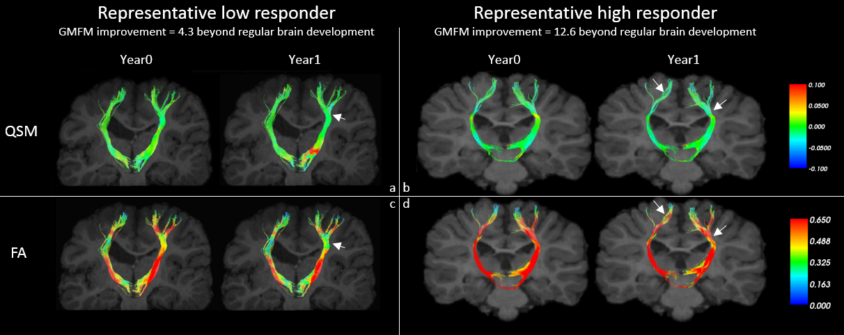
Shown in Fig. 1 are results from two representative subjects (a low responder with moderate motor improvement and no significant connectivity increase, and a high responder with significant motor improvement and connectivity increase), demonstrating corresponding increases in CST FA and diamagnetism (likely due to improved myelination) from year 0 to year 1 in the high responder.
As shown in our results, tract-based FA analysis allowed for quantitation of WM tracks connecting regions of interest in CP patients, while tract-based QSM analysis can be used to infer the degree of myelination of these axonal tracts7, which is integral to the transmission of impulses along functional neural pathways. Importantly, the fact that high responders with increased brain connectivity showed the local diamagnetic susceptibility change implies that improved myelination may be a plausible cause for the increased brain connectivity. This indicates that the function recovery as seen in the high responders does not necessarily indicate axonal regeneration from the stem cell infusion. Our findings support recent literature revealing cell-dosage-dependent positive outcomes in models of CNS injury, despite a paucity of donor cell engraftment into recipient brains.
Conclusion
We have developed a tract-based FA and QSM analysis approach and demonstrated its initial promise in providing a comprehensive assessment for both axonal and myelin microstructure. Further effort is underway to further carry out group-based correlational analysis to better characterize the impact of cell therapy on potential axonal growth and myelin repair and functional recovery.Acknowledgements
No acknowledgement found.References
[1] Englander, ZA, Pizoli, CE, Batrachenko, A, Sun, J, Worley, G, Mikati, MA, Kurtzberg, J, Song, AW, Diffuse reduction of white matter connectivity in cerebral palsy with specific vulnerability of long range fiber tracts, NeuroImage Clin. 2:440-7, 2013.
[2] Englander ZA, Sun J, Laura C, Mikati MA, Kurtzberg J, Song AW. Brain structural connectivity increases concurrent with functional improvement: evidence from diffusion tensor MRI in children with cerebral palsy during therapy. Neuroimage Clin. 7:315-324, 2015.
[3] Drobyshevsky A, Cotten CM, Shi Z, et al. Human Umbilical Cord Blood Cells Ameliorate Motor Deficits in Rabbits in a Cerebral Palsy Model. Developmental neuroscience. 2015.
[4] Kurtzberg J KB, Stephens C, Snyder EY. Umbilical cord blood cells engraft and differentiate in neural tissues after human transplantation. Biol Blood Marrow Transplant. 9:128a, 2003.
[5] Oishi, K et al. tlas-based whole brain white matter analysis using large deformation diffeomorphic metric mapping: application to normal elderly and Alzheimer's disease participants. NeuroImage. 46:486–499, 2009.
[6] Avants BB, Tustison NJ, Song G, Cook PA, Klein A, Gee JC. A reproducible evaluation of ANTs similarity metric performance in brain image registration. Neuroimage. 54(3):2033-2044, 2011.
[7] Argyridis, I, Li, W, Johnson, GA, Liu, C, Quantitative magnetic susceptibility of the developing mouse brain reveals microstructural changes in the white matter, NeuroImage 88: 134-142, 2014.
Figures