0495
Fast quantitative 31P MRI using prior information from 1H MRI1German Cancer Research Center (DKFZ), Heidelberg, Germany, 2University Hospital Erlangen, Erlangen, Germany
Synopsis
Phosphorus-containing biomolecules play a crucial role in the energy metabolism of the human body. Compared to hydrogen, the in vivo phosphorus MR signal is four orders of magnitude lower. In this study, a conventional gridding reconstruction applied on the phosphocreatine signal of the human calf was compared to a constraint iterative approach, which uses prior knowledge from hydrogen MRI data. For both reconstructions, different acquisition times were tested and phosphocreatine concentrations in the gastrocnemius and soleus muscles were estimated.
Purpose
Phosphorus (31P)-containing biomolecules play a crucial role in the energy metabolism of the human body. Compared to hydrogen (1H), the in vivo 31P MR signal is four orders of magnitude lower 1. In this study, a conventional gridding reconstruction applied on the phosphocreatine (PCr) signal of the human calf was compared to a constraint iterative approach, which uses prior knowledge from 1H MRI data 2. For both reconstructions, different acquisition times were tested and PCr concentrations in the gastrocnemius and soleus muscles were estimated.Methods
Calf muscles of three healthy volunteers (2 male, 1 female, 24-29 y/o,
BMI 19-25) were examined on a 7T whole body MR system (Magnetom 7T, Siemens
Healthcare) using a double-resonant (31P/1H) quadrature birdcage
coil (Rapid Biomed) with an inner diameter of 26cm. A 3D radially-sampled and
density-adapted 3 31P balanced steady-state
free-precession (bSSFP) sequence 4 (Fig. 1) was employed
with the following parameters: TR=7.21ms, TE1=2.34ms,
TE2=4.87ms, nominal α=30°, TRO=1ms, 1000 projections, A0=3.7mT/m, t0=0.5ms,
1cm isotropic resolution and 2, 4 ,8, 16, 32, 64 averages leading to
acquisition times of TA=14s, 29s, 58s, 2min, 4min, 8min. Due to the distinct
chemical shift of 31P-containing molecules, frequency selective
excitation was performed by a Gaussian RF pulse with a bandwidth of 3.5ppm (420Hz)
full width at half maximum (FWHM) to differentiate PCr 5. Six
reference tubes with a diameter of 2.8cm and 11.4cm length were filled with PCr
concentrations of 10, 20, 30, 40, 50, 60mM in distilled water and placed next
to the calf muscle in order to enable quantification of the 31P
signal. The in vivo concentrations of gastrocnemius and soleus muscles were
determined in both, gridding and iteratively reconstructed images, from a
linear fit to the data.
The signal-to-noise ratio (SNR) was enhanced by accumulating the magnitudes of the acquisitions from each
contrast (Fig. 2A-C). Additionally, the accumulated images were
oversampled 8 times and a Hamming filter was used to reduce Gibbs ringing and
to further increase the SNR for the gridding reconstructed images (Fig. 2D). To
represent the anatomy of the calf, 1H FLASH images
(TR=8.1ms, TE=4.9ms, nominal α=10°, TA=6min, BW=500Hz/px, 1mm isotropic
resolution) were acquired.
Furthermore, the 31P images were iteratively reconstructed (Fig.
2G) using prior knowledge in form of a binary mask (Fig. 2F) from 1H
data (Fig. 2E) 6. The binary mask M only comprises muscle
tissue in which PCr is expected. Hence, non-zero pixel intensities of the 31P
data outside the object, originating from noise or artifacts, are suppressed. Therefore,
the reconstructed image is obtained by minimizing the objective function $$f(\pmb{x})=\frac{1}{2}\parallel\pmb{A}\cdot\pmb{x}-\pmb{y}\parallel_{2}^{2}+\tau\parallel\pmb{M}\cdot\pmb{x}\parallel_{2}^{2} ,$$ where A denotes the system matrix, describing the imaging process
that maps the image vector x on the corresponding raw data
vector y. The regularization is weighted with a constant factor τ to enable manual adjustment. While the
first term ensures data consistency by including a squared L2-norm,
the second part of the objective function describes prior knowledge of the
image in terms of the regularization. The iterative reconstruction was
performed with 300 iterations and a weighting factor of τ=1 7.
Results
Fig. 3 illustrates 31P images obtained with the gridding and
iterative reconstructions. Images obtained by the gridding reconstruction
require a minimum acquisition time of 2min to yield appropriate quality,
whereas images reconstructed iteratively with the 1H MRI constraint
are applicable with an acquisition time of 29s.
Fig. 4 shows the signal-to-noise ratio (SNR) over the PCr concentration for different acquisition times. The
mean concentrations obtained for the calf muscles are 20% lower for the
iterative reconstruction and 35% for the gridding reconstruction compared to
published data (homogeneous CPCr=33±8mM 1).
Fig. 5 illustrates the superposition of anatomical 1H images
with physiological 31P acquisitions. While for gridding
reconstructed images tissue boundaries are blurred, the iterative
reconstruction emphasizes sharp tissue boundaries. Furthermore, partial volume effects as well as Gibbs ringing
artifacts are reduced with the constraint iterative approach.
Discussion
The PCr concentration estimated in the soleus compared to the gastrocnemius might be lower, since images were not corrected for B1-inhomogeneity. This also might result in a bias of the measured absolute concentrations. Furthermore, the voxel size is rather large causing partial volume effects, which could be reduced at tissue transitions by the iterative reconstruction.Conclusion
In this work, 31P/1H images of the human calf were examined with signal-efficient acquisition techniques and innovative reconstruction algorithms. The use of an iterative reconstruction implementing prior knowledge from 1H imaging allows for a reduction of the acquisition time by a factor of 4, while still yielding appropriate image quality, and thus enabling dynamic studies.Acknowledgements
This work was funded by the Helmholtz Alliance ICEMED - Imaging and Curing Environmental Metabolic Diseases, through the Initiative and Networking Fund of the Helmholtz Association.References
1 Parasoglou P, Xia D, Chang G, Regatte RR. 3D-mapping of
phosphocreatine concentration in the human calf muscle at 7T: Comparison to 3T:
Imaging of Phosphocreatine in the Human Calf Muscle at 3T and 7T. Magn. Reson.
Med. 2013;70:1619-1625. doi: 10.1002/mrm.24616.
2 Ajraoui S, Parra-Robles J, Wild JM. Incorporation of prior
knowledge in compressed sensing for faster acquisition of hyperpolarized gas
images. Magn. Reson. Med. 2013;69:360-369. doi: 10.1002/mrm.24252.
3
Nagel AM, Laun FB, Weber M-A, Matthies C, Semmler W, Schad
LR. Sodium MRI using a density-adapted 3D radial acquisition technique. Magn.
Reson. Med. 2009;62:1565-1573. doi: 10.1002/mrm.22157.
4 Johnson KM, Fain SB, Schiebler ML, Nagle S. Optimized 3D
ultrashort echo time pulmonary MRI. Magn. Reson. Med. 2013;70:1241-1250. doi:
10.1002/mrm.24570.
5 Rink K, Berger MC, Korzowski A, Breithaupt M, Biller A,
Bachert P, Nagel AM. Nuclear-Overhauser-enhanced MR imaging of 31P-containing
metabolites: multipoint-Dixon vs. frequency-selective excitation. Magn. Reson. Imaging 2015;33:1281-1289.
doi: 10.1016/j.mri.2015.07.017.
6 Gnahm C, Bock M, Bachert P, Semmler W,
Behl NGR, Nagel AM. Iterative 3D projection reconstruction of 23Na data
with an 1H MRI constraint. Magn. Reson. Med. 2014;71:1720-1732. doi:
10.1002/mrm.24827.
7 Rink K, Berger MC, Benkhedah N, Gnahm C, Bachert P, Nagel
AM. 31P MR imaging applying a radial bSSFP data acquisition and 1H constraint
iterative reconstruction. In Proc: ISMRM 2016, 3938.
Figures
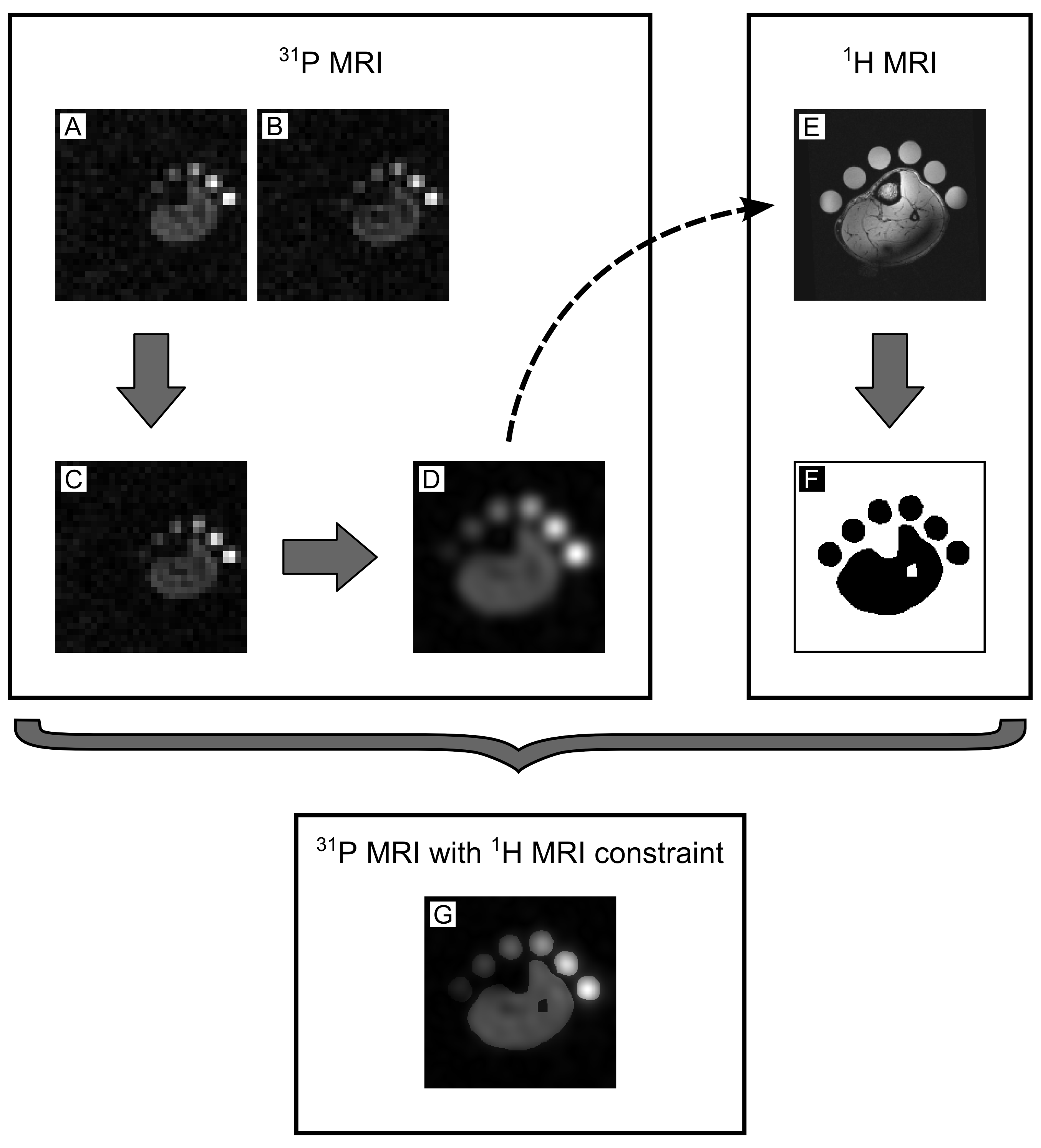
Fig. 2: Conceptual workflow to obtain iteratively reconstructed 31P
images:
(A) First contrast of the 31P acquisition (gridding reconstruction).
(B) Second contrast of the 31P acquisition (gridding reconstruction).
(C) Accumulated 31P image (gridding reconstruction) with a native
resolution of 1cm3 (matrix size 32×32×32px3).
(D) Gridding-reconstructed and 8 times oversampled 31P image (matrix
size 256×256×32px3) applying a Hamming filter.
(E) 1H FLASH acquisition utilized as anatomical reference and
registered on the oversampled 31P image.
(F) Binary mask employed as prior knowledge by serving as a 1H MRI
constraint.
(G) Iteratively reconstructed 31P image with 1H MRI
constraint.