0494
Multi-transmit proton MRI combined with high power body transmit and receive array for 7 Tesla phosphorus imaging1University Medical Center Utrecht, Utrecht, Netherlands, 2MRCoils, Zaltbommel, Netherlands, 3University of Oxford Centre for Clinical Magnetic Resonance Research (OCMR), Oxford, United Kingdom
Synopsis
We have integrated a body RF coil tuned at the 31P frequency outside the patient tube in a 7T MR system for homogeneous B1 transmit and combined it with a 8 channel 31P receive array for optimal sensitivity. Imaging and B0 shimming is done with radiative antennas integrated in the receiver setup. With this setup, large field of view imaging and broadband 31P MRSI with high SNR can be obtained.
Introduction
Clinical use of ultra-high field 31P MRSI is limited because surface transceivers with a small field of view and very inhomogeneous RF field are usually employed. To obtain a homogeneous flip angle distribution with such a setup, high SAR demanding adiabatic pulses are necessary, which is suboptimal for SNR. Here we show the use of a whole body coil for uniform RF 31P transmit at 7T, driven with a retuned 25kW RF amplifier and combined with an eight channel receive array for 31P MRSI. Imaging and shimming are done with a multi-transmit array of fractionated dipole antennas merged into the 31P receiver setup. The setup is tested with a large field of view on the legs of a healthy volunteer showing high quality proton and phosphorus imaging that can be obtained in the same setup within the same scan session.Methods
A detunable 24-rod birdcage phosphors body coil was designed with an inner diameter of 600 mm and a shield of 640 mm (MR Coils)1. The body coil is mounted around the patient tube of a 7tesla MRI system (Philips Healthcare) and is driven by a 3tesla 25KW RF amplifier (MKS China) tuned to the phosphors frequency at 7tesla (120.6MHz) via a quadrature hybrid. The 8-channel body-array receiver was taken from an obsolete 3T MRI system and retuned to 120.6MHz. In addition, four fractionated dipole antennas2 were merged to the array and driven with 4 of the 8 transmitters of the multi-transmit setup of the Philips system (Figure 1). B1+ calibration using a flip angle series (TR=10s) was obtained from a small sphere with Pi surrounded by a large phantom that mimicked the patient load to the coil. In-vivo proton MRI acquisitions were obtained from the legs of a healthy male volunteer. B1+ phase shimming was applied prior to obtain the T1 weighted MRI scans of the legs and image based B0 shimming was applied. 3D 31P MRSI was obtained with a flip angle of 20 degrees and TR of 150ms in a scan time of 7 minutes. Data of the receivers was combined using WSVD3. The spectra were then fitted using an open-source, Matlab spectroscopy fitting tool4.Results
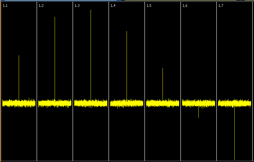
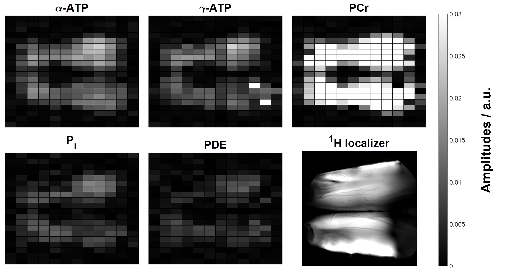
After replacing the 7T patient tube with the integrated 31P RF bodycoil, the same clear bore size was maintained and no effects of eddy currents, RF coupling to 1H RF coils, nor spikes were observed in the MRI scans run over the last month in our institute. Consequently, the body coil remains in the 7T system without compromising any scans. The flip angle series showed that 20kW of RF power was needed to obtain 16uT (Figure 2). With the Ernst angle of the MRSI of 20 degrees a pulse duration of 200us could be obtained. The 4 fractionated dipole antennas were sufficient to provide MRI at a large field of view as shown in figure 3. The reconstructed images of PCr, ATP, Pi, and PDE are well matched to the MRI.Discussion
The B1 efficiency and level of the body coil driven with 20kW of RF power matches closely to the B1 levels obtained at 3T from the same MRI vendor. Having the transmit coil integrated behind the covers of the MRI system significantly simplifies setting up 31P experiments. As the 1H transceiver elements could be integrated into the housing of a commercially available clinical 3T array, the setup for 1H and 31P MRI is as simple and patient comfortable as a traditional clinical MRI. The old 3T RF amplifier had to be tuned to the lower frequency of 120.6MHz compared to 128Mhz, which could be accomplished by adapting the inductors at the end stage of the amplifier. Combining multi transmit for 1H with 31P exams was essential for obtaining good background MRI and shimming capabilities.Conclusion
High power phosphorus body transmit was successfully combined with multi receiver arrays and multi-transmit array to obtain optimal proton and phosphorus MRI at large field of views. The integrated setup is useful for studies that require optimal metabolic MRI combined with good proton MRI.Acknowledgements
CTR is funded by the Wellcome Trust [098436/Z/12/Z]References
1. Whole-body radiofrequency coil for (31) P MRSI at 7 T. Löring J, van der Kemp WJ, Almujayyaz S, van Oorschot JW, Luijten PR, Klomp DW. NMR Biomed. 2016 Jun;29(6):709-20. doi: 0.1002/nbm.3517.
2. The fractionated dipole antenna: A new antenna for body imaging at 7 Tesla. Raaijmakers AJ, Italiaander M, Voogt IJ, Luijten PR, Hoogduin JM, Klomp DW, van den Berg CA. Magn Reson Med. 2016 Mar;75(3):1366-74. doi: 10.1002/mrm.25596.
3. Receive array magnetic resonance spectroscopy: Whitened singular value decomposition (WSVD) gives optimal Bayesian solution. Rodgers CT, Robson MD. Magn Reson Med. 2010 Apr;63(4):881-91. doi: 10.1002/mrm.22230.
4. Linewidth Constraints in Matlab AMARES using per-Metabolite T2 and per-Voxel Delta B0. Purvis LAB, Clarke WT, Biasiolli L, Robson MD, Rodgers CT. In the Proceedings of the 22rd Annual Meeting of ISMRM, Milan, Italy, 2014. p. 2885.
Figures
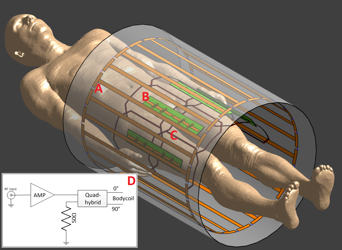
Figure 1: A: detunable 24-rod birdcage phosphors body coil, B: 4 fractionated dipole antennas, C: 8-channel body-array receiver, D: schematic of the RF amplifier combined with a quadrature hybrid.