0472
Anisotropy of inhomogeneous Magnetization Transfer (ihMT) in White Matter1Aix Marseille Univ, CNRS, CRMBM, Marseille, France, 2Division of MR Research, Beth Israel Deaconess Medical Center, Harvard Medical School, Boston, MA, United States
Synopsis
Inhomogeneous magnetization transfer (ihMT) is a new endogenous contrast mechanism that has been proposed for imaging myelinated tissues. The dipolar interaction underlying the ihMT effect is intrinsically anisotropic, exhibiting the well-known (3cos²θ -1) angle dependency. Here we report experimental evidence of the anisotropy of ihMT in white matter and we derive a realistic theoretical model combining the angular dependency of the myelin lineshape and dipolar-order RF saturation theory.
Introduction
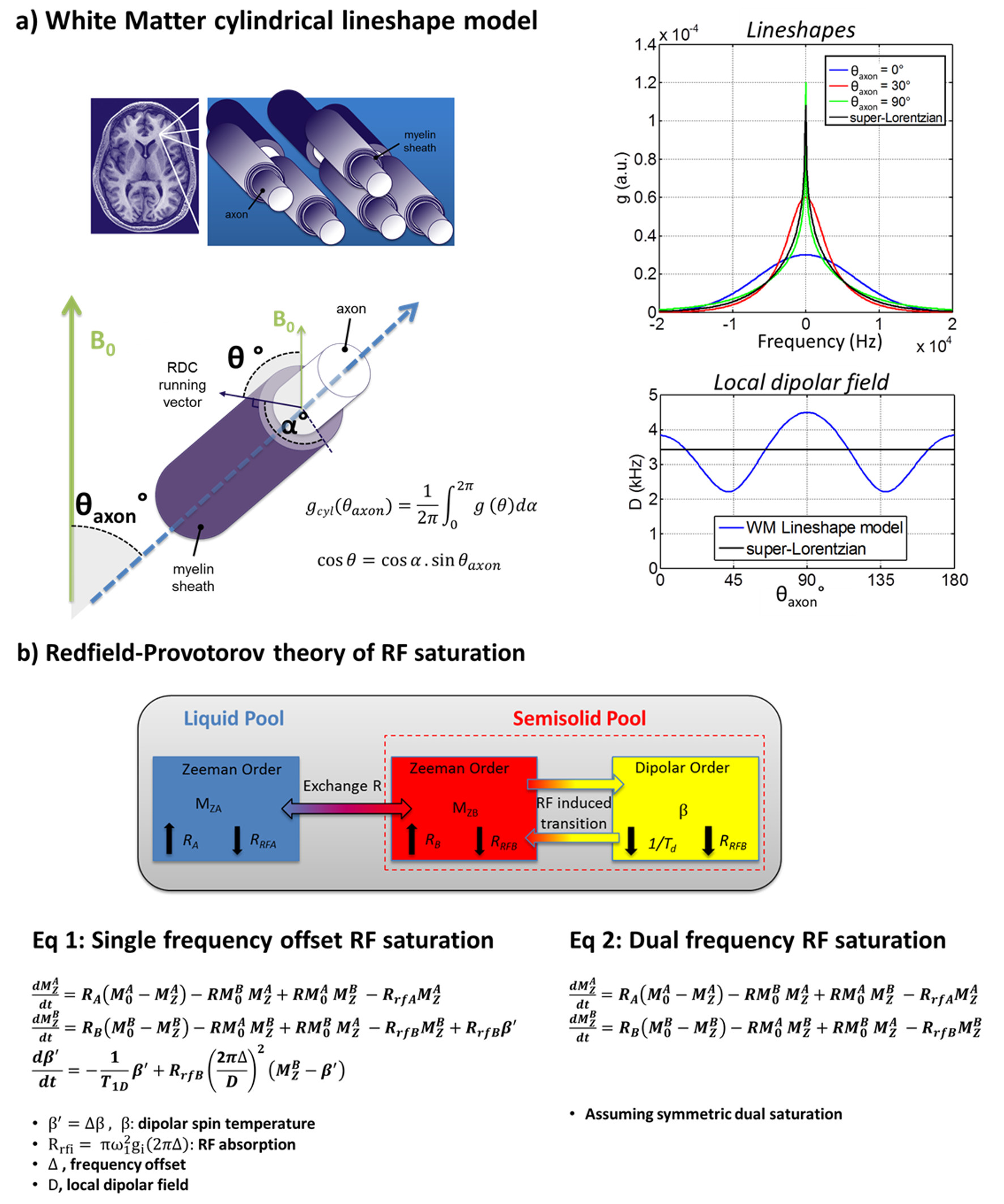
Inhomogeneous magnetization transfer (ihMT) is a new endogenous contrast mechanism that has been proposed for imaging myelinated tissues (1,2). It relates to inhomogeneous, asymmetric, RF saturation behavior of dipolar-broadened macromolecular lines (3). The underlying dipolar interaction is intrinsically anisotropic, exhibiting the well-known (3cos²θ -1) angle dependency, θ being the angle between the residual dipolar coupling vector and the static magnetic field B0 (Fig.1). Angular dependency of conventional MT parameters, mainly the macromolecular transverse relaxation time T2b, has been observed in white matter (WM) and described in terms of the underlying liquid crystalline lipid structure of myelin and the corresponding lineshape (4). Similar angular dependency would be expected for ihMTR because of its unique sensitivity to dipolar order, but have not yet been fully explored. In the current study we report experimental evidence of the anisotropy of ihMT in WM and we derive a realistic theoretical model combining the angular dependency of the myelin lineshape and dipolar-order RF saturation theory.Theory
IhMT is interpreted as a dipolar order effect and described according to the Redfield-Provotorov theory of RF saturation. The ihMT model combines thermodynamic models corresponding to the single- and dual- frequency offset saturation scenarios involved in ihMT detection (5) (Fig.1b). Here, following Pampel et al. (4), the cylindrical symmetry of the lipid myelin structure was considered to model the WM lineshape, gcyl (Fig.1a). The local dipolar field D, is related to the second moment of the lineshape gcyl, and is thus also angular dependent. IhMT ratio simulations were performed by numerical integration of Eq.1 and Eq.2 (Fig.1) assuming 2s continuous saturation, using Matlab (The MathWorks Inc., Natick, MA, USA), and calculated as 2x(MZA_Single- MZA_Dual)/M0. Myelinated WM fibers were first assumed to be mono-directional. Significant angular dispersion of WM bundles was then introduced by averaging the simulation results ± δ° around the nominal θaxon value (Fig.1)Materials and Methods
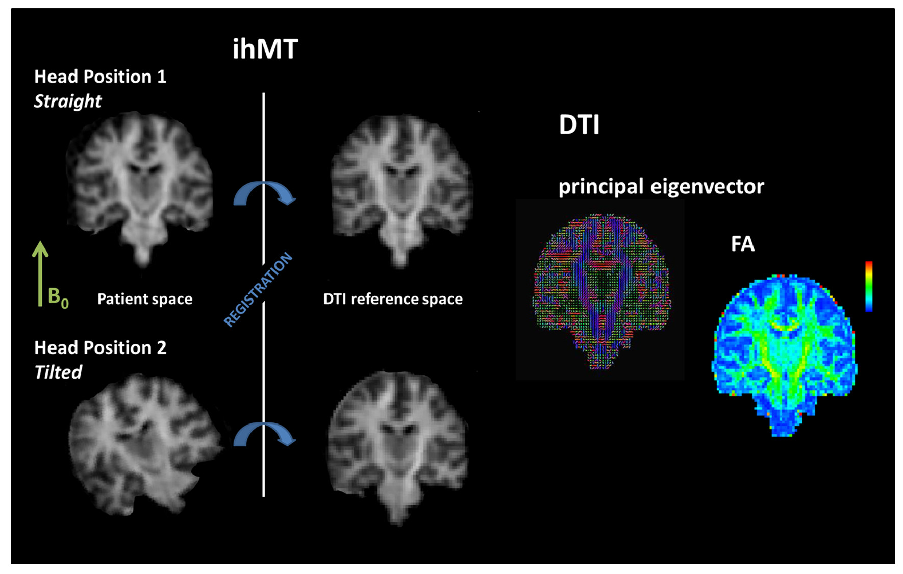
Clinical experiments were performed on a whole body 1.5T MRI system (Avanto, Siemens Healthcare, Erlangen, Germany) and a 12-channel receive-only head coil to evaluate sensitivity of ihMT to the WM fiber orientation. Whole brain ihMT imaging was performed using a 3D ihMT-GRE sequence, consisting of short train of saturation pulses interleaved with gradient echo readouts (6) (6 Hann-shaped pulses, pulsewidth 0.5ms, pulse repetition time 1ms, B1rms = 4.1μT, Δf=±7kHz, TR/TE=19/4.8ms). The main WM fiber orientation θaxon was measured as the principal DTI eigenvector orientation with 64 directions and b = 1000s/mm². The angular dependency of ihMT was assessed by repeating the same ihMT protocol before and after titling the subject’s head from B0 axis by approximately 25° (Fig.2).
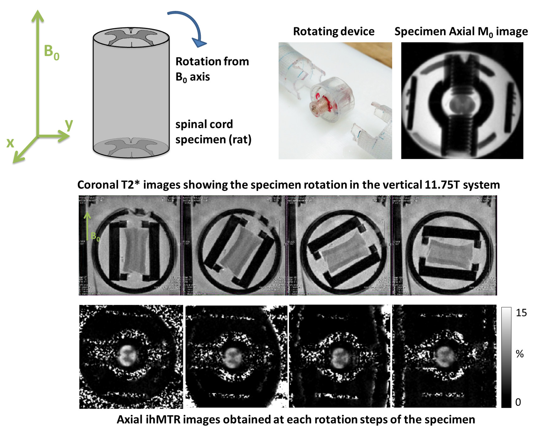
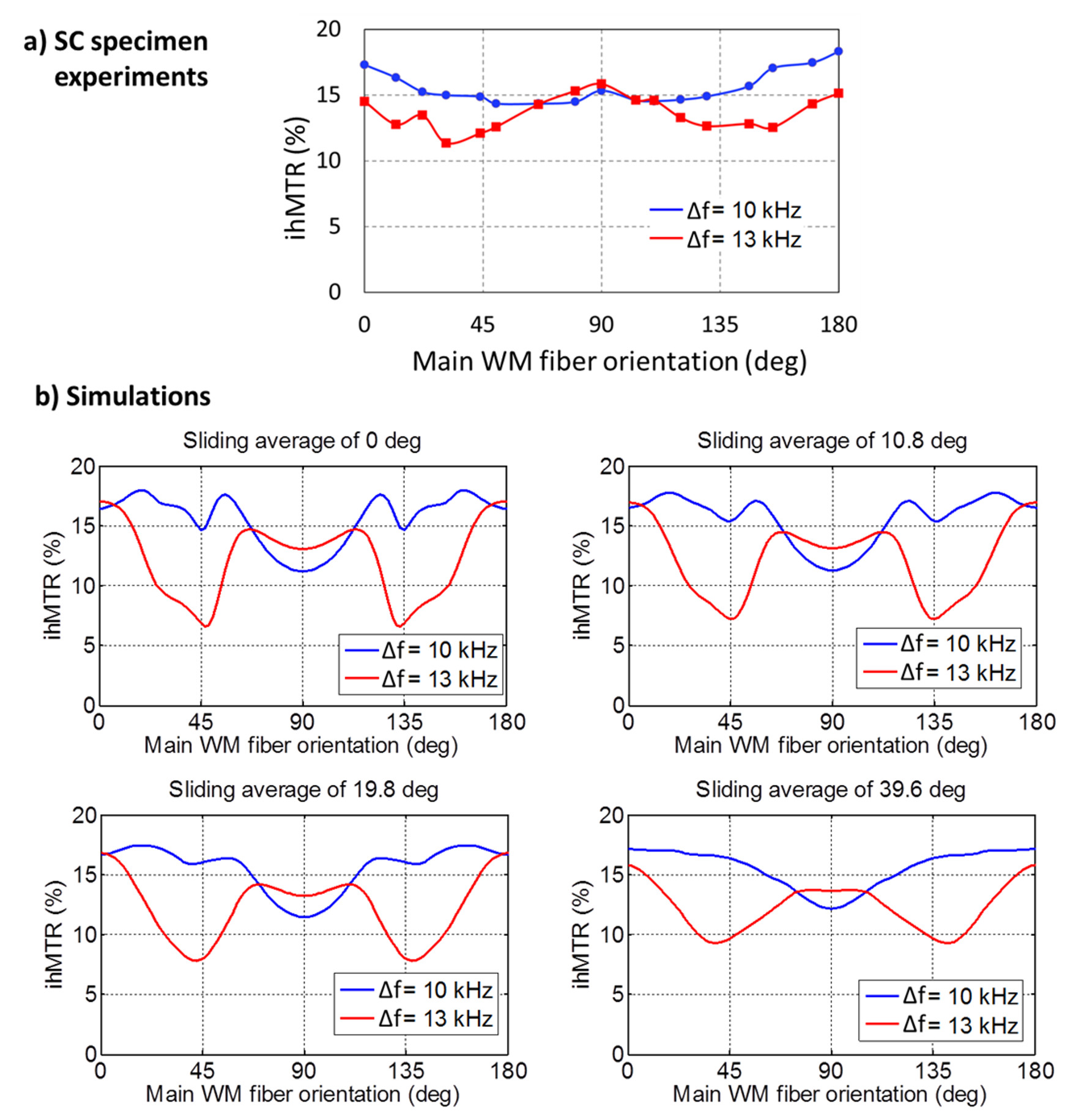
Ex vivo experiments were performed on a rat spinal cord (SC) specimen at 11.75T (Bruker 500 vertical system). The specimen was placed in a custom MRI compatible rotating device (Fig.4) allowing free rotation around the X-axis. Axial ihMT prepared HASTE acquisitions were acquired using a pulsed ihMT approach (1ms Hann-shaped pulses, repeated every time 1.3ms, B1rms = 6.6μT during 0.9s, Δf=±10kHz and ±13kHz) for a rotation range [0-180°] starting from the SC aligned with B0. IhMTR was measured in WM areas of the SC.
Results and Discussion
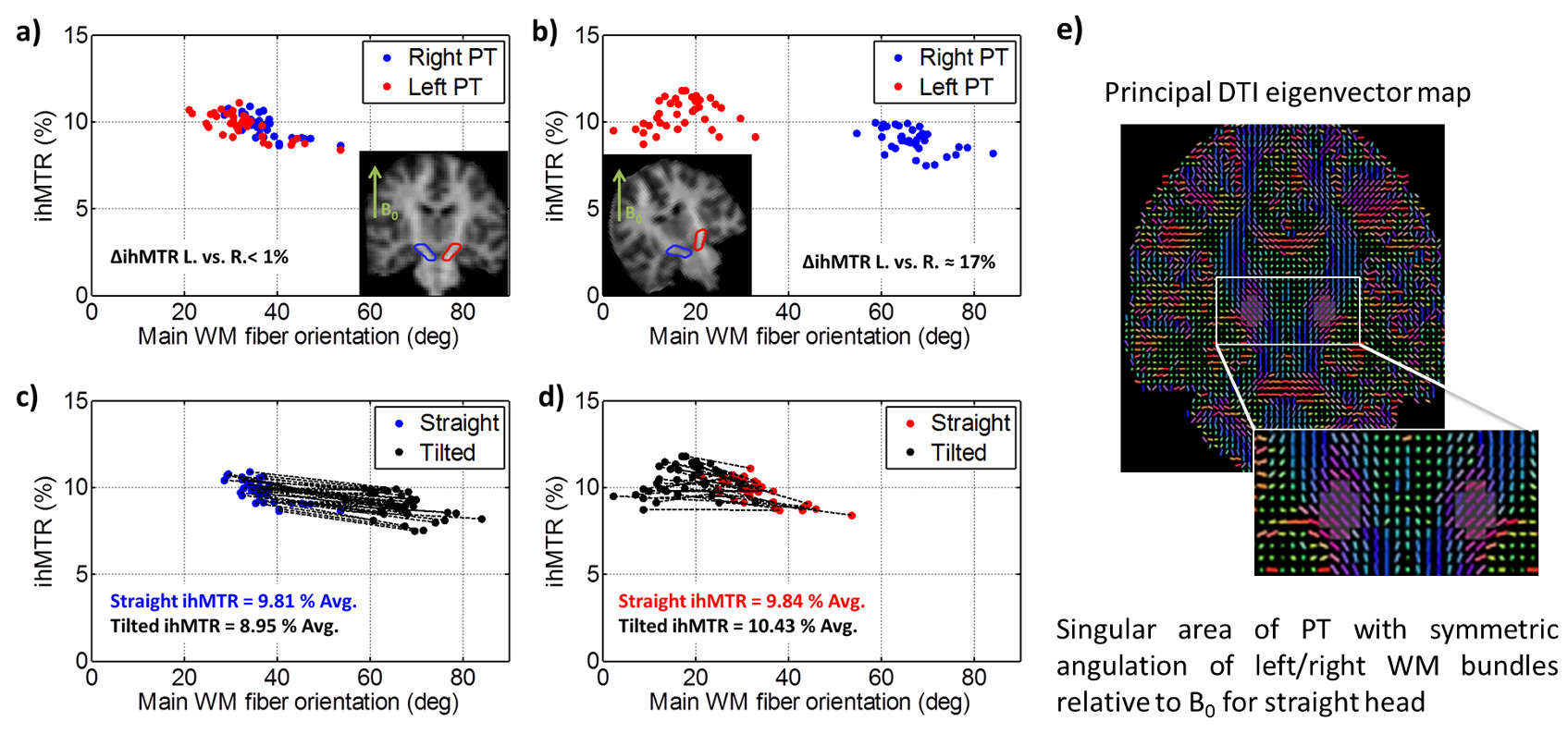
Fig. 2 illustrates the in vivo human protocol with both straight- and tilted- head configurations. The resulting ihMT ratios measured in a singular area of Pyramidal Tract (PT) are reported on Fig. 3 as a function of the main WM fiber orientation. For the straight head configuration, both right and left PT areas have similar orientation and ihMTR measured in these areas are equal within 1% margin (Fig.3a). Conversely the tilted head configuration reveals relative difference of ihMTR between right and left area of about 17% (Fig.3b), further demonstrating the sensitivity of ihMT for WM anisotropy. Fig. 5a shows the ihMTR of the SC specimen as a function of the main WM fiber orientation and confirmed the anisotropic behavior of ihMTR. IhMTR simulation accounting for the angular dependency of the myelin lineshape (Fig. 5b) demonstrates relatively sharp features characteristic of WM orientation that are partially smoothed out when averaging over angular dispersion. Good agreement with experiment was obtained for dispersion in the range of ± 10° to ± 20°.Conclusion
IhMT anisotropy has been explored in human WM and in a SC specimen. For human applications noticeable effects have been observed. Although these may not bias ihMT metrics when subject positioning within the magnet is carefully maintained, it may complicate absolute comparison of ihMTR measured in WM tracts exhibiting different orientations, such as PT and Corpus Callosum. SC specimen experiments confirmed the ihMT anisotropy effect and ihMT modeling properly account for the observed effects.Acknowledgements
This study was supported by the ARSEP foundation 2015. V.H.P. received support from A*MIDEX grant (n°ANR-11-IDEX-0001-02) funded by the French Government “Investissements d’Avenir” program. The authors thank Pr. M. Guye, E. Soulier, P. Viout, L. Pini and V. Gimenez for volunteer management.References
1. Varma G, Duhamel G, de Bazelaire C, Alsop DC. Magnetization Transfer from Inhomogeneously Broadened Lines: A Potential Marker for Myelin. Magn. Reson. Med. 2015;73:614–622.
2. Girard OM, Prevost VH, Varma G, Cozzone PJ, Alsop DC, Duhamel G. Magnetization transfer from inhomogeneously broadened lines (ihMT): Experimental optimization of saturation parameters for human brain imaging at 1.5 Tesla. Magn. Reson. Med. 2015;73:2111–2121.
3. Korb J-P, Maruani J. Pulse saturation behavior of inhomogeneously broadened lines. I. Slow spectral diffusion case. Phys. Rev. B 1981;23:971–975.
4. Pampel A, Müller DK, Anwander A, Marschner H, Möller HE. Orientation dependence of magnetization transfer parameters in human white matter. NeuroImage 2015;114:136–146.
5. Varma G, Girard OM, Prevost VH, Grant AK, Duhamel G, Alsop DC. Interpretation of magnetization transfer from inhomogeneously broadened lines (ihMT) in tissues as a dipolar order effect within motion restricted molecules. J. Magn. Reson. 2015;260:67–76.
6. Girard OM, Le Troter A, Varma G, Prevost VH, Guye M, Ranjeva J-P, Alsop DC, Duhamel G. Whole Brain inhomogeneous MT using an ihMT prepared 3D GRE sequence at 1.5T. In: Proceedings of the ISMRM. Toronto, Canada; 2015. p. 3356.
Figures