0471
Combining inhomogenous Magnetization Transfer (ihMT) with multi-point Dixon (mDixon) for myelin imaging with efficient fat suppression1Radiology, University of Texas Southwestern Medical Center, Dallas, TX, United States, 2Advanced Imaging Research Center, University of Texas Southwestern Medical Center, Dallas, TX, United States, 3Philips Healthcare, Gainesville, FL, United States, 4Radiology, Division of MR Research, Beth Israel Deaconess Medical Center, Harvard Medical School, Boston, MA, United States
Synopsis
Inhomogeneous
magnetization transfer (ihMT) imaging is a novel enhanced magnetization
transfer technique which has recently been applied in human brain and spinal
cord. Spinal cord applications of ihMT can especially benefit from a robust fat
suppression to help with reducing of the strong fat signal from the large voxel
size used by this method. Here we introduce a pulsed ihMT-prepared 3D SPGR
sequence with multi-echo Dixon acquisition for a robust fat suppression of the
ihMT images. The ihMT multi-echo Dixon method is shown to provide an excellent fat
and water separation without a compromise of the observable ihMT effect.
Introduction
Inhomogeneous magnetization transfer (ihMT) imaging is an enhanced magnetization transfer technique, which employs subtraction of images obtained with dual off-resonance frequency saturation (alternating positive and negative), as opposed to the conventional technique of a single off-resonance frequency saturation1,2,3. This method has been shown to produce a higher white/gray matter contrast compared to conventional MT methods. This contrast is thought to be originating from dipolar order effects in myelinated tissues4,5,6. The ihMT method has been applied in human brain and cervical spinal cord1,2,4,7,8. Spinal cord applications of ihMT can especially benefit from robust fat suppression to help with reducing of the strong fat signal from the large voxel size used by this method. Saturation-based fat suppression methods are however not practical for ihMT because they can create unwanted MT effects, as well as an increase in scan time and specific absorption rate (SAR). The multi-echo Dixon method on the other hand can provide separate fat and water images without inducing unwanted MT effects9. In this study, we propose a 3D gradient echo based ihMT sequence with multi-echo Dixon acquisition, which allows for a robust acquisition of ihMT in regions proximal to fatty tissue.Methods
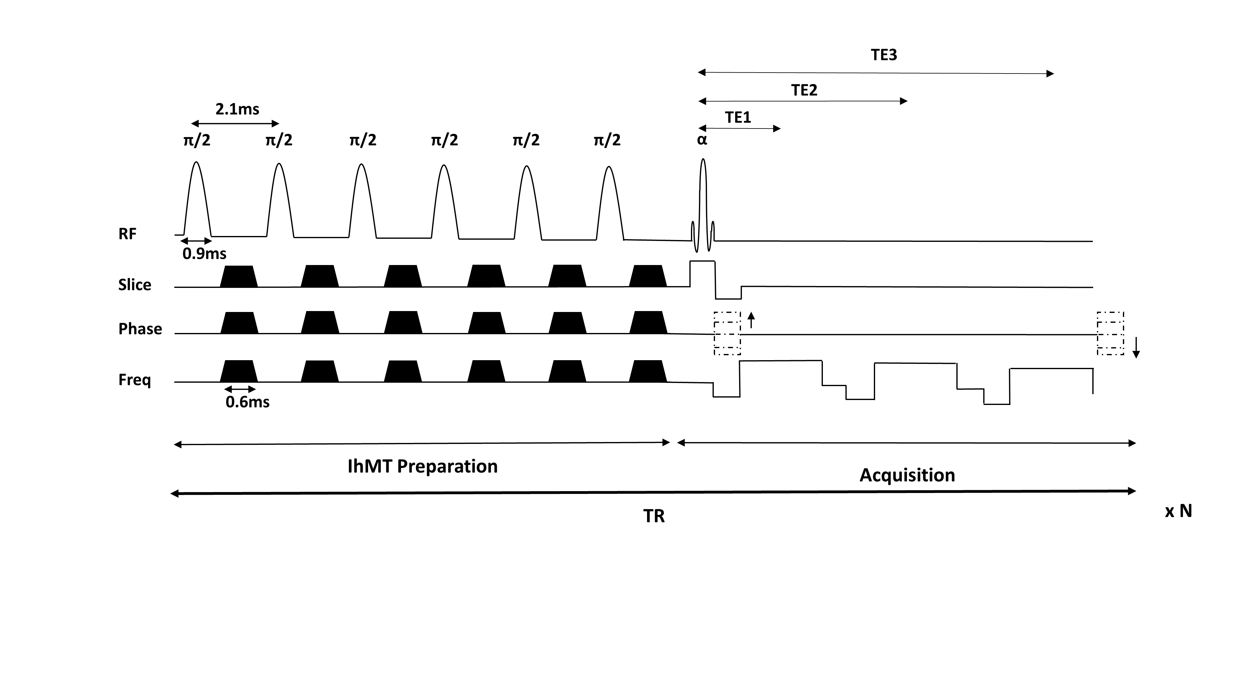
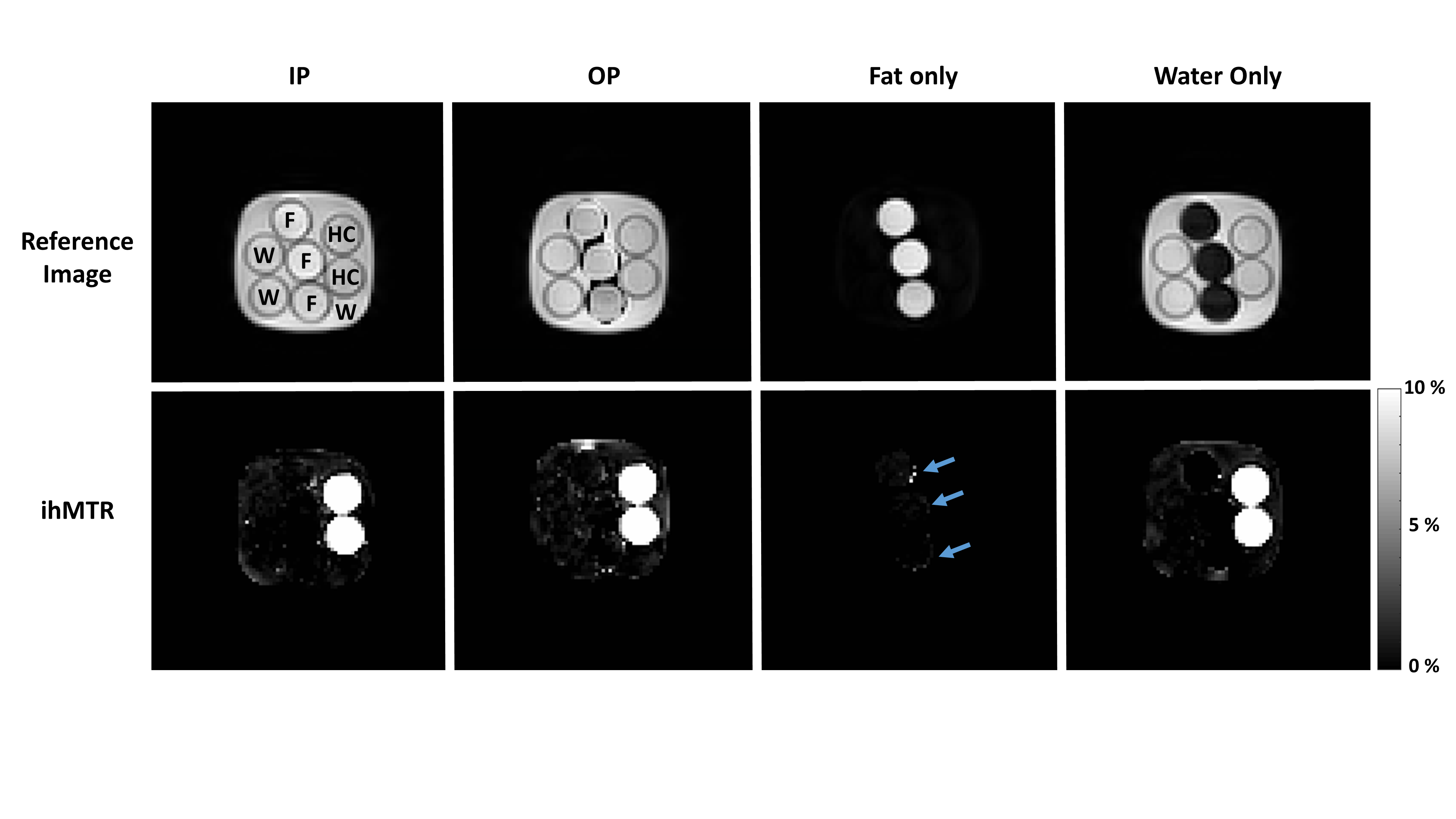
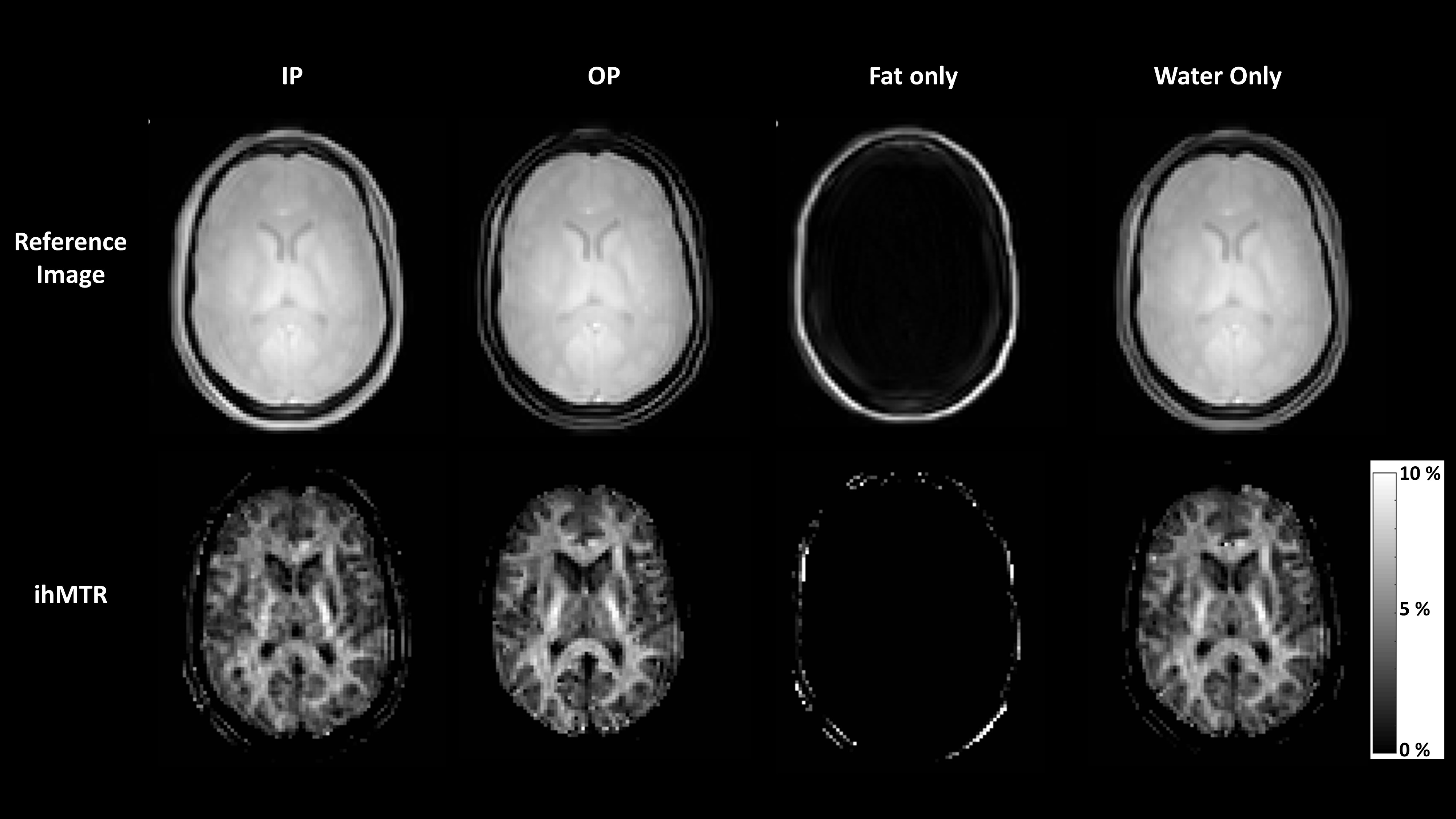
All experiments were performed on a 3T Ingenia Philips MRI scanner (Philips Healthcare, Best, The Netherlands) equipped with a multitransmit body coil and a 32-channel receive head-coil. The sequence was first tested on a phantom consisting of water (W), oil (F) and commercial hair conditioner (HC) (to mimic the lamellar structure of myelinated tissues1). The sequence was then applied on the brain of two healthy volunteers (all female, age: 26.0 ± 1.4 years). The study adhered to the local Institutional Review Board guidelines and a written informed consent was obtained from the volunteers. 3D ihMT SPGR mDixon acquisition: A pulsed ihMT-prepared 3D spoiled gradient echo (SPGR) sequence was applied with three-point Dixon acquisition. The ihMT preparation was repeated for each repetition time and employed 6 Hann-shaped RF pulses (pulse duration = 0.9 ms, flip angle = 90°, frequency offset (Δf) = ±7 kHz, RF phase cycling of 117°), each followed by a gap of 0.3 ms, dephasing gradients of 0.6 ms duration in 3 directions (slice, phase, frequency) and another gap of 0.3 ms, as shown in Figure 1. A reference image (M0) and 4 separate ihMT images (frequency offsets: +7 kHz (MT+), -7 kHz (MT-), alternating ±7 kHz (MT+-) and alternating -+7kHz (MT-+)) were obtained per echo. The imaging parameters for the three-point Dixon SPGR were the following: flip angle (α)=10°, TR/TE/ΔTE=70/1.5/1.0ms, unipolar echo acquisition, FOV=220x220x50mm3, resolution=2.5x2.5mm2, slice thickness=5mm and acquisition time=2 min 33 s. Post-processing: For all experiments, in-phase (IP), out-phase (OP), fat-only (F) and water-only (W) images were reconstructed by the standard manufacturer on-line processing. The ihMT ratio (ihMTR) was calculated for IP, OP, F, and W conditions separately, according to the following equation: ihMTR =(MT+ + MT- - MT+- - MT-+)/M0. Regions of interest (ROIs) were chosen from the HC phantom and from the white matter of human volunteers to compare the ihMTR obtained from IP, OP and water images.Results and Discussion
Figures 2 and 3 show excellent fat and water separation on the reference (M0) images of phantom and human brain. As expected, the HC phantom appears only on ihMTR images from IP, OP, and W reconstructions (Figure 2). No fat signal is observed in the ihMTR water-only image in the phantom. Some residual ihMTR is observed on the ihMTR fat-only image in voxels along fat-water interfaces (shown with blue arrows on Figure 2). Mean ihMTR values calculated from an ROI within the HC phantom were very similar for IP, OP, and W reconstructions (Table in Figure 4). As expected, the mean ihMTR values calculated from the ROIs within the human brain white matter were similar between IP, OP, and W conditions due to the lack of fatty tissue in human brain (Figure 4). The mean ihMTR values are in line with the white matter ihMTR values from reported in the literature10-11.Conclusions
A pulsed ihMT-prepared 3D SPGR sequence with multi-echo Dixon acquisition is feasible and results in robust fat suppression of the ihMT images without any increase in scan time or SAR, and without a compromise of the observable ihMT effect.Acknowledgements
No acknowledgement found.References
1. Varma, G., et al. Magnetization transfer from inhomogeneously broadened lines: A potential marker for myelin. Magn. Reson. Med. 2015; 73: 614–622.
2. Girard, O. M., et al. Magnetization transfer from inhomogeneously broadened lines (ihMT): Experimental optimization of saturation parameters for human brain imaging at 1.5 Tesla. Magn. Reson. Med. 2015; 73: 2111–2121.
3. Swanson S. D., et al. Molecular, dynamic, and structural origin of inhomogeneous magnetization transfer in lipid membranes. Magn. Reson. Med. 2016. DOI:10.1002/mrm.26210.
4. Varma, G. et al. Interpretation of magnetization transfer from inhomogeneously broadened lines (ihMT) in tissues as a dipolar order effect within motion restricted molecules. J. Magn. Reson. 2015; 260: 67–76.
5. Duhamel et al. in Proceedings of ISMRM 2016: p2909.
6. Manning et al. in Proceedings of ISMRM 2016: p305.
7. Girard O.M., et al. Magnetization transfer from inhomogeneously broadened lines (ihMT): improved imaging strategy for spinal cord applications Magn. Reson. Med. 2016. DOI:10.1002/mrm.26134
8. Taso M., et al., Tract-specific and age-related variations of the spinal cord microstructure: a multi-parametric MRI study using diffusion tensor imaging (DTI) and inhomogeneous magnetization transfer (ihMT), NMR in Biomedicine, 2016; 29: 817-832.
9. Smith A.K., et al., Incorporating dixon multi-echo fat water separation for novel quantitative magnetization transfer of the human optic nerve in vivo. Magn. Reson. Med. 2016. DOI:10.1002/mrm.26164
10. Varma et al. in Proceedings of ISMRM 2013: p4224.
11. Varma et al. in Proceedings of ISMRM 2014: p3334.
Figures