0465
Deactivation responses induced by acupuncture are associated with changes in GABA concentrations: a BOLD fMRI and MEGA-PRESS 1H-MRS Study1Guang An Men Hospital, China Academy of Chinese Medical Sciences, Beijing, People's Republic of China, 2Peking Union Hospital, Peking Union Medical University, Beijing, People's Republic of China, 3Beijing Institute of Technology, Beijing, People's Republic of China, 4Siemens Healthcare, MR Collaboration, CA, United States, 5Siemens Healthcare, MR Collaboration NE Asia, Beijing, People's Republic of China
Synopsis
This study investigated the neurological mechanism that mediates the deactivation of the medial prefrontal cortex induced by acupuncture. Gamma-aminobutyric acid (GABA) concentrations before and immediately after acupuncture stimulation in healthy volunteers were assessed with MEGA-PRESS 1H-MRS. We found that GABA concentrations in the medial prefrontal cortex decreased significantly after acupuncture stimulation compared with control stimulation with Von Frey sensory. Analysis of the task-functional magnetic resonance image (fMRI) acquired during acupuncture showed deactivation in the same area. These results demonstrated that the decreased blood oxygenation level dependent (BOLD) response induced by acupuncture was associated with inhibitory effects.
PURPOSE
Previous fMRI studies have found that the medial prefrontal cortex showed deactivation during or after acupuncture; however, the underlying neuromechanism was not elucidated. This study aimed to investigate the neurological basis of the acupuncture-evoked deactivation of the medial prefrontal cortex with assessments of gamma-aminobutyric acid (GABA) concentrations before and after acupuncture, and also to analyze the blood oxygenation level dependent (BOLD) responses during acupuncture.METHODS
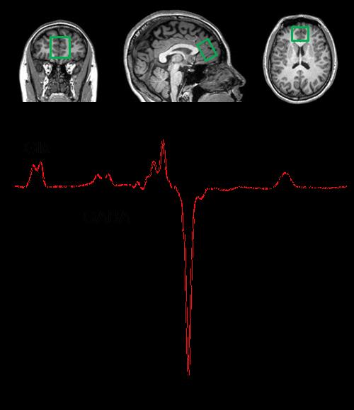
This study was enrolled forty healthy adult volunteers. Each subject received three blocks of stimuli during the acupuncture procedure with asteelness needle (diameter 0.30mm, Huatuo, China) inserted into the right LI4 (Point Hegu) acupoint. Each block of stimuli included a 30-second task (needle rotating, 1Hz) period, and the subsequent block of stimuli was separated by a 120-second interval (needle in place) from the previous one. For control purposes, the volunteers received von Frey sensory stimulation on the surface of the right LI4 acupoint on the other day in a week. The same licensed acupuncturist administered the acupuncture and Von Frey sensory throughout the study. MR data were collected on a MAGNETOM Skyra 3T MR scanner (Siemens, Erlangen, Germany) with a 20-channel head-neck coil. The MRI exam included MPRAGE for localization and a prototype MEGA-PRESS MR spectroscopy (MRS) sequence. The MRS scan was performed in the bilateral medial prefrontal cortex before the task and immediately after the acupuncture stimulus (Fig. 1). The sequence parameters of MEGA-PRESS were as follows: voxel size = 35 x 30 x 25 mm3 (F >> H, R >> L, A >> P), TR = 2000 ms, TE = 68 ms, averages = 128, edit pulse frequency 1.9 ppm, edit center frequency 4.7 ppm. During the acupuncture stimulus, BOLD signals were acquired with the following parameters: TR = 750 ms, TE = 30 ms, flip angle = 90°, 43 slices, slice thickness = 3 mm, distance factor = 0%, FOV = 210 ×210 mm2, matrix = 70×70. The MRS data were analyzed with the LCModel. BOLD data was processed by SPM8 with a standard task fMRI post-processing pipeline. The results of BOLD fMRI were shown in t-values (p < 0.05, cluster size > 5). Activation was defined as an average t-value greater than 3 within the ROI, and deactivation, less than -3.RESULTS
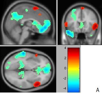
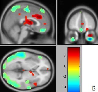
We analyzed the t-map of each subject and found that out of the 40 subjects, 27 showed strong deactivation (t < -3), 12 showed activation, and one showed no signal change in the medial prefrontal cortex during the acupuncture stimulus. In the 27 subjects who showed deactivation, GABA concentration decreased in the same ROI, and the average decrease measured 0.00165 ± 0.00444 mmol/L. In contrast, the average GABA concentration was 0.00063±0.00326 mmol/L in paired controls (p = 0.031). Furthermore, 20 of the subjects who showed deactivation during acupuncture also showed deactivation during control stimulation. A decrease in GABA concentration was observed in the same region, which measured 0.00104±0.00390 mmol/L. The comparison between the two groups was significant (p =0.036).DISCUSSION
The medial prefrontal cortex, a part of the default mode network, is a key area for mood regulation including ACC . Acupuncture is thought to have inhibitory effects in this region as previous studies have found a predominantly negative BOLD response to acupuncture; thus, acupuncture can be effectively used as a treatment for analgesia, mood modulation in insomnia, depression, and anxiety. In this study, we observed a main trend of decreased GABA concentration that was associated with a negative BOLD response for acupuncture. These results suggested that acupuncture-evoked deactivation was mediated by GABAergic inhibitory interneurons.CONCLUSION
The negative BOLD response in the medial prefrontal cortex induced by acupuncture may be associated with neural inhibition effects.Acknowledgements
This study was funded by the National Natural Science Foundation of China (Grant 81273674 and Grant 81303056).References
1. Kathleen K.S. Hui, Ovidiu Marina, Jiliang Fang ,Kenneth K. Kwong, and Bruce R. Rosen.et al. (2009): Acupuncture mobilizes the brain’s default mode and its anticorrelated network in healthy subjects. Brain Research. 1287:84-103.
2. Jiliang Fang, Zhen Jin, Yin Wang, Kathleen K.S. Hui,et al. (2009): The salient characteristics of the central effects of acupuncture needling: limbic–paralimbic–neocortical network modulation. Hum. Brain Mapp. 30 (4), 1196–1206.
3. Georg N, Martin W, Rolf FS,et al(2007): GABA concentrations in the human anterior cingulate cortex predict negative BOLD responses in fMRI. Nature Science.10(12):1515-1517.
Figures