0454
Field Map Combination Method for Multiple-Acquisition bSSFP1Electrical Engineering, Stanford University, Stanford, CA, United States
Synopsis
FIESTA-C/CISS enables the reconstruction of banding-free bSSFP images, but residual ripple remains in the combined images. Also, in the heart, flow near a stop-band may cause a component image to include significant contributions from out-of-slice spins; this hyper-intense signal persists in images combined using conventional methods. Here, we use a B0 map to combine FIESTA-C/CISS cardiac cine images. Knowledge of B0 enables us to only include pass-band signal in the final image and exclude stop- and bright-bands. In phantom and in vivo studies, the proposed method had less ripple and greatly reduced flow artifacts compared to maximum-intensity projection and root-sum-of-squares.
Purpose
Multiple-acquisition balanced steady-state free precession (bSSFP) enables the reconstruction of bSSFP images without signal nulls in the presence of off-resonance. However, the resulting combined images have residual ripple.1 In addition, if multiple-acquisition bSSFP is used in the heart, through-plane flow near a phase-cycle’s stop-band may cause that component image to include significant contributions from out-of-slice spins;2 these regions of hyper-intense signal persist in the maximum-intensity projection (MIP) or root-sum-of-squares (SOS) combined images. Other combination methods partially mitigate flow artifacts, but require at least four component images.3 This doubles the scan time relative to FIESTA-C/CISS, which acquires two.4 In this work, we illustrate the feasibility of using a field map to combine cardiac cine datasets with only two phase-cycles to achieve good ripple reduction and flow-artifact suppression with limited scan-time penalty.Methods
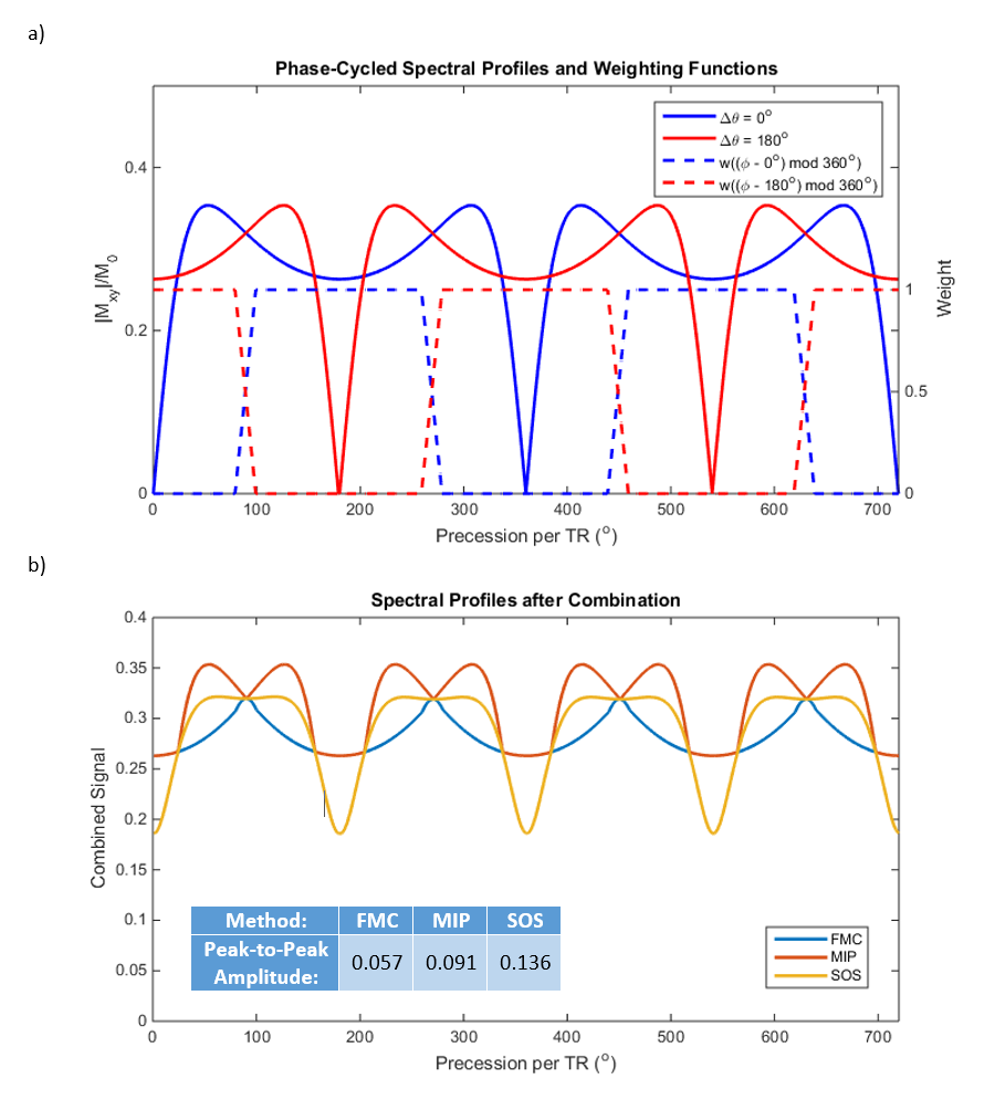
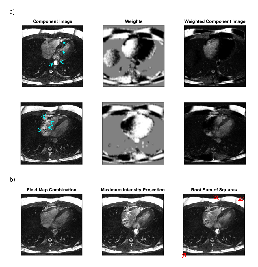
The magnitude of the bSSFP signal for flip angle α, precession per TR φ, and RF phase-cycling Δθ can be approximated by $$$\|M_{xy}\|/M_0 = (\cot{(\beta/2)} + T1/T2 \cdot \tan{(\beta/2)})^{-1}$$$ where $$$\beta = 2\arctan{(\tan{(\alpha/2)}/\cos{((\phi - \Delta\theta - 180)/2)})}$$$.5 For T1/T2 = 2 and α = 35o, 0o and 180o phase-cycling result in the spectral profiles in Figure 1(a). If the main field map is known, $$$\phi = \Delta B_0 \cdot TR$$$ and, based on the voxel’s φ, the phase-cycled components can be weighted to emphasize pass-band signal and deemphasize stop-band and bright-band signal. Since the most severe flow artifacts occur near the stop-band,2 we hypothesized that such a field map combination method would substantially reduce contributions from out-of-slice spins in the final reconstructed image.
The combined image mc is calculated as $$m_c(x, y) = \frac{\sum_{n = 1} ^ {N} \|m_n(x, y)\|w((\phi(x, y)-\Delta\theta_n) \mod 360^o)}{\sum_{n = 1} ^ {N} w((\phi(x, y)-\Delta\theta_n) \mod 360^o)}$$ where N is the number of phase-cycles, mn is the nth component image, and w is a weighting function. We used a trapezoidal weighting function $$$w(\phi) = rect((\phi-180)/180) * rect(\phi/20)$$$ to smoothly merge the pass-bands of the two phase-cycles while avoiding as much of the stop- and bright-bands as possible (Figure 1). In regions where the intensity was less than 10% of the maximum magnitude, the magnitude mean of the component images was used instead of relying on the noisy field map estimate.
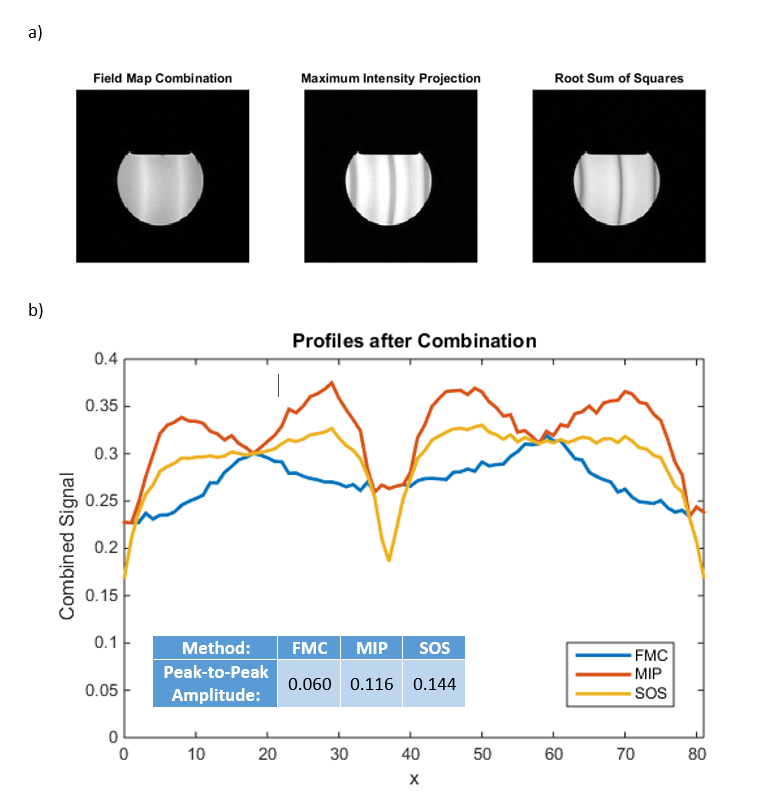
The proposed method was compared with root-sum-of-squares and MIP. FIESTA-C/CISS cine images of a phantom and four volunteers were acquired at 1.5 T with 60o nominal flip angle, 3.4 ms TR, 1.4 ms TE, 24x24 cm2 FOV, 1.25x1.43 mm2 resolution, and 6-8 mm slice thickness. With effective acceleration of R=2, the temporal resolution was 40.8 ms. The X or Y linear gradients were deliberately offset from the optimal shim to create increased off-resonance.
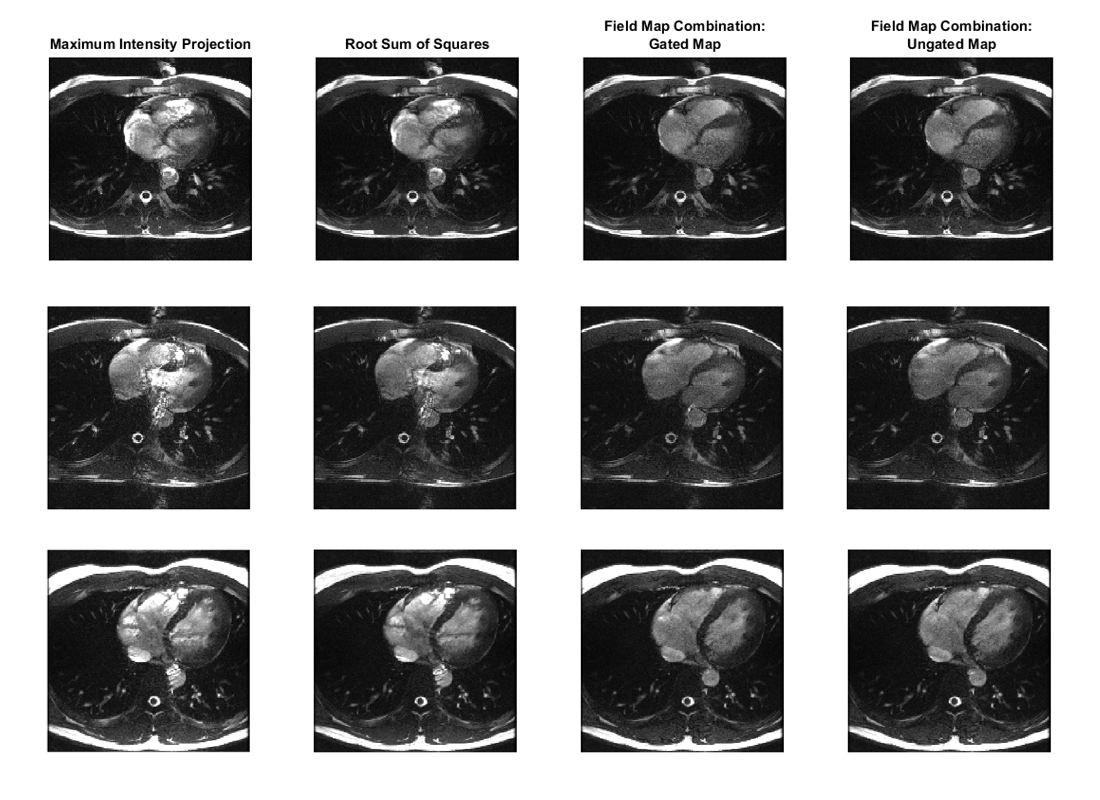
Breath-held field maps were acquired during diastole with a flow-compensated GRE sequence with 20o nominal flip angle, 10.0 ms TR, 4.0 ms TE1, 1.7 ms ΔTE, and matched spatial parameters. In addition, free-breathing, ungated field maps were acquired to assess if the resulting field-map-combined bSSFP images are comparable to those reconstructed using the breath-held, gated maps. The ungated field map acquisition was ~2 s, while the phase-cycled cine was ~16 s and the gated field mapping sequence was ~14 s.
Results
The peak-to-peak amplitude of the residual ripple is smaller with field map combination than with MIP or SOS for both the theoretical spectral profiles (Figure 1(b)) and phantom data (Figure 2). This suggests that the method achieves good banding reduction.
In vivo, the weights based on the B0 map mask out the flow artifacts in the component images (Figure 3). As a result, for all subjects imaged, the field map combinations have substantially reduced hyper-intense regions and transient-related artifacts relative to the MIP and SOS combinations (Figure 4). Furthermore, the reconstructions using free-breathing, ungated maps are similar to those using breath-held, cardiac-gated maps.
Discussion and Conclusion
We provided proof-of-concept of a combination method for multiple-acquisition bSSFP that, unlike most existing methods, is intended to exclude signal outside of the pass-band, even if it is bright. Simulation, phantom, and in vivo results indicate that using a main field map to combine two phase-cycles yields less residual ripple and greatly suppresses flow artifacts relative to using the FIESTA-C/CISS data alone. For cardiac cine imaging, field map combination is feasible with minimal extra scan-time and no additional breath-holding since free-breathing, ungated field maps can be used. In addition, this suggests that the method is relatively insensitive to cardiac and respiratory motion, although the potential for artifacts from misregistration should be investigated. It may also be possible to acquire the field map during the cine scan itself, e.g., using BMART.6Acknowledgements
We would like to thank the Fannie and John Hertz Foundation, NSF Graduate Research Fellowship Program, and GE Healthcare for their support.References
1. Bangerter NK, Hargreaves BA, Vasanawala SS, Pauly JM, Gold GE, Nishimura DG. Analysis of multiple-acquisition SSFP. Magn Reson Med 2004;51:1038–1047.
2. Markl M, Pelc NJ. On flow effects in balanced steady-state free precession imaging: pictorial description, parameter dependence, and clinical implications. J Magn Reson Imag 2004;20:697-705.
3. Fischer A, Hoff MN, Ghedin P, Brau ACS. Banding-artifact free bSSFP cine imaging using a Geometric Solution approach. In Proceedings of the 24th Annual Meeting of ISMRM, Singapore, 2016. 1827.
4. Chavhan GB, Babyn PS, Jankharia BG, Cheng HL, Shroff MM. Steady-state MR imaging sequences: physics, classification, and clinical applications. RadioGraphics 2008;28(4):1147–1160.
5. Zun Z, Nayak KS. Graphical derivation of the steady-state magnetization in balanced SSFP MRI. In Proceedings of the 14th Annual Meeting of ISMRM, Seattle, Washington, USA, 2006. 2410.
6. Baron CA, Nishimura DG. B0 mapping using rewinding trajectories. Magn Reson Med 2016. doi: 10.1002/mrm.26391.
Figures