0448
Real-time 3D cardiac MRI using through-time radial GRAPPA and GPU-enabled reconstruction pipelines in the Gadgetron framework1Biomedical Engineering, Case Western Reserve University, Cleveland, OH, United States, 2School of Medicine, Case Western Reserve University, Cleveland, OH, 3Radiology, University Hospitals, Cleveland, OH
Synopsis
An accelerated 3D cardiac dataset (8 partitions; in-plane spatial resolution: 2.34 x 2.34 mm2; partition thickness = 8 mm) can be reconstructed using through-time radial GRAPPA and GPU-enabled pipelines in the Gadgetron framework in approximately 515.5 ms. This work may enable real-time, 3D imaging in the heart with on-line reconstruction.
Purpose
MR-guided interventional procedures often rely on 2D real-time imaging to visualize anatomy and devices. Imaging guidance may include 2 or 3 slices of different views to represent the 3D anatomy. Real-time 2D images and tracked device positions may also be overlaid on 3D volumes acquired before a procedure. However, it may be difficult with these 2D methods to view target tissues or devices that move in and out of the imaging planes, and to view the relative positions of different tissues and devices that are not in the same plane. Three-dimensional real-time imaging may enable continuous visualization of all anatomies within the field of view, and provide current relative positions of target tissues and devices, even if positioned in different slices at the same time.
Non-Cartesian data acquisition coupled with parallel imaging methods such as through-time GRAPPA have been shown to enable data acquisition that is fast enough for real-time cardiac imaging1,2. Additionally, graphics cards have been used to speed up image reconstruction to be as fast as data acquisition, particularly for 2D imaging using radial GRAPPA3. This work combines through-time radial GRAPPA and GPU-enabled reconstruction pipelines for real-time data collection and reconstruction of 3D cardiac images.
Methods
Experiments were performed on a 3T scanner (Skyra, Siemens Medical Solutions, Erlangen, Germany). A radial FLASH sequence was used to collect a 3D short-axis volume centered over the ventricles with the following parameters: 8 partitions/volume; 25% partition oversampling; partition thickness = 8 mm; field-of-view = 300 x 300 mm2; matrix size = 128 x 128; in-plane resolution = 2.34 x 2.34 mm2; flip angle = 5°; TR/TE = 3.22/0.73 ms; bandwidth = 1115 Hz/pixel; number of coils = 34 using the spine array and a body array. An acceleration factor of 9 (16/144 radial projections) was tested, leading to an acquisition time of 515.2 ms per 3D volume. Twenty fully-sampled calibration volumes were collected for the radial GRAPPA reconstruction prior to collection of the accelerated dataset.
Reconstruction was performed offline using the Gadgetron framework4. The computer used to perform the reconstructions has a 4GB Nvidia Quadro K4200 graphics card; an 8 core, 2.4GHz Intel Xeon E5-2630 processor; and 32GB (4 x 8GB) of 2133MHz DDR4 RAM. Two pipelines were constructed in C++ and CUDA. The first calculates the non-Cartesian GRAPPA weights using through-time, through-partition data for the calibration on the CPU. Eight repetitions and 10 partitions were used in this work with a through-time GRAPPA segment size of 8 readout points and 1 projection. The second pipeline accepts the undersampled data, loads the saved GRAPPA weights to the GPU, and performs GRAPPA reconstruction on the entire volume. The NUFFT was used to grid the data using a GPU-enabled toolbox that is part of the Gadgetron framework5. A sum-of-squares coil combination was used. A coil compression step was added to the beginning of each pipeline to reduce the number of coils to 12.
Results
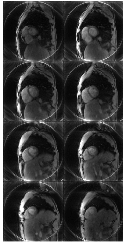
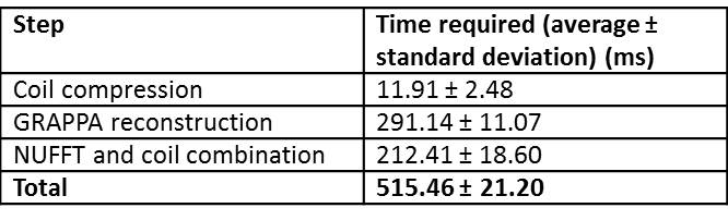
A reconstructed volume is shown in Figure 1. Data for one undersampled volume was acquired in approximately 515.2 ms. Data reconstruction was performed in approximately 515.5 ms per volume. The time required to perform the most time-consuming steps in the reconstruction process are shown in Table 1. The remaining steps, such as buffering data, were difficult to time on the Gadgetron but are negligible in comparison to the listed steps.Discussion
This real-time reconstruction framework can be used to collect a full volume of data in one heartbeat, and to reconstruct that volume before completing acquisition of the following volume in the next heartbeat. Although reconstruction is not yet as fast as data acquisition, it can be performed in a single heartbeat, before the next volume of data is available for reconstruction. Through-time radial GRAPPA is well-suited to parallelized reconstructions because each unacquired data point is estimated independently of other points, the method is not iterative, and it does not rely on temporal sparsity (which would require the entire dataset to be collected before reconstruction can begin). Once a weight set has been calculated, the same weight set can be used for multiple repetitions as long as the volume position and orientation do not change. Future work will include installing this framework at the scanner for on-line reconstruction. This setup will also allow evaluation of overhead time required to transfer data and images between the scanner and a reconstruction computer.Conclusion
Real-time data acquisition and reconstruction of 3D cardiac images is possible using through-time radial GRAPPA and GPU-enabled reconstruction pipelines in the Gadgetron framework.Acknowledgements
Siemens Healthcare, R01EB018108, NSF 1563805, R01DK098503, and R01HL094557.References
1. Wright K, Lee G, Ehses P, et al. Three-dimensional through-time radial GRAPPA for renal MR angiography. J Magn Reson Imaging. 2014;40(4):864-874.
2. Barkauskas K, Rajiah P, Ashwath R, et al. Quantification of left ventricular functional parameter values using 3D spiral bSSFP and through-time non-Cartesian GRAPPA. J Cardiovasc Magn Reson. 2014;16(1).
3. Saybasili H, Herzka D, Seiberlich N, et al. Real-time imaging with radial GRAPPA: Implementation on a heterogeneous architecture for low-latency reconstructions. Magn Reson Imaging. 2014;32(6):747-58.
4. Hansen M, Sorensen T. Gadgetron: An open source framework for medical image reconstruction. Magn Reson Med. 2013;69(6):1768-1776.
5. Sorensen T, Schaeffter T, Noe K, et al. Accelerating the nonequispaced fast Fourier transform on commodity graphics hardware. Med Imaging, IEEE Trans. 2008;27(4):538-547.
Figures