0397
One-year aerobic exercise increases regional cerebral blood flow in anterior cingulate cortex: a blinded, randomized trial in patients with amnestic Mild Cognitive Impairment1Advanced Imaging Research Center, UT Southwestern Medical Center, Dallas, TX, United States, 2Institute for Exercise and Environmental Medicine, Texas Health Presbyterian Hospital, Dallas, TX, United States, 3Department of Neurology and Neurotherapeutic, UT Southwestern Medical Center, Dallas, TX, United States, 4Department of Psychiatry, UT Southwestern Medical Center, Dallas, TX, United States, 5Department of Internal Medicine, UT Southwestern Medical Center, Dallas, TX, United States, 6Department of Radiology & Radiological Science, Johns Hopkins University School of Medicine, Baltimore, MD, United States
Synopsis
Amnestic mild cognitive impairment (MCI) represents the early stage of Alzheimer’s disease (AD). Much research has focused on preventing the inevitable decline of MCI to AD. Aerobic exercise is considered a viable choice, and is shown to improve cognitive function in MCI. We focus on understanding the mechanisms that lead to this improvement. Pseudo-continuous-arterial-spin-labeling (PCASL) was used to assess resting cerebral blood flow (CBF) in two MCI groups. One group performed aerobic exercise, while another non-aerobic stretching. CBF was measured before and after training. CBF increase in the anterior-cingulate-cortex (ACC) was the proven mechanism that improves cognitive function in MCI.
Purpose
Individuals with amnestic mild cognitive impairment (MCI) are at the early stage of Alzheimer’s disease (AD), and have a high risk of following an inevitable cognitive and physical decline to AD. Much research in the past decade has focused on preventing this inexorable decline to AD, but disappointing negative outcomes from several clinical trials of amyloid vaccines has led us to research other alternatives. Aerobic exercise may be a low cost and effective approach for AD prevention. Exercise is shown to improve cardiorespiratory fitness and cognitive function in older adults with and without[1-3] MCI[4-6]. The mechanisms that lead to beneficial effects of aerobic exercise training on neurocognitive function is not well understood, and is the topic of this investigation. We hypothesized that aerobic exercise improves cerebrovascular function, which leads to brain function improvement. We used Pseudo-Continuous-Arterial-Spin-Labeling (PCASL) MRI to measure resting cerebral blood flow (CBF) in MCI volunteers. Non-aerobic forms of exercise such as stretching, was used as control to compare the beneficial effects of aerobic exercise.Methods
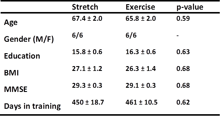
24 MCI participants were recruited and randomly assigned to perform either aerobic exercise, or non-aerobic stretching. Informed consent was obtained using IRB-approved protocol. Both groups were age, gender, education, BMI, and clinically matched (Table1). The aerobic exercise group was allowed to perform aerobic exercise and was trained to maintain their heart rate within a prescribed range, calculated using maximal oxygen uptake (VO2max) test, done on a treadmill[7]. The stretch group performed a stretch and balance routine that focused on their upper and lower body, and were trained to maintain their heart rate below prescribed range. Both groups began training at a frequency of 3 sessions per week, 30 minutes per session, and intensity was gradually increased over time. Experiment: Resting CBF was measured in a Philips 3T MRI scanner, using a PCASL sequence with parameters: TR/TE=4300/14ms, label duration=1650ms, post label delay=1525ms, 29, 5mm thick slices, duration 5min 40s. CBF was measured in all participants at the start and end of the training. Data processing: CBF maps were generated using a perfusion kinetic model[8]. For each participant, the CBF map before training was subtracted from that after training (Post-Pre), to obtain a difference map. The resulting CBF maps were then compared between groups to assess changes in CBF due to aerobic exercise compared to stretch (Exercise>Stretch). Cardiorespiratory fitness and cognitive function were also assessed in all participants.Results and Discussion
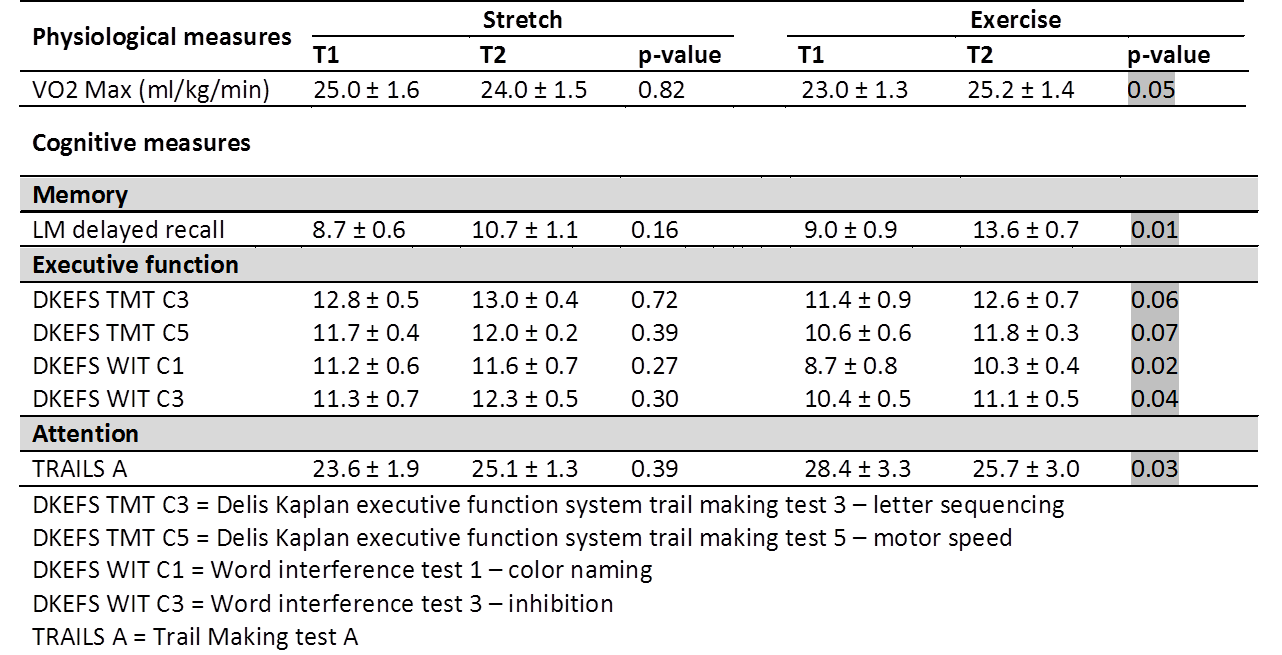
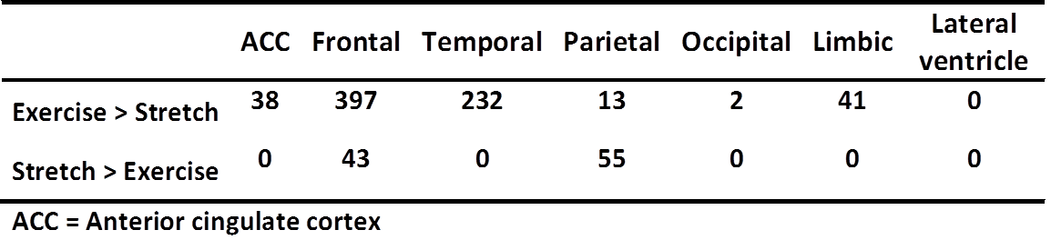
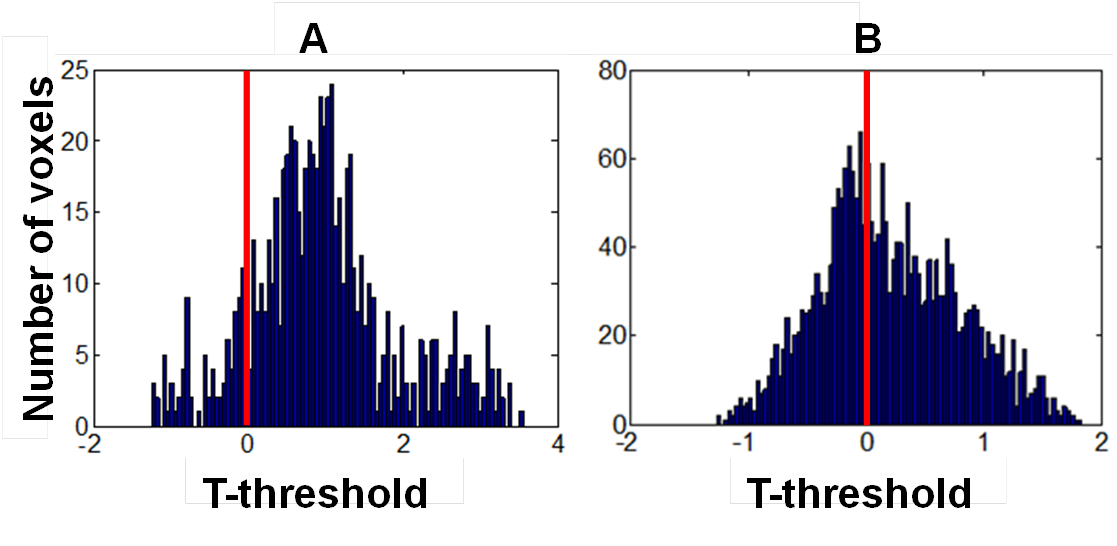
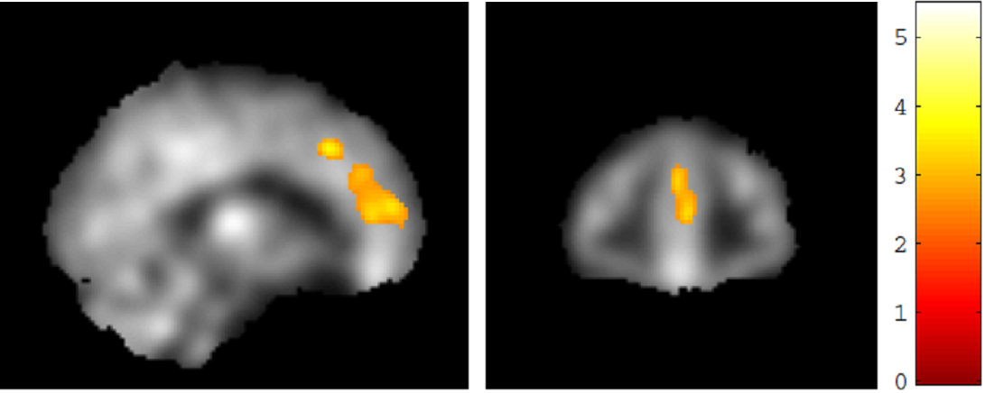
VO2 max, the maximum amount of oxygen consumed during peak exercise and an index of cardiorespiratory fitness, was significantly higher (p<0.05) in the exercise group, while the stretch group did not show any significant difference (Table2). Improvements on measures of episodic memory, cognitive flexibility, and attention were seen in the aerobic exercise group alone (Table2). From the two-sample t-test which compared “Post-Pre CBF” between exercise and stretch groups, we counted the number of significantly positive (T>2.82, p<0.005) and significantly negative (T<-2.82, p<0.005) voxels in the major brain regions (Table3). All regions except the Parietal lobe show more number of voxels with increases in CBF in the exercise group compared to the stretch group, especially in anterior cingulate cortex (ACC), frontal and temporal lobes. We also plotted the histogram of T-scores of all voxels in each region. Results for the ACC are shown in Figure 1A, which clearly revealed a skew towards positive values (i.e. CBF increases with exercise relative to stretch group). Figure 1B shows results in a control region, lateral ventricle, which is not expected to show change in CBF with exercise, confirming that the histogram in lateral ventricle is centered about ‘0’. A voxel-wise regression analysis was also performed to correlate changes in CBF to changes in LM delayed recall scores. Both exercise and stretch groups were combined in this analysis. Results revealed that improvement in LM delayed recall scores correlated with CBF increase in the ACC and medial frontal gyrus (BA6) (Figure2). This finding is similar to the increase in CBF in the ACC seen in older adults after 12 weeks of aerobic exercise training[1]. The ACC along with the medial frontal gyrus, represent a critical node in working memory[9, 10], and is involved in monitoring of memory[11] and allocation of attention supporting memory[9]. Thus, it appears that aerobic exercise improves memory function by augmenting ACC activity.Conclusions
Aerobic exercise improved cardiovascular and cognitive functions. The salutary effects in brain function were shown to be mediated by increases in CBF in the ACC and the frontal lobe, which are a critical node to working memory and attention function.Acknowledgements
We would like to thank all MCI volunteers that participated in this study. We would also like to thank NIH for funding.References
1. Chapman SB, Aslan S, Spence JS, et al. Shorter term aerobic exercise improves brain, cognition, and cardiovascular fitness in aging. Front Aging Neurosci 2013;5:75.
2. Thomas BP, Yezhuvath US, Tseng BY, et al. Life-long aerobic exercise preserved baseline cerebral blood flow but reduced vascular reactivity to CO2. J Magn Reson Imaging 2013;38:1177-1183.
3. Colcombe S, Kramer AF. Fitness effects on the cognitive function of older adults: a meta-analytic study. Psychol Sci 2003;14:125-130.
4. Baker LD, Frank LL, Foster-Schubert K, et al. Effects of aerobic exercise on mild cognitive impairment: a controlled trial. Arch Neurol 2010;67:71-79.
5. Geda YE, Roberts RO, Knopman DS, et al. Physical exercise, aging, and mild cognitive impairment: a population-based study. Arch Neurol 2010;67:80-86.
6. Varela S, Ayan C, Cancela JM, Martin V. Effects of two different intensities of aerobic exercise on elderly people with mild cognitive impairment: a randomized pilot study. Clin Rehabil 2012;26:442-450.
7. Levine BD, Stray-Gundersen J. "Living high-training low": effect of moderate-altitude acclimatization with low-altitude training on performance. J Appl Physiol (1985) 1997;83:102-112.
8. Chalela JA, Alsop DC, Gonzalez-Atavales JB, Maldjian JA, Kasner SE, Detre JA. Magnetic resonance perfusion imaging in acute ischemic stroke using continuous arterial spin labeling. Stroke 2000;31:680-687.
9. Kondo H, Morishita M, Osaka N, Osaka M, Fukuyama H, Shibasaki H. Functional roles of the cingulo-frontal network in performance on working memory. Neuroimage 2004;21:2-14.
10. Engstrom M, Landtblom AM, Karlsson T. Brain and effort: brain activation and effort-related working memory in healthy participants and patients with working memory deficits. Front Hum Neurosci 2013;7:140.
11. Risius UM, Staniloiu A, Piefke M, et al. Retrieval, monitoring, and control processes: a 7 tesla FMRI approach to memory accuracy. Front Behav Neurosci 2013;7:24.
Figures