0391
Optimal control B1-robust T2 preparation1GE Global Research, Munich, Germany
Synopsis
T2 is an important MR imaging contrast, including visualizing edema, myocarditis, separating coronary lumen from myocardium, as well as cerebrospinal fluid from gray and white matter. For diagnostic accuracy, it is important to achieve uniform T2-weighting throughout the imaging field-of-view. This is often not achievable because of non-uniform B1, especially at ultra-high magnetic field. This limitation was overcome by numerical optimization of a T2 preparation module. The optimal control T2 preparation pulse achieved good T2 contrast despite ±40% B1 variation and was scalable to achieve different T2 weighting times. Evaluation was done with Zero-TE imaging in the human brain at 3T.
Purpose
To design a T2 preparation module, achieving T2-weighted Mz-preparation robust to variation in B1 and B0 using a single, low-amplitude RF pulse followed by a gradient crusher.Methods
The RF pulse amplitude and phase were numerically optimized based on the Bloch equations under the presence of relaxation, B1 variation, and B0 offsets. The desired target state of longitudinal magnetization was: Mz,t=exp(-TEeff/T2), where TEeff is the desired effective echo time. The cost function weights deviations of the actual final state from the desired state: φ=(Mz-Mz,t)2. Numerical optimization was performed on a Linux desktop computer using a custom made gradient descent algorithm1. Optimization parameters were pulse duration T=52ms, TEeff=40ms, B1=4.8-11.2µT (±40%, 5 increments), B0=±250Hz (401 increments), T2=30-456ms (15 increments), T1=1000ms.
Existing techniques apply 180° refocusing pulses in between a pair of 90° excitation and -90° tipup pulses using rectangular, composite, or adiabatic RF pulses, and followed in the end by a crusher2,3. The conventional T2 preparation module consisted of hard pulses with 2 180° refocusing pulses using phase cycling (“MLEV2”). MLEV2 was compared to the optimized pulse (“T2opt”) on a Discovery MR750w 3.0T (GE Healthcare) in phantoms with similar T1 (496-516ms) and different T2 (49, 89, and 151ms) while experimentally modifying B1 and B0, as well as in two healthy volunteers (ZTE, 3D radial trajectory, 3° flip, 64 spokes per segment, 500ms recovery time, ±15.6kHz rBW, scan time 3min9s). Parametric maps were calculated based on a pair of ZTE images with TEeff 20 and 60ms (T2,est=-40ms/log(A60ms/A20ms), where A is the image matrix). T1-weighted images were obtained by repeating ZTE with flip angles 6° and 1° (T1w=A6°/A1°). Images were aligned by rigid registration (NIfTI, NIH) and masking was based on thresholding the magnitude image.
Results
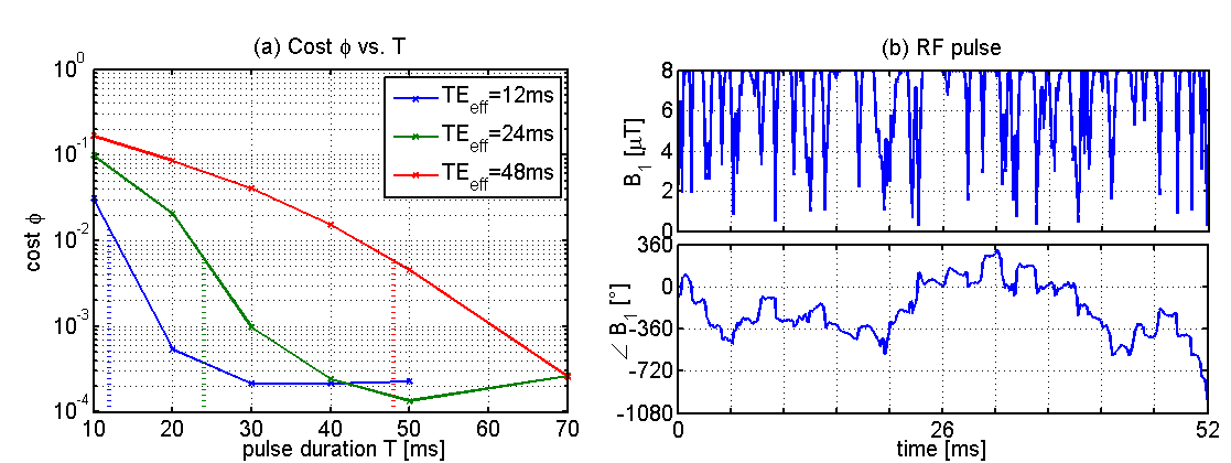
The cost depending on different effective echo times TEeff and RF pulse durations T (Fig. 1(a)) visualizes that good T2-weighting can only be achieved when T>TEeff. This is an expected result, as the RF field cannot modify relaxation and the desired T2 contrast can only be achieved by keeping magnetization in the transverse plane for the duration of TEeff. T2opt does not resemble the typical (+90)-180-180(-90) pulse sequence (see RF waveform in Fig. 1 (b)).
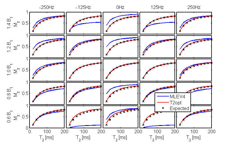
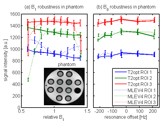
Fig. 2 shows the performance of T2opt in Bloch equation simulation, for different B1 variation and B0 resonance offsets. The T2opt pulse achieves the desired T2 preparation within the range of B1 and B0, while MLEV4 experiences large deviations, especially for 60% B1 and ±125Hz B0. These findings were confirmed by signal intensity evaluation in the phantom (Fig. 3).
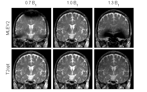
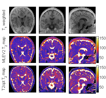
In vivo T2opt showed B1-robustness, while MLEV2 was sensitive to B1 (Fig. 4). To generate parametric T2 maps the T2opt pulse was scaled to T=26 and 78ms, achieving TEeff=20 and 60ms, respectively.
Discussion
We presented an optimized T2 preparation pulse (T2opt) which achieved desired T2 weighting in the presence of ±40% B1 variation and ±250Hz B0 off-resonance. The pulse had a low nominal maximum B1 amplitude (8µT), was designed for TEeff=40ms and was successfully scaled to achieve 20 and 60ms T2 weighting. As the RF bandwidth scales with the pulse duration it would be preferred to generate a library of T2opt pulses for different TEeff. B1-robust T2 preparation enabled parametric imaging with ZTE: T1w and T2 contrasts were obtained by an 8min scan. Due to the very high B1 robustness the method can be adapted to 7T imaging. Additionally, the scan is silent when the gradient crusher is derated.Acknowledgements
The authors gratefully acknowledge assistance of and fruitful discussions with Xin Liu.References
1 Khaneja N, Reiss T, Glaser SJ, et al. Optimal control of coupled spin dynamics: design of NMR pulse sequences by gradient ascent algorithms J Magn Reson, 2005, 172, 296-305.
2 Nezafat R, Stuber M, Pettigrew RI, et al. B1-Insensitive T2 Preparation for Improved Coronary Magnetic Resonance Angiography at 3 T. Magn Reson Med, 2006, 55, 858-864.
3 Jenista ER, Rehwald WG, Kim RJ, et al. Motion and flow insensitive adiabatic T2 -preparation module for cardiac MR imaging at 3 Tesla. Magn Reson Med, 2013, 70, 1360-1368.
Figures