0390
A single-channel universal SPINS pulse for calibration-free homogeneous excitation without PTX1Imaging Sciences and Biomedical Engineering, King's College London, London, United Kingdom, 2UMC Utrecht, Utrecht, Netherlands
Synopsis
A universal single-channel pulse was created that improves excitation homogeneity at 3T, for low flip angles. This method does not require subject specific calibration nor a PTX system, thus making it widely applicable. Though even better homogeneity could be achieved through the use of PTX or subject-specific pulse design, in our study the universal single-channel pulse always outperformed the quadrature mode excitation. This could be used to create more uniform contrast in MP-RAGE imaging.
Introduction
Parallel transmit (PTX) technology is a proven facilitator to mitigate the high magnetic field strength-related inhomogeneity of the radiofrequency (RF) field1. RF pulse design using PTX requires measurement of the RF (B1+) and static (B0) magnetic field properties, and subsequent calculations to provide subject-specifical optimized transmit pulses. Gras and co-workers2 proposed the concept of universal pulses: one solution of PTX RF pulses that can be applied on an entire population class without subject-specific calibration. We took this generalization even further by considering homogeneity-correcting RF pulses without PTX at 3T. A single-channel universal excitation pulse could benefit a wider range of MRI scans since PTX is not always available and SAR management is not yet routine with PTX as it is with single channel operation.Methods
All experiments were performed at 3T using an 8-channel TEM PTX body coil of which the quadrature mode is similar to that of a whole body birdcage coil3. An 8-channel head coil was used for signal reception.
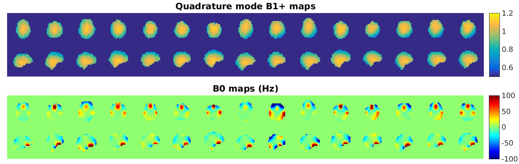
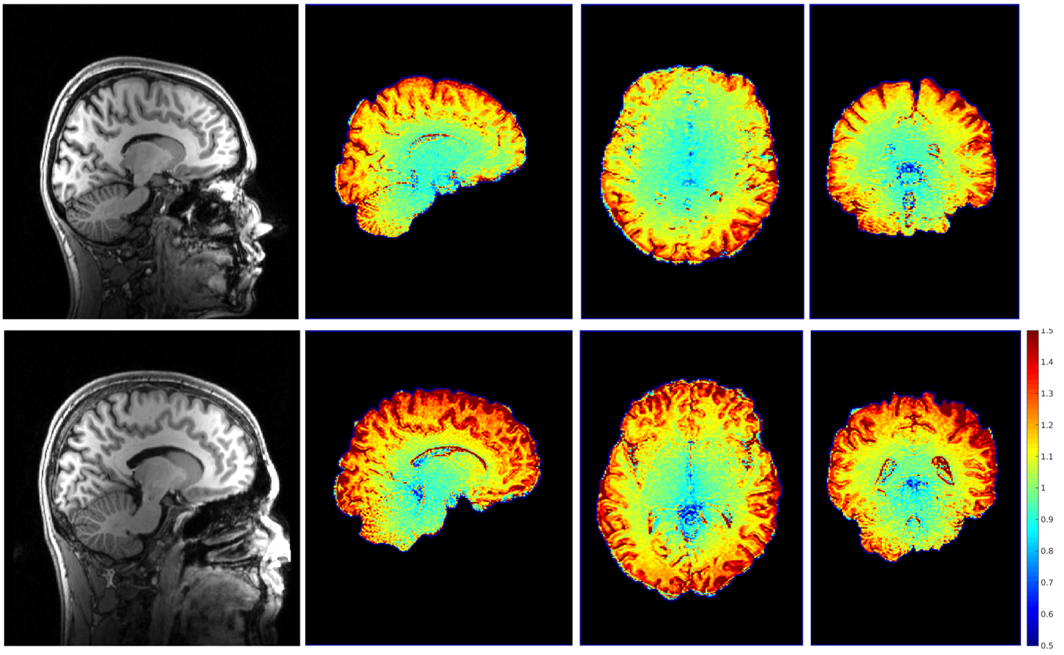
A training data set was created from B1+ and B0 field maps acquired in nine volunteers (Figure 1). B0 maps were measured with ΔTE = 2.3 ms, B1+ using linear combinations of channels4 and AFI5. The maps were masked using the Brain Extraction Tool6 and converted to a common coordinate system.
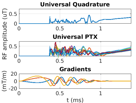
Universal pulses were calculated using SPINS7 from the combined data by concatenating the encoding matrices from each subject. This was done 1) for driving the system in quadrature mode (all TX channels equal) and 2) for PTX (different pulses per channel). All designs used a fixed, pre-measured k-space trajectory.
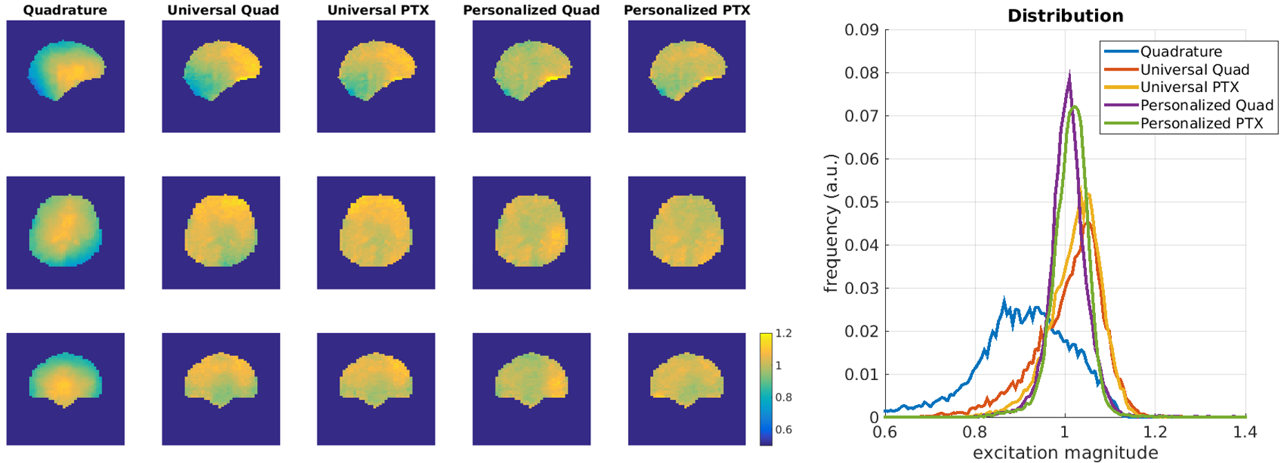
In six additional subjects (the control group), field maps were collected and personalized SPINS pulses designed. On each subject we measured the excitation magnitude from universal and personalised SPINS pulses for both single and parallel transmission. Excitation magnitude of each candidate pulse was measured by dividing a spoiled gradient echo scan (SPGR) at 1˚ flip angle by an SPGR using a standard quadrature pulse, multiplied by the quadrature-mode B1+ map.
MP-RAGE8 images were collected from two subjects comparing quadrature (standard) excitation with the single channel universal SPINS pulse. In this sequence, not achieving the requested flip angle will reduce the contrast between tissue types. The TR between excitation pulses was extended from 7.2ms in the original protocol8 to 7.5 ms to fit the SPINS pulses of length 1.37 ms (FA = 8˚). In all scans with PTX pulses the system-calculated global SAR was restricted to 10% of the 3.2 W/kg limit, following local policy to control local SAR.
Results
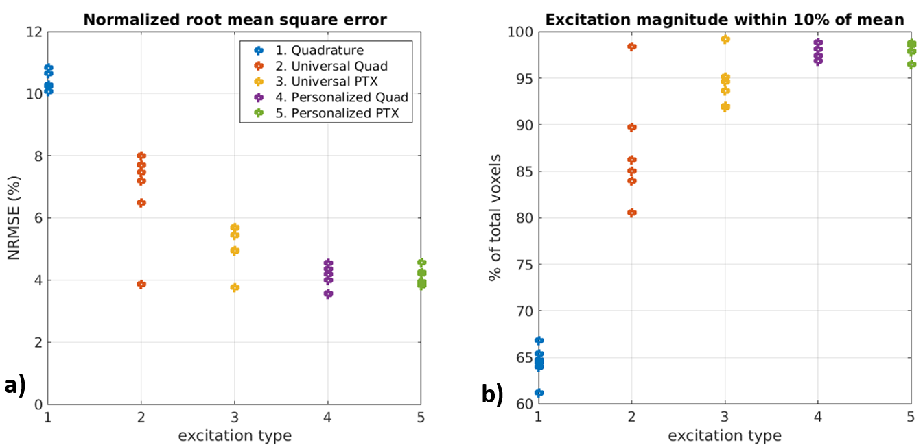
Figure 1 shows the different field maps measured in all subjects, and Figure 2 shows the universal SPINS pulses computed from the training data. Figure 3 shows SPINS excitation magnitudes in one subject, demonstrating improved uniformity. This is confirmed for all subjects when considering the NRMSE (Figure 4a) and the percentage of voxels with less than 10% deviation from the mean (Figure 4b) – this additional measure was computed because the histograms (Figure 3) are not Gaussian. In all cases the universal single channel pulses perform better than normal quadrature excitation, and personalised pulses perform better still, though PTX does not significantly improve the performance of the personalized single-channel pulses.
Figure 5 shows MP-RAGE images using the universal single channel pulse and ratios of these to images acquired in quadrature mode to visualise signal recovery. Increased signal levels can be observed in the periphery, where quadrature mode typically underachieves (Figure 1).
Discussion & conclusion
Single channel universal SPINS pulses could be used without calibrations. In our experiments they always improved the spread in flip angles. The distribution of flip angles is also changed, in particular SPINS pulses reduce the number of pixels with very low values, seen in the periphery of the brain. MP-RAGE images showed corresponding increased signal level at the brain periphery, and a tissue specific signal difference indicating a change in contrast. Such pulses could improve brain segmentation methods using a T1-weighted volume images as input.
The method used to generate the universal pulses could be improved by including a larger cohort of training data – the solution presented here could be viewed as the starting point for this. Individual optimization using only a single channel was found to perform very well (as well as PTX), suggesting personalized SPINS pulses are generally applicable to any (PTX) MRI system. However this would require run time optimization that most scanners currently cannot do as standard.
Acknowledgements
No acknowledgement found.References
1. Padormo, F., Beqiri, A., Hajnal, J. V. & Malik, S. J. Parallel transmission for ultrahigh-field imaging. NMR Biomed. n/a-n/a (2015). doi:10.1002/nbm.3313
2. Gras, V., Vignaud, A., Amadon, A., Bihan, D. Le & Boulant, N. Universal Pulses?: A New Concept for Calibration-Free Parallel Transmission. 0, 1–9 (2016).
3. Vernickel, P. et al. Eight-channel transmit/receive body MRI coil at 3T. Magn. Reson. Med. 58, 381–389 (2007).
4. Malik, S. J., Larkman, D. J. & Hajnal, J. V. Optimal linear combinations of array elements for B 1 mapping. Magn. Reson. Med. 62, 902–909 (2009).
5. Yarnykh, V. L. Actual flip-angle imaging in the pulsed steady state: a method for rapid three-dimensional mapping of the transmitted radiofrequency field. Magn. Reson. Med. 57, 192–200 (2007).
6. Smith, S. M. Fast robust automated brain extraction. Hum. Brain Mapp. 17, 143–155 (2002).
7. Malik, S. J., Keihaninejad, S., Hammers, A. & Hajnal, J. V. Tailored excitation in 3D with spiral nonselective (SPINS) RF pulses. Magn. Reson. Med. 67, 1303–1315 (2012).
8. Jack, C. R. et al. The Alzheimer’s Disease Neuroimaging Initiative (ADNI): MRI methods. J. Magn. Reson. Imaging 27, 685–691 (2008).
Figures