0384
High resolution imaging by phase encoded xSPEN MRI1Department of Chemical Physics, Weizmann Institute of Science, Rehovot, Israel, 2Department of Electrical Engineering and Computer Sciences, University of California, Berkeley, CA, United States
Synopsis
We have recently introduced cross-term SPatiotemporal ENcoding (xSPEN), a technique with exceptional resilience to field heterogeneities. Like other single-shot methods, however, xSPEN’s resolution and SNR are intrinsically limited. This study explores a multi-scan, phase-encoded extension of xSPEN, which improves sensitivity while increasing resolution along both the phase-encoded and the slice-selection dimensions simultaneously. This reflects xSPEN’s unusual kernel whereby a y-axis can be sampled by a z-gradient and viceversa. Furthermore, as each phase-encoded xSPEN scan provides an entire 2D image, each low-resolution xSPEN scan in the set may be used to correct motions leading to very high definition 3D MRI capabilities.
Purpose
To improve the resolution and SNR of xSPEN imaging1 by extending it to a multi-scan, phase-encoded version, whose suitable processing leads to the fast acquisition of high resolution 3D NMR images with good sensitivity and motion compensation capability.Methods
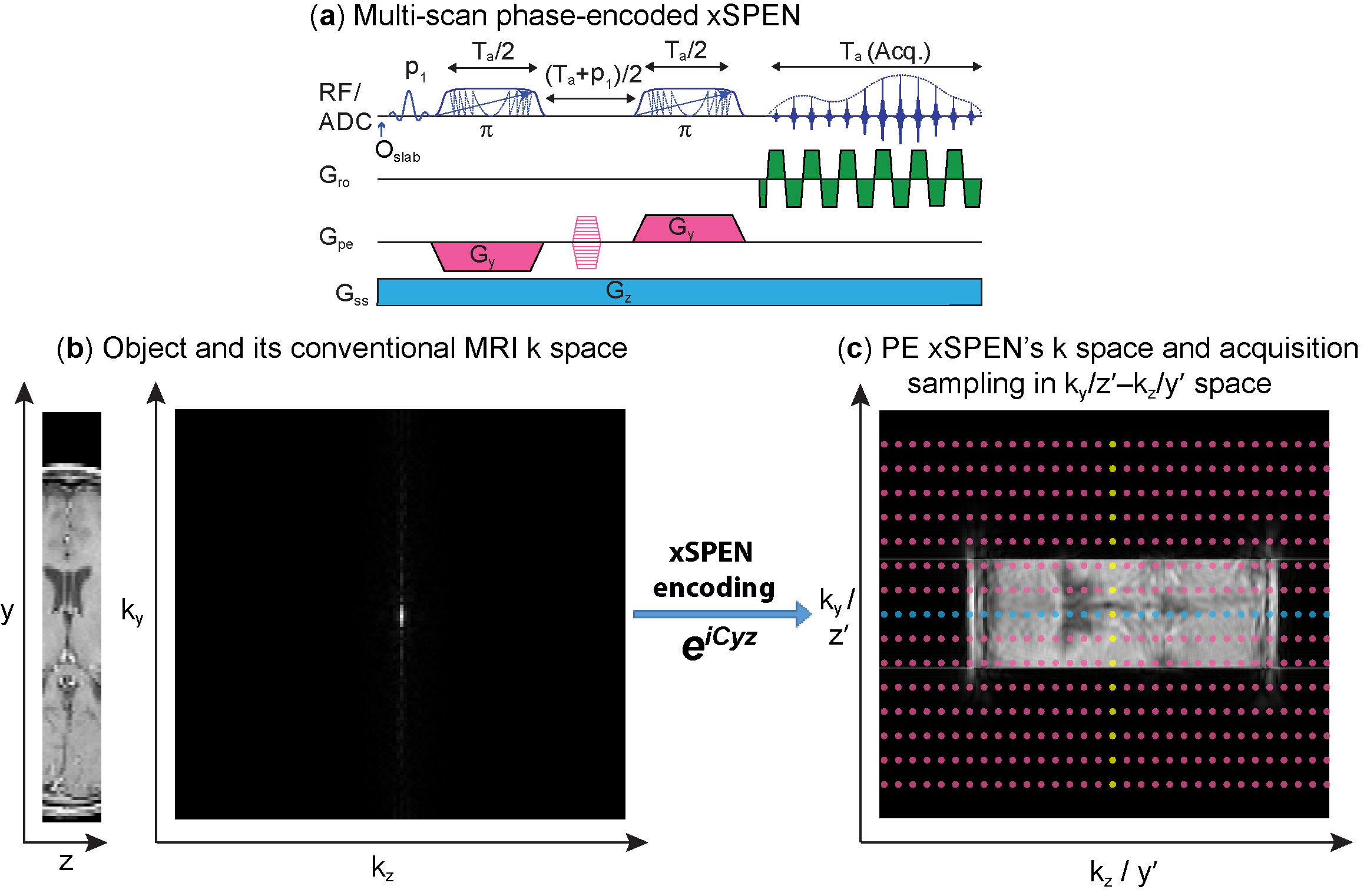
xSPEN is a single-shot imaging technique using frequency-swept pulses under the action of bipolar ±Gy and constant Gz gradients, to impart a hyperbolic phase eiCyz modulation; assuming a uniform object across the slice thickness Δz, subsequent readout by kz=γGzt provides what we referred to as a low-resolution y′=kz/C profile.1 Resolution of this y-axis profile is given by the time-bandwidth product Q of the frequency-swept encoding pulses;1 here we seek to improve this resolution by performing additional ky encodings in independent scans (Fig. 1a).
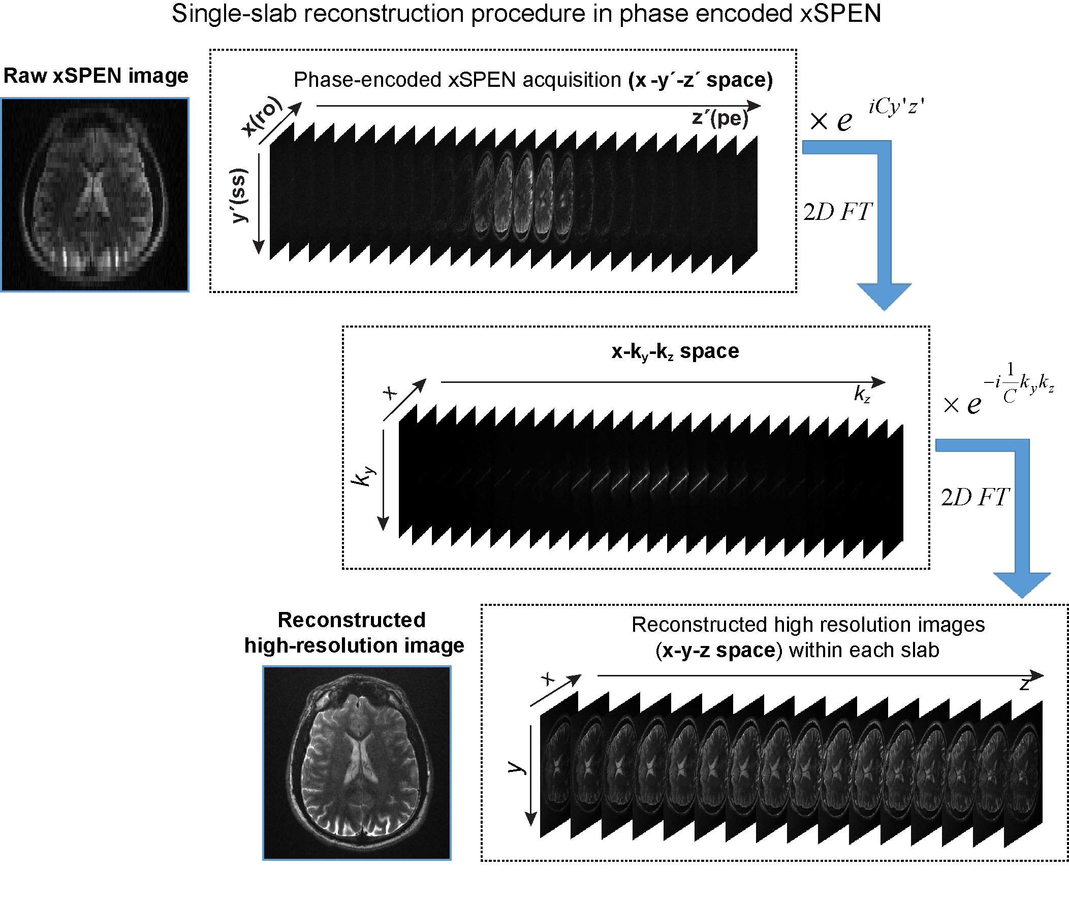
The bidimensional eiCyz kernel encoding spins in xSPEN leads to a unique situation, whereby the k-space sampling is spread from the well-localized situation of traditional MRI (Fig. 1b) into a 2D box-like shape (Fig. 1c). Single-shot xSPEN1 only captures the kz /y′ axis for ky=0 (Fig. 1c, blue dots); conversely, should one collect signals under the action of Gy (kz=0), the outcome would be a ky/z′ image (Fig. 1c, yellow dots). While this 2D kernel underlies xSPEN’s uniqueness, it is also the main source of SNR loss of these single-shot implementations. This study remedies this by introducing a ky-encoding that captures extended regions in k-space (Fig. 1c, magenta dots), improving SNR by Fourier multiplexing and resolution along the y-axis by probing higher kymax values. Furthermore, by exploring the z′= ky/C profile, these ky-acquisitions lift the need to assume uniform slices, augmenting the z-axis resolution by a factor ≤Q. Taking into account that xSPEN’s EPI-like ±kx oscillations also yield a third, readout domain,2 suitable reconstruction of such data yields high in-plane resolution as well as ultrahigh though-plane resolution within the excited slab. To extract this information a suitable analysis of the data in its unusual kz/y′-ky/z′ space is needed. Figure 2 summarizes one of the reconstruction procedures that based on such analysis we developed for this phase-encoded version of xSPEN. Remarkable aspects of this new imaging approach are that (i) the k-space kernel provided by the hyperbolic phase allows sampling along ky to break the stringent Δky=1/FOVy criterion, (ii) that sampling along ky can actually extend beyond kymax by Q/FOVy, and (iii) that the single-shot 2D nature underlying the phase encoded approach allows identification and elimination of motional artifacts.
Results and Discussion
Experimental tests of the proposals in Figs. 1 and 2 were carried out on an ex-vivo rat brain (Agilent 7T) and on a human volunteer (Siemens 3T).
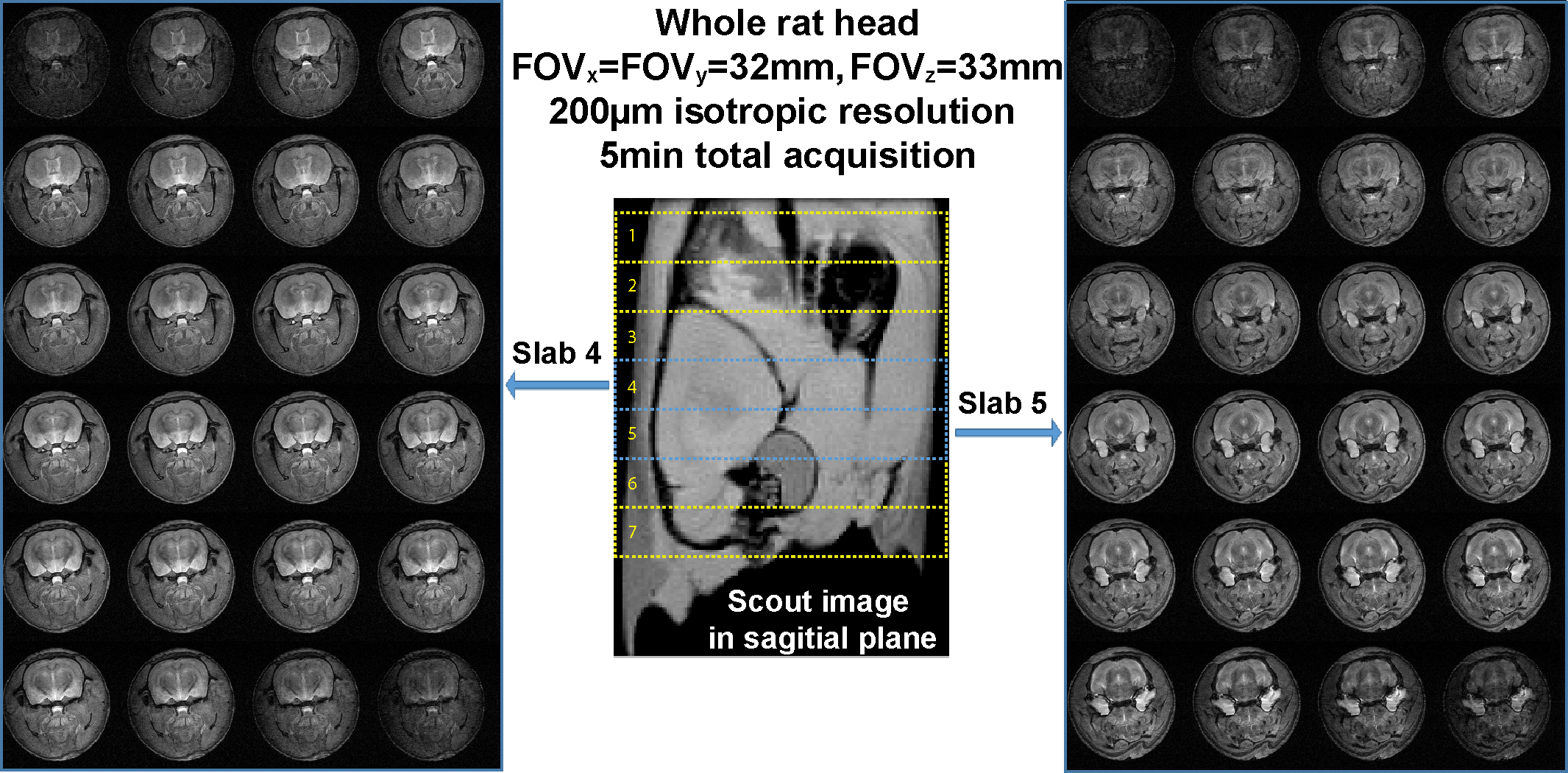
Figure 3 shows results collected at 200µm isotropic resolution for a whole rat brain in 5min. Each of the 7 excited slabs was 4.8mm and frequency swept pulses with Q=24 were used, leading to a 200µm slice-dimension resolution. With a Δky=1/FOVy step and 136 ky phase-encodes this led to kymax = 160/FOVy, i.e. to a 200µm resolution across a FOVy =32mm. Reconstructed z-slices from slabs 4 and 5 are shown in Fig. 3’s left and right panels. As almost all the energy associated to the object’s original k-space is captured and used by this experiment, its SNR is similar to a conventional Fourier imaging experiment with the same resolution.
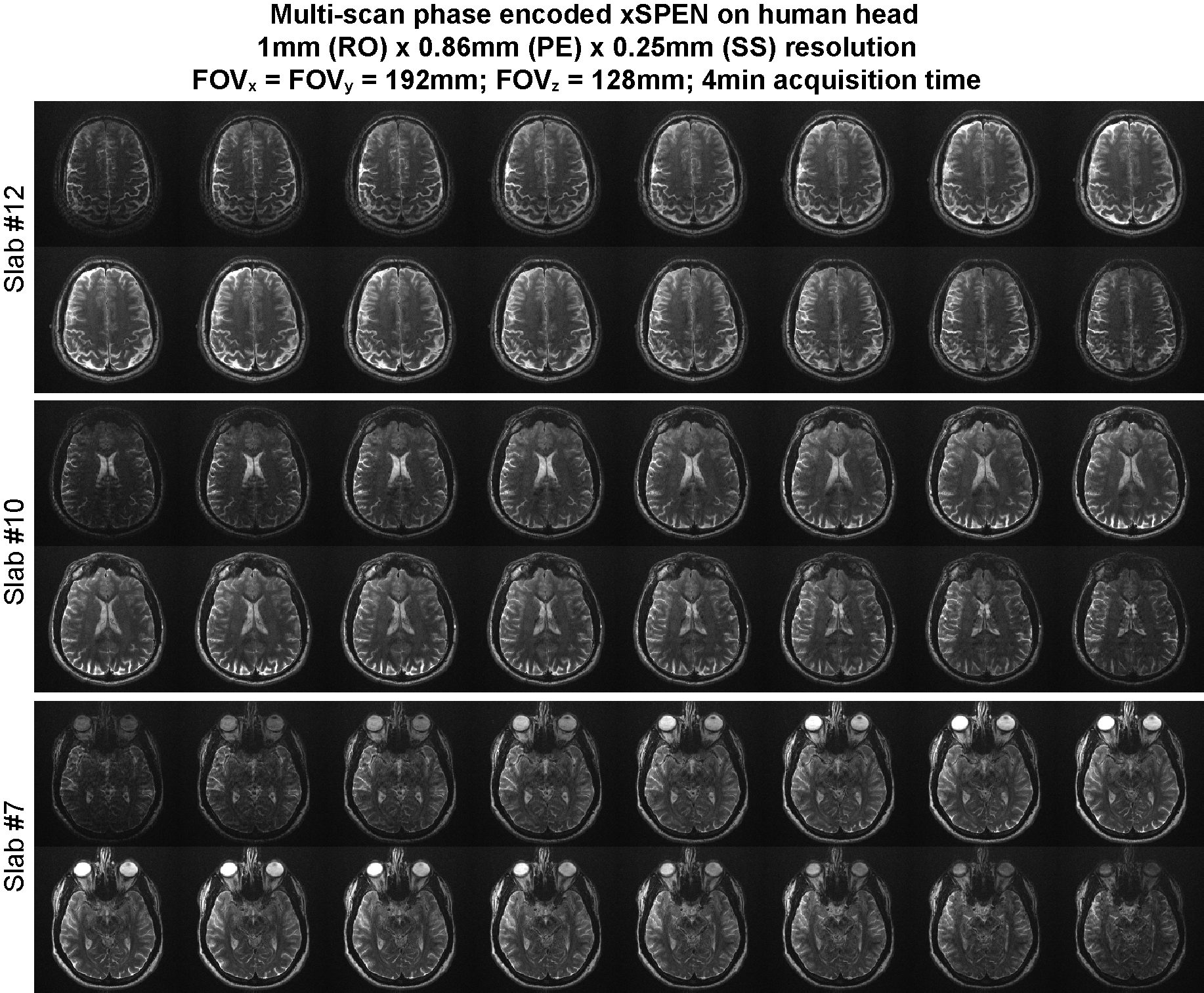
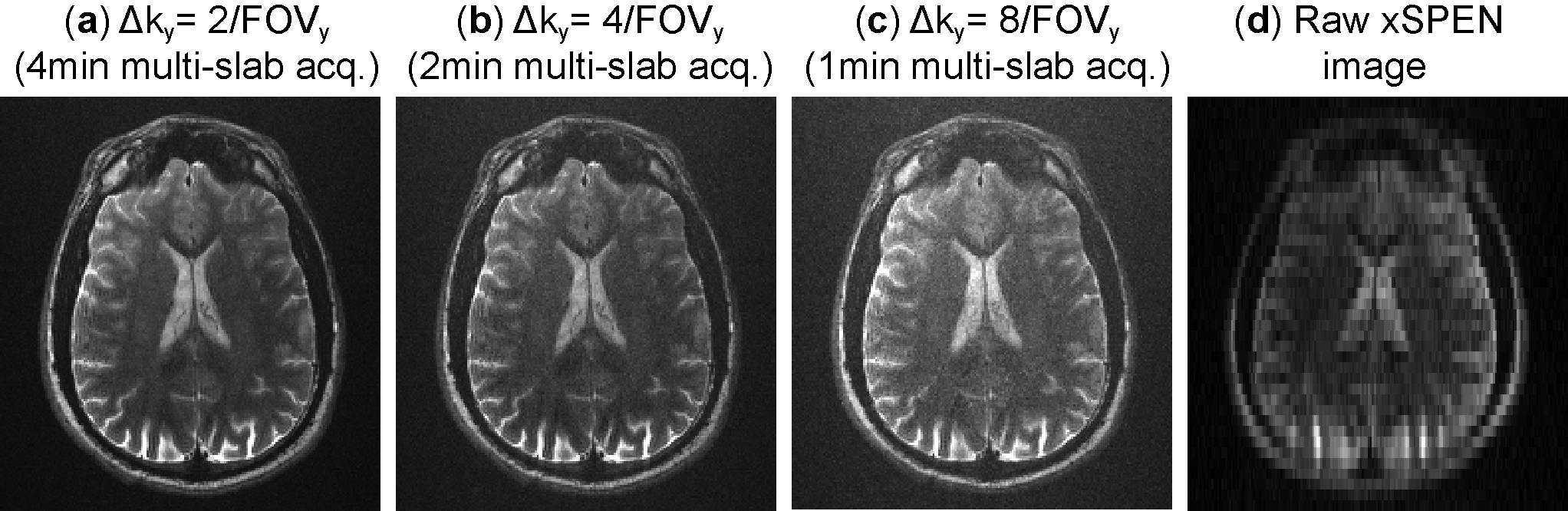
Figure 4 illustrates a 4min phase-encoded xSPEN acquisition on a human volunteer. A Δky=2/FOVy step –twice as large as needed for normally encoding a FOVy=192mm– was used, and the number of ky phase-encodes was 96. When considering frequency swept pulses with Q=32 this spreads kymax to 224/FOVy; after reconstruction this lead to a resolution Δy=0.85mm. Given the 8mm slabs and the Q=32 value used the reconstructed resolution along z was 0.25mm; however, the non-dense sampling along ky also means that there will be a gap of 0.25 mm between the resolved z-slices. This loss of information is not here visible owing to the final thinness and denseness of the z-slices. In fact, Figure 5 illustrates how acquisition times can be shorten even further by undersampling along the phase encoding dimension with steps Δky= 4/FOVy, 8/FOVy, at no expense in the in-plane resolution (despite SNR degradation owing to the fewer scans). Only center slices from slab 10 are shown in Fig. 5.
Conclusions
A multi-scan acquisition and reconstruction xSPEN strategy was introduced, capable of improving both in-plane and through-plane resolutions. By relying on a Fourier processing full sensitivity is restored, while its special kernel permits the fast acquisition of very high resolution, motion-compensated 3D images. Further improvements including compressed sensing and parallel acquisitions are also feasible.Acknowledgements
This work was funded by the Israel Science Foundation grant 795/13, ERC-2014-PoC grant # 633888, and the Kimmel Institute of Magnetic Resonance (Weizmann Institute). ZZ thanks Israel’s Council of Higher Education as well as the Koshland Foundation for postdoctoral fellowships. ML acknowledges a Visiting Faculty Program Fellowship (Weizmann). We are also grateful to Dr. Sagit Shushan (Wolfson Medical Center) and to the Weizmann MRI team (Edna Furman-Haran, Fanny Attar and Nachum Stern).References
1 Zhang Z. Seginer A., Frydman L. Single scan MRI with exceptional resilience to field heterogeneities. Magn Reson Med; doi:10.1002/mrm.26145 2 Mansfield P. Multi-planar image-formation using NMR spin echoes. J Phys C: Solid State Phys 1977;10:L55-L58. 3 Barth, M., F. Breuer, P. J. Koopmans, D. G. Norris and B. A. Poser. Simultaneous multislice (SMS) imaging techniques. Magn Reson Med 2016;75: 63-81. 4 Griswold, M. A., P. M. Jakob, R. M. Heidemann, M. Nittka, V. Jellus, J. Wang, B. Kiefer and A. Haase., Generalized autocalibrating partially parallel acquisitions (GRAPPA). Magn Reson Med 2002;47: 1202-1210.Figures