0376
Rotator Cuff Tendon Assessment Using Magic-Angle Insensitive 3D Ultrashort Echo Time Cones Magnetization Transfer (UTE-Cones-MT) Imaging and Modeling1Department of Radiology, University of California, San Diego, La Jolla, CA, United States, 2Radiology Service, VA San Diego Healthcare System, San Diego, CA, United States
Synopsis
The rotator cuff tendon (RCT) is the primary dynamic stabilizer of the glenohumeral joint. However magic angle effect decrease the sensitivity of MRI in assessment of RCT. The purpose of our study is to utilize the 3D ultrashort echo time Cones sequence with magnetization transfer preparation (UTE-Cones-MT) and two-pool quantitative MT modeling to assess the RCT.
Introduction
The rotator cuff tendon (RCT) is the primary dynamic stabilizer of the glenohumeral joint. RCT dysfunction can lead to abnormal joint kinematics through loss of force couples, deterioration of shoulder function, and ultimately cartilage degeneration and cuff arthropathy. Magnetic resonance imaging (MRI) is widely considered as the gold standard for evaluation of the RCT. Although high accuracy has been shown for the assessment of tearing, conventional MRI demonstrates a low sensitivity of 13-50% for RCT tendinopathy compared with surgical findings with exceedingly poor interobserver reliability1. A major confounding factor for the assessment of tendon is the magic angle effect. Unlike other tendons, such as the Achilles, which have a more constant orientation in clinical imaging scenarios, the RCT orientation is varied and always courses through the magic angle. Such large signal intensity changes may exceed those produced by disease. Recently the ultrashort echo time magnetization transfer(UTE-MT) technique with two-pool modeling has shown promise as a clinically compatible quantitative technique which is resistant to the magic angle effect2. However, to date, evaluation has only been performed using 2D UTE sequences on Achilles tendons. The purpose of our study is to utilize the 3D ultrashort echo time Cones sequence with magnetization transfer preparation (UTE-Cones-MT) and two-pool quantitative MT modeling to assess the RCT.
Methods
Specimens: Four rotator cuff tendons were harvested from two human cadaveric shoulder specimens (mean age 86 years). Cuff tendons cut at the myotendinous junction and sharply dissected from the greater tuberosity, measuring approximately 4.5 cm in length (medial-lateral dimension) as shown in figure 1. No cuff tears were appreciated upon dissection. Protocol: Tendons were imaged on a 3T clinical scanner (Signa HDx, GE Healthcare, Milwaukee, WI) using a surface coil in five different orientations. Specifically, the long-axis of the tendons were oriented 0°, 30°, 55°, 70°, and 90° relative to the main magnetic field (B0). Imaging included a 2D-CPMG sequence (TR=800 ms, TE=7, 14, 22, 29, 36, 43, 50, 57 ms, FOV=8 cm, matrix=128x128, slice thickness=4 mm). The 3D UTE-Cones-MT sequence which consisted of a MT preparation using of a Fermi shaped RF pulse was used for acquisition (TR=100 ms, TE=32 μs, FA=7°, FOV=8cm, matrix=128x128, slice thickness=3mm) with five spokes per MT pulse. In total three MT powers (300°, 550° and 750°) and five MT frequency offsets (2, 5, 10, 20 and 50 kHz) were used for a total of 15 different MT datasets. T1 was measured with the 3D UTE-Cones acquisition with the same spatial resolution and a series of TRs (6, 15, 40, 60, 80 ms) and a fixed flip angle of 25°. Image Processing and Analysis: Analysis was performed using MATLAB (Mathworks, 2015b, Natick, MA). Two-pool UTE-Cones-MT modeling and parameter mapping were performed on the RCT samples. Regions of interest (ROIs) analysis were performed and mean and standard deviation of macromolecular proton fraction, T2 relaxation time, exchange rates and water longitudinal relaxation were calculated.Results
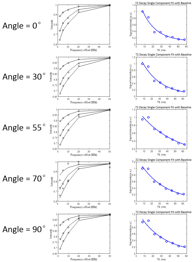
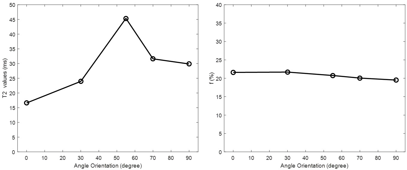
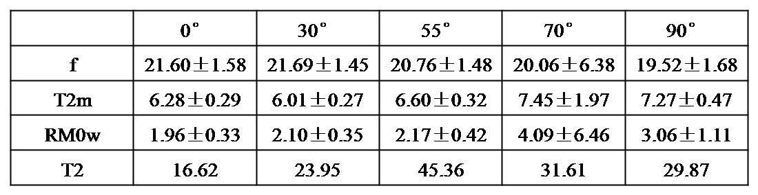
Figure 2 shows the results of both UTE-MT modeling and multiple-TE T2 fitting for a selected sample. The measured T1 value of this sample was 648 ms. The results for the five angle orientations between fiber direction and B0 are shown in the different rows. The two-pool UTE-MT modeling provides excellent fitting (all residuals were less than 0.6%) of signal changes over five MT powers and five off-resonance frequencies. Figure 3 shows graphs of the T2 values derived by fitting multiple-TE data and macromolecular proton fractions in a selected sample. Using the CPMG sequence, T2 ranged from 16.6 ms to 45.4 ms at 0° to 55° relative to B0, representing a 173% increase. However, macromolecular fraction remains relatively consistent ranging from 19.5 to 21.7%, representing an 11% difference. Several other UTE-MT modeling parameters, including T2 value of macromolecular protons (T2m) and exchange rate from macromolecular protons to water protons (RM0w) also demonstrated minimal angular dependence(Table 1). These results suggest that f, T2m and RM0w can be used as magic angle insensitive biomarkers of the RCT.
Discussion
The UTE-MT technique with two-pool modeling shows promise as a clinically compatible technique that is resistant to the magic angle effect. This method may be particularly useful for evaluation of the RCT since this tissue always courses through the magic angle in the clinical scenario. In addition, the UTE-MT sequence provides information on the macromolecular proton pool that cannot be directly obtained by other methods, including regular UTE techniques.Acknowledgements
The authors acknowledge grants from the VA Clinical Science R&D Service (Merit Award I01CX001388), NIH (1R01 AR062581-01A1, 1R01 AR068987-01), National Natural Science Foundation of China (81501463, 81671853), Natural Science Foundation of Guangdong Province, China (Grant No. 2014A030310360), Guangdong Innovative Research Team Program of China (2011S013).References
[1] Robertson PL, Schweitzer ME, Mitchell DG, Schlesinger F, Epstein RE, Frieman BG, Fenlin JM. Rotator cuff disorders: interobserver and intraobserver variation in diagnosis with MR imaging. Radiology. 1995;194(3):831-5
[2] Ma YJ, Shao H, Du J, Chang EY. Ultrashort echo time magnetization transfer (UTE-MT) imaging and modeling: magic angle independent biomarkers of tissue properties. NMR Biomed. 2016 Nov;29(11):1546-1552.
Figures