0346
Evaluation of Portal Vein System in patients after liver transplantation by Unenhanced MR Angiography Using Spatial Labeling with Multiple Inversion Pulses Sequence and by CT portography1Tongji Hospital, Tongji Medical College, Huazhong University of Science and Technology, Wuhan, People's Republic of China
Synopsis
Unenhanced MR angiography with spatial labeling with
multiple inversion pulses (SLEEK) is a reliable method for depicting portal
vein system in patients with liver transplantation compared with computed
tomographic portography (CTP) results. In a study of 20 patients who underwent liver
transplantation, we found that there was excellent correlation between SLEEK and CTP in
presenting the diameter of portal vein. Unenhanced MRA using SLEEK is relatively inexpensive and
is not associated with renal complications. It can be as a good choice for
screening portal vein system in patients with liver transplantation, especially
in patients with renal insufficiency.
Objectives
The objective of this study was to evaluate the diagnostic performance of unenhanced MR Angiography using spatial labeling with multiple inversion pulses sequence (SLEEK) in comparison with CT portography in the detection of Portal Vein System in patients with liver transplantation.Introduction
Unenhanced MR angiography with spatial labeling with multiple inversion pulses (SLEEK) sequence (Fig. 1) has made substantial progress and is effectively used for visualization of transplant renal vascular anatomy and complications (1). Although SLEEK and routine inflow inversion recovery renal MR angiography [2]were basically the same on the principle of imaging, SLEEK technique is used to highlight the importance of the preparation of multiple space selective inversion recovery pulses, with which blood flow could be labeled in a more flexible way. The purpose of this prospective study was to evaluate the ability to depict portal vein system with unenhanced MR angiography by using SLEEK.Materials and Methods
22 patients, 21 men and 1 women (mean age 44.3 years; age range, 15–51 years). Unenhanced MRA using SLEEK was performed on a 1.5-T MRI system for assessing portal vein system in 22 patients with liver transplantation. Then all patients underwent 16-slice CT portography within 1–4 days. The ability to present the portal vein system and to reveal portal vein system disease with SLEEK was evaluated by two experienced radiologists and was compared with CT portography results using a joint reading performed in consensus.Results
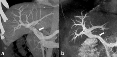
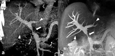
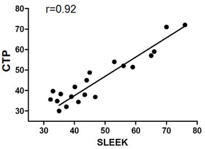
22 patients with liver transplantation underwent SLEEK MRA. A total of 20 portal veins were successful assessed, including 13 no significant stenoses (Fig. 2), 7 with significant stenoses (>50% narrowing) (Fig. 3). Nineteen of the 20 patients were performed end-to-end anastomosis between the donor’s and recipient’s portal veins. One of the 20 patients was performed end-to-end anastomosis between the donor’s portal vein and recipient’s inferior vena cave. There was excellent correlation between SLEEK and CT portography in presenting the diameter of portal vein (R = 0.92; p < 0.05) (Fig. 4). SLEEK was superior to CT portography in revealing the third- and fourth-order segmental branches in the hepatic parenchyma (p < 0.05). SLEEK has the advantage of avoiding interference from ribs, arterial and venous system enhancement.Discussion and conclusion
In this study, the preliminary data from our study demonstrate that SLEEK sequence was capable of displaying transplant portal vein system anatomy and complications. Our results show that consistently high-quality images can be obtained by using SLEEK, which enabled visualization of even small branches within the transplant liver parenchyma. However, because the signal of the portal vein depends on the cardiac output of the patient, a suboptimal blood-suppression TI may lead to poor signal-to-noise ratio and vessel depiction. We did not obtain an additional scout image, which may be helpful in evaluating the flow velocity in the aorta to optimize the blood-suppression TI; thus, a larger study for full comparative evaluation of diagnostic performance is necessary. The SLEEK has a comparable ability in demonstrating portal vein system in patients with liver transplantation as well as CT portography does. It can provide helpful information for surgeons to make an accurate postoperative assessment. Unenhanced MRA using SLEEK is relatively inexpensive and is not associated with renal complications. It can be as a good choice for screening portal vein system in patients with liver transplantation, especially in patients with renal insufficiency.Acknowledgements
We thank ShaofaWang, Zhihui Wang, and Nan Wang, whose important contributions to this study were indispensableto its success.References
1. Tang H, Wang Z, Wang L, Hu X, Wang Q, Li Z, Li J, Meng X, Wang Y, Hu D. Depiction of Transplant Renal Vascular Anatomy and Complications: Unenhanced MR Angiography by Using Spatial Labeling with Multiple Inversion Pulses. Radiology2014 Mar 3:131800.
2. Parienty I, Rostoker G, Jouniaux F, Piotin M, Admiraal-Behloul F, Miyazaki M. Renal artery stenosis evaluation in chronic kidney disease patients: nonenhanced time-spatial labeling inversion-pulse three-dimensional MR angiography with regulated breathing versus DSA. Radiology2011 May;259(2):592-601.
Figures


