0305
AMoCo, a software package for prospective motion correction1Max Planck Institute for Biological Cybernetics, Tübingen, Germany, 2IMPRS for Cognitive and Systems Neuroscience, University of Tübingen, Tübingen, Germany, 3Department of Biomedical Magnetic Resonance, University of Tübingen, Tübingen, Germany
Synopsis
Long scan time makes MRI prone to subject motion which can result in image artifacts. Here we introduce a library for advanced motion correction (AMoCo) for Siemens platforms which can be embedded in any sequence and enables connecting to any tracking device. The library is programmed in a modular way that allows user to customize the correction procedure. The library is integrated with EPI, GRE, and FLASH sequences and tested with various tracking devices.
Introduction
Typically, long spatial encoding durations in anatomical imaging as well as repetitive acquisitions in functional experiments makes MRI prone to subject motion which can result in image artifacts 1, false activations 2 or even can make it necessary to repeat measurements. The gold standard to degrade motion induced artifacts is prospective motion correction: A tracker measures the object´s position in real-time, calculates the most recent position of the object and sends it back to scanner in order to update imaging coordinates 3. Here we introduce a library for advanced motion correction (AMoCo) for Siemens platforms which can be embedded in any sequence and enables connecting to any tracking device. The main function of the software is to receive the measured object position and to update the measurement parameters dealing with acquisition. The library also comprises wide comprehensive tools to facilitate performing advanced correction and evaluation of motion.Method
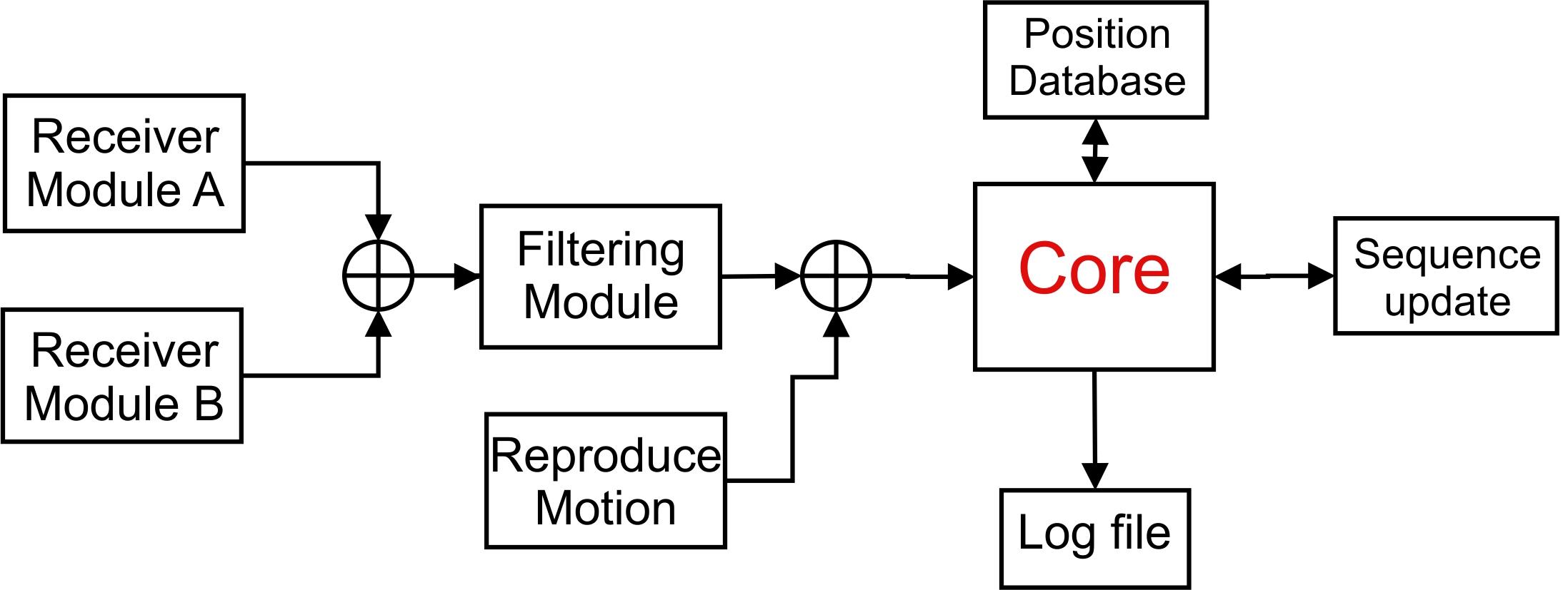
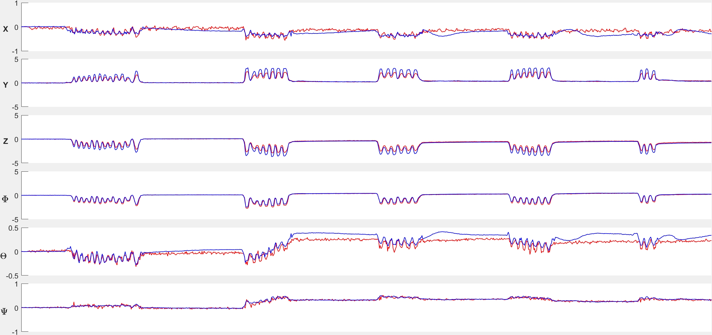
The developed C++ library AMoCo is compatible with Siemens MR Scanners and designed for the IDEA sequence development environment. Figure 1 illustrates the workflow of how AMoCo interfaces with motion trackers and the scanner. AMoCo has a modular approach, in other words, the user can redesign and replace library functions without interfering with other parts. This enables the utilization of motion data from any arbitrary source. Currently it supports communication with three main prospective approaches (i.e. optical tracking system, FID navigators, and field sensors) as well as reading from text files for motion simulation. A dedicated log file containing motion information in 6 degrees of freedom (DOF) in respect to the imaging volume center is produced for each measurement. AMoCo can receive data from two distinct motion trackers simultaneously, and creates separate log files for each for subsequent comparison (Fig. 2). In the case of corrupted motion data, AMoCo can switch automatically to the next motion tracker, if present. This can be beneficial particularly for optical trackers when the marker is out of the camera’s field of view. For performance analysis of the motion correction, the option of reproducing the motion artifact is included in the library based on the method presented in 4. In order to improve the accuracy in case of noisy tracking data, additional filtering can be employed. Another feature of the presented library is the assignment of a unique ID to reference head positions. This allows, for example, to compensate for motion in and between different sessions and therefore long-term studies with a minimum of interpolation errors due to retrospective motion correction.Results
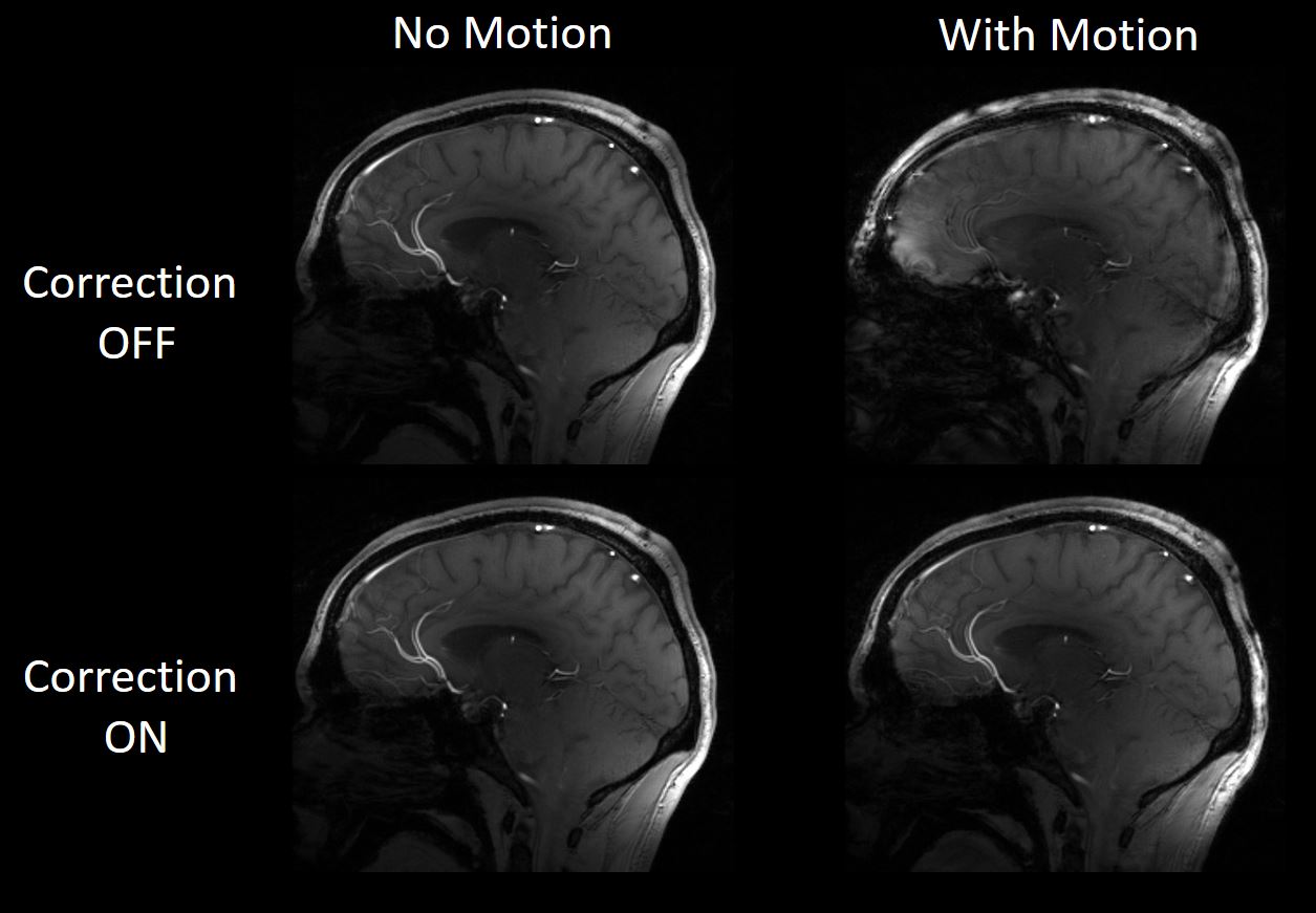
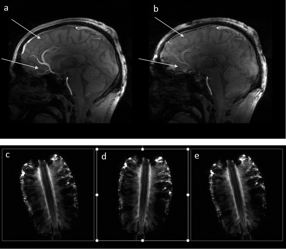
The test of correct functionality was performed with simulation of motion on a phantom and in-vivo. Figure 3 demonstrates the results of scans where an optical tracking camera 5 was used as source. During the measurements with motion, the subject was asked to move his head slightly in random directions. Figure 4a and 4b show how motion artifacts can be reproduced to compare corrected and ‘uncorrected’ images. Figure 4c to e show three successive scans. The phantom was translocated slightly between the first and the second scan. Before the start of the third scan, AMoCo adjusted the volume to the original place (the first scan) and thereby compensated for intra and inter-scan motion.Discussion & Conclusion
AMoCo is a motion correction library designed for Siemens MR scanners and can easily be implemented in a wide range of sequences. The library considers all aspects of working with a real-time operation, scanner hardware and all synchronization issues. It has the ability to integrate different motion correction technologies in a single library, which makes it possible to compare their individual performance with very high temporal precision. This may help to improve the accuracy of tracking methods, to debug tracking errors and to optimize data filtering, if required. In addition, an approach was presented of saving an individual reference positions for each subject in order to reduce the amount of co-registration needed in long-term studies and thereby increasing the overall spatial accuracy of such data.Acknowledgements
No acknowledgement found.References
1. R. Van de Walle, I. Lemahieu, and E. Achten, “Magnetic resonance imaging and the reduction of motion artifacts: review of the principles.,” Technol. Health Care, vol. 5, no. 6, pp. 419–35, Dec. 1997.
2. K. J. Friston, S. Williams, R. Howard, R. S. Frackowiak, and R. Turner, “Movement-related effects in fMRI time-series.,” Magn. Reson. Med., vol. 35, no. 3, pp. 346–355, 1996.
3. J. Maclaren, M. Herbst, O. Speck, and M. Zaitsev, “Prospective motion correction in brain imaging: A review,” Magn. Reson. Med., vol. 69, no. 3, pp. 621–636, 2013.
4. M. Herbst, J. MacLaren, C. Lovell-Smith, R. Sostheim, K. Egger, A. Harloff, J. Korvink, J. Hennig, and M. Zaitsev, “Reproduction of motion artifacts for performance analysis of prospective motion correction in MRI,” Magn. Reson. Med., vol. 71, no. 1, pp. 182–190, 2014.
5. J. Maclaren, B. S. R. Armstrong, R. T. Barrows, K. A. Danishad, T. Ernst, C. L. Foster, K. Gumus, M. Herbst, I. Y. Kadashevich, T. P. Kusik, Q. Li, C. Lovell-Smith, T. Prieto, P. Schulze, O. Speck, D. Stucht, and M. Zaitsev, “Measurement and Correction of Microscopic Head Motion during Magnetic Resonance Imaging of the Brain,” PLoS One, vol. 7, no. 11, pp. 3–11, 2012.
Figures