0304
On the impact of real-time motion and B0 correction during 3D-MRSI measurements in Parkinson’s, Mild Cognitive Impairment and young/elderly controls1High Field MR Centre, Medical University of Vienna, Vienna, Austria, 2Christian Doppler Laboratory for Clinical Molecular MR Imaging, Vienna, Austria, 3Biomedical Center Martin, Division of Neurosciences, Jessenius Faculty of Medicine in Martin, Comenius University in Bratislava, Martin, Slovakia, 4Martinos Center for Biomedical Imaging, Massachusetts General Hospital, Harvard Medical School, Boston, MA, United States, 5Institute of Experimental Endocrinology, Biomedical Research Center, Slovak Academy of Sciences, Bratislava, Slovakia
Synopsis
Presence of motion during MRSI acquisition and scanner instabilities affect the localization accuracy of the measurement and consequential quality of the data. We determined the extent of inter-acquisition head movement, frequency and B0 shim changes during approximately 20 min MEGA-edited MRSI scan with integrated vNav in 4 different groups of subject, with significantly larger amount of head motion in Parkinsons's patients, Mild Cognitive Impairment and elderly controls comparing to young healthy volunteers. With real-time motion, B0 correction and reacquisition we obtained satisfactory data quality in all groups of subjects, which makes it a valuable tool for spectral quality accurance.
Purpose
Magnetic resonance spectroscopic imaging (MRSI) can map the spatial distribution of several brain metabolites. The major limitation of MRSI is its relatively long scan time, which gives rise to motion-induced artifacts and scanner instabilities that consequently affect the localization accuracy of the measurement and quality of the data1. In particular children, elderly people or patients are more prone to unprompted movement. In the presented work we determined the extent and bias due to head motion and scanner instabilities during ~20 min long MEGA-edited MRSI scans in 4 different subjects groups (i.e. young controls (YCon), elderly controls (OldCon), Parkinson´s disease (PD) patients and Mild Cognitive Impairment (MCI)) using an MRSI sequence with integrated EPI volumetric navigator (vNav)2,3.Methods
All measurements were performed on a 3T TIM Trio MR scanner (Siemens, Erlangen, Germany) consisting of a) an AutoAlign localizer ensuring the same inter-subject alignment of the VOI/FOV in the head; b) 3D T1-weighted MRI (MPRAGE) for localization purpose and c) spiral-encoded 3D-MRSI MEGA-editing sequence with LASER localization and integrated 3D multi-shot EPI-based vNav3 performing real-time updates in localization (rotation and translation), frequency and first-order B0 shims within the same repetition time (TR). The sequence parameters were: TR/TE=1600/68 ms; nominal resolution 8cc, matrix size 14×14×12 interpolated to 16×16×16; 16 acquisition-weighted averages. Moreover immediate selective reacquisition of corrupted data was applied if head translation or rotation between two TR was larger than 0.8 mm and 0.8° respectively, but not exceeding 50% of total scan time. More details about the vNav setup were published previously2.
After Institutional Review Board approval, data were obtained from 156 subjects divided into 4 groups: a) 33 YCon-s (16F/17M; age:30±5y), b) 34 OldCon-s (19F/15M; age:66±7 y), c) 45 PD-s (19F/26M; age:66±9y) and d) 37 MCI-s (28F/9M; age:74±5y). In 7 scans (3 PD,4 MCI) >50% of reacquisitions would have been necessary. Thus, the acquisitions were interrupted and the datasets excluded from the study due to inability to perform necessary reacquisitions within the granted time. All spectroscopic data were processed automatically using an in-house developed software tool4 using MATLAB (MathWorks, USA), Bash (Free Software Foundation, USA), MINC (MINC tools, Canada) and LCModel (Provencher, Canada). Statistical analysis was performed with SPSS software (Version 21, USA).
Results
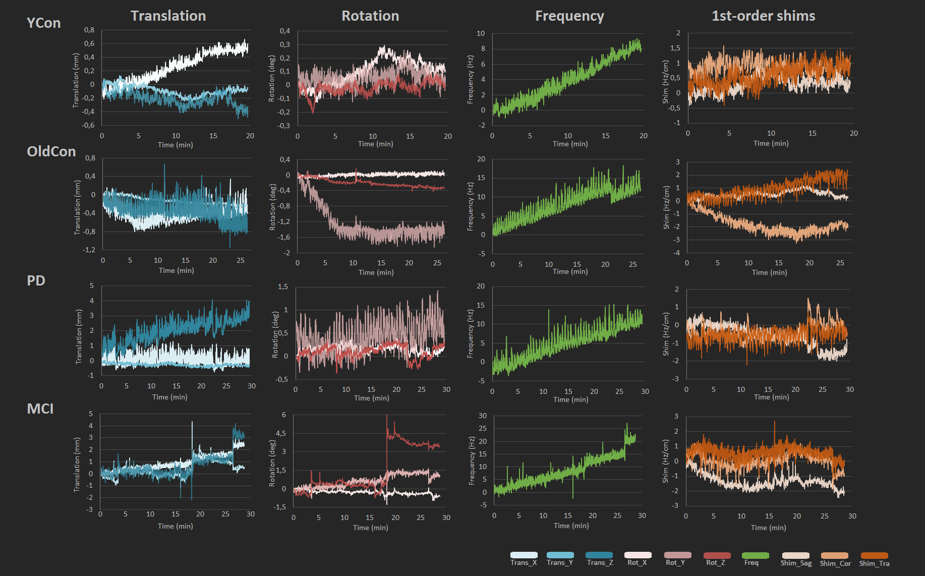
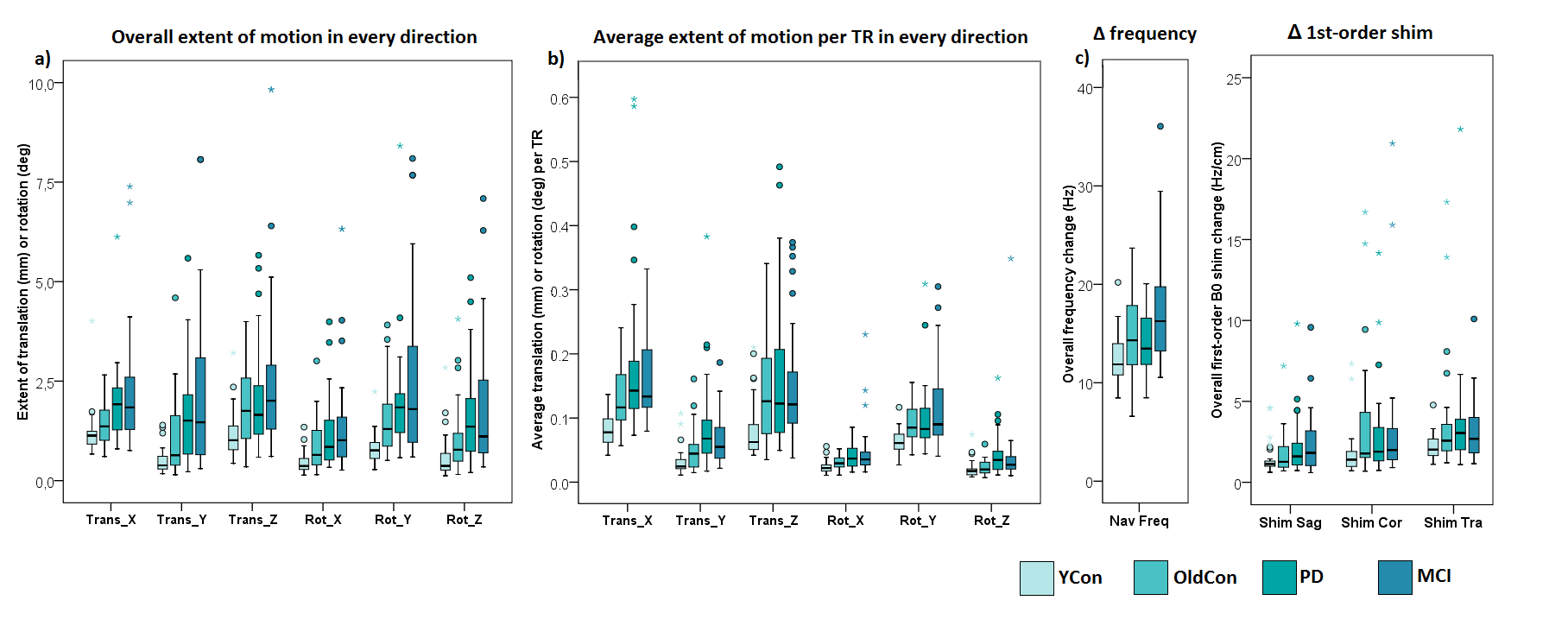
Figure 1 displays representative records of head translation, rotation; frequency change and first-order shim terms measured by vNav for each group. Boxplots in Figure 2 show a) amount of translation/rotation in each direction during the measurement; b) average translation/rotation within TR and c) overall frequency and B0 first-order shim changes of all subjects. The predominant movement directions were y-rotation in all 4 groups and z-translation in OldCon and MCI group. For YCon and PD groups the most prevalent translation was along x-axis.
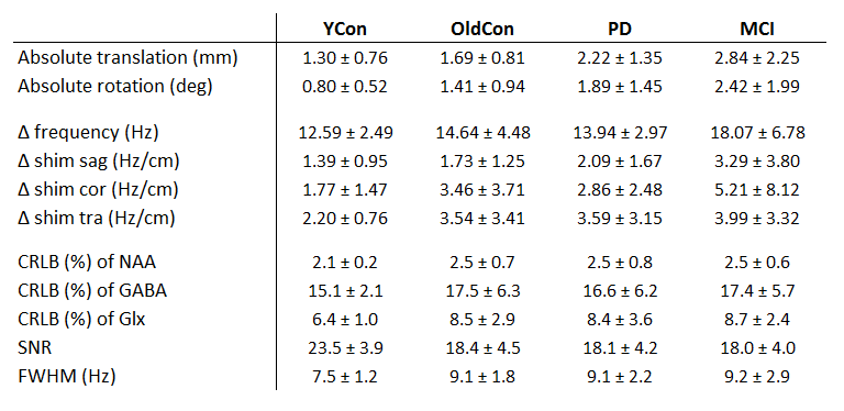
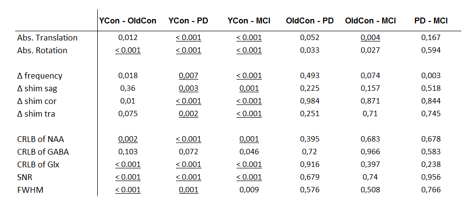
Statistical analysis showed 2.2/1.7-fold larger amount of absolute head translation in MCI/PD comparing to YCon (both p<0.001) and 1.7-fold larger amount of translation in MCI comparing to OldCon (p=0.004). In terms of absolute head rotation during the MRSI scan we found significantly larger amount of head rotation in OldCon (1.8-fold), PD (2.4-fold) and MCI (3-fold) groups when comparing to YCon (all p<0.001). Overall frequency and B0 shim changes were significantly larger in MCI and PD in comparison to YCon (Table 1,2).
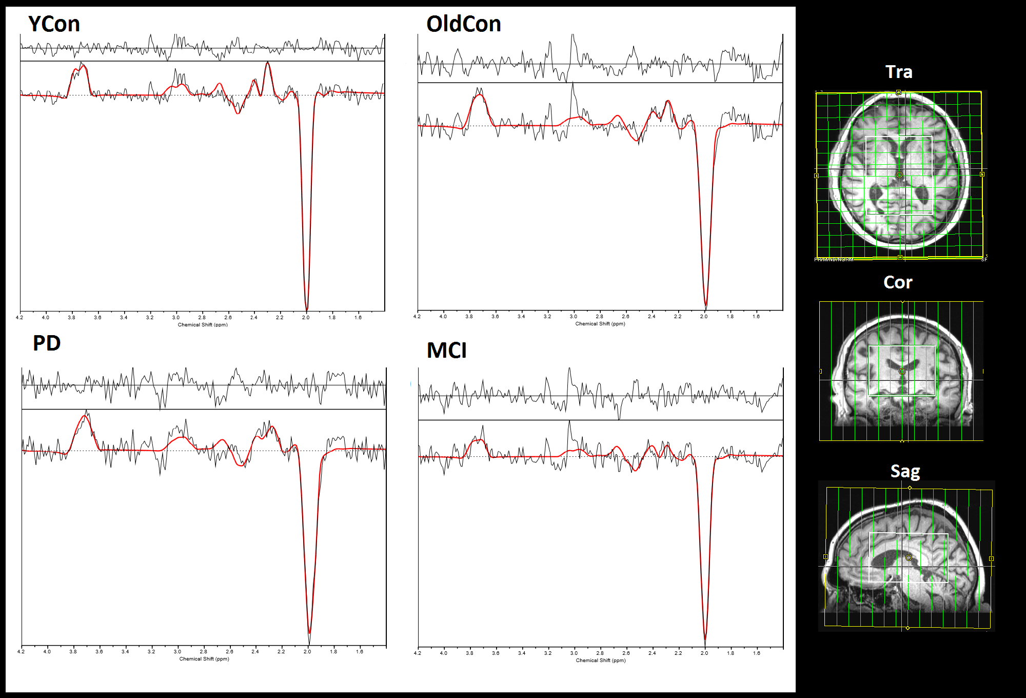
Representative spectra for each group are shown in Figure 3. An ROI consisting of 12 voxels was defined in occipital lobe and average CRLB of GABA, Glx and NAA; FWHM and SNR were calculated based on LCModel output (Table 1). In the YCon group we found significantly lower values of FWHM, CRLBs of NAA and Glx and higher SNR in comparison to OldCon, PD and MCI (p-values displayed in Table 2).
Discussion
The presence of motion during measurement has a substantial impact on the data quality, in particular during long MRSI acquisitions. We found significantly larger amount of head motion in groups of elderly people and patients comparing to young healthy subjects. The predominant movement directions in the scanner coordinate system were rotation around y-axis and translation along z- or x-axis, respectively. With real-time motion, B0 correction and reacquisition of the corrupted data we obtained satisfactory spectral quality in all groups of subjects (CRLBs<20%, SNR>15), with significantly better results in young controls.Conclusion
This work assessed the extent of motion during ~20 min long MRSI measurements in 4 groups of subjects with various tendencies of head movement. The implementation of prospective motion and B0 shim corrections improves the quality of the data, as was shown also previously, and therefore is a valuable tool for spectral quality assurance, especially when head pose cannot be assumed to be constant.Acknowledgements
This study was supported by the Austrian Science Fund (FWF): KLI-61 and the FFG Bridge Early Stage Grant #846505.References
1. Kreis R. Issues of spectral quality in clinical 1 H-magnetic resonance spectroscopy and a gallery of artifacts. NMR BIOMED 2004;17:361–81.
2. Bogner W, Hess AT, Gagoski B, et al. Real-time motion- and B0-correction for LASER-localized spiral-accelerated 3D-MRSI of the brain at 3 T. NeuroImage 2014;88:22-31.
3. Bogner W, Gagoski B, Hess AT, et al. 3D GABA imaging with real-time motion correction, shim update and reacquisition of adiabatic spiral MRSI. NeuroImage 2014;103:290-302.
4. Považan M, Strasser B, Hangel G, et al. Proc. Intl. Soc. MRM 2015;23:1973.
Figures