0301
Motion correction in volumetric brain imaging based on DISORDER: Distributed and Incoherent Sample Orders for Reconstruction Disentanglement using Encoding Redundancy1Division of Imaging Sciences and Biomedical Engineering, King’s College London, London, United Kingdom, 2Electrical Engineering Department, COMSATS Institute of Information Technology, Islamabad, Pakistan
Synopsis
The DISORDER framework for motion tolerant reconstruction in parallel volumetric brain imaging synergistically combines distributed and incoherent sample orders with a joint retrospective motion estimation and reconstruction technique based on encoding redundancy provided by coil arrays. DISORDER is fully data-based, does not make use of external sensors or acquisition of navigators, does not require data rejection, and can be applied to different sampling schemes and imaging modalities. In-vivo application of DISORDER has shown robustness against extreme and continuous motion in low resolution images and moderate and continuous motion in standard and high resolution images as well as slightly improved contrast properties in high resolution motion images without deliberate motion.
Purpose
Tolerance against motion is desirable in volumetric brain imaging for preventing artifacts1 and performing high resolution studies2. In3 we presented a fully data-driven reconstruction method for retrospective head motion correction that does not use external sensors or acquisition of navigators and can be applied to any sampling scheme. Here, we show that the performance of this method can be greatly boosted by using optimized orderings of $$$\mathbf{k}$$$-space traversals, creating a framework to correct for head motion on a variety of spatio-temporal scales and image modalities.Methods
Retrospective motion corrected brain reconstruction for parallel MRI using a rigid model is posed as $$$(\hat{\mathbf{x}},\hat{\boldsymbol{\theta}})=argmin_{\mathbf{x},\boldsymbol{\theta}}f(\mathbf{x},\boldsymbol{\theta})$$$, with $$$f(\mathbf{x},\boldsymbol{\theta})=\|\mathbf{A}\boldsymbol{\mathcal{F}}\mathbf{S}\mathbf{T}_{\boldsymbol{\theta}}\mathbf{x}-\mathbf{y}\|_2^2$$$, where $$$\mathbf{y}$$$ denotes the measurements, $$$\mathbf{x}$$$ the image to be reconstructed, $$$\mathbf{S}$$$ the coil sensitivity matrix, $$$\mathbf{T}_{\boldsymbol{\theta}}$$$ the rigid transformation matrix described by the motion parameters $$$\boldsymbol{\theta}$$$, $$$\boldsymbol{\mathcal{F}}$$$ the Fourier transform matrix, and $$$\mathbf{A}$$$ the applied sampling matrix. This problem is solved by alternating between reconstructing $$$\mathbf{x}$$$ for a fixed $$$\boldsymbol{\theta}$$$ (least-squares) via conjugate gradient4 and estimating $$$\boldsymbol{\theta}$$$ for a fixed $$$\mathbf{x}$$$ (maximum likelihood) via Newton-type methods3. Its basic assumption is that the image acquisition can be temporally decomposed into a set of motion states $$$s\in\mathcal{S}$$$ for which motion is assumed to be negligible; i.e., for a given set of acquired samples $$$\mathbf{A}_s$$$ the motion state of the imaged structure is modelled by $$$\boldsymbol{\theta}_s$$$, which may correspond to all or only part of an acquired shot in multi-shot sequences.
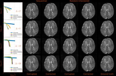
The simulations in3 showed that global convergence of the aligned reconstruction is strongly sensitive to the traversal order. Here, simulations are extended to gain evidence of optimal Cartesian sampling ordering in volumetric imaging, where measurements cover a 2D phase-encode space. Mimicking this undersampled 2D space and looking for concise conclusions, the simulations investigate the ability to retrieve 2D rotational motion for sequential (standard manufacturer sampling), regular-checkered, random-checkered, and purely random $$$\mathbf{k}$$$-space traversals (Figs. 1 and 2). The non-sequential orderings proposed are aimed at maximizing the sensitivity to motion induced inconsistencies by sampling a diverse set of harmonics in a short period of time.
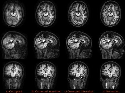
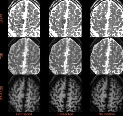
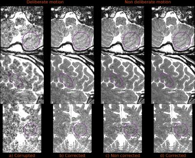
These schemes were implemented on a Philips 3T Achieva for in-vivo application in virtually any 3D sequence. Testing demonstrated that fully random sampling resulted in intrusive artifacts attributed to Eddy currents, whereas random-checkered zig-zag patterns were largely free of such problems5 while allowing matched contrast with standard sampling and presenting interesting properties to resolve motion (see caption of Fig. 1). Three experiments, using a standard 32-channel coil, are covered here: a) low resolution / extreme motion, where a volunteer was asked to perform extreme and continuous motion of the head during the whole 1.5mm isotropic MP-RAGE scan (Fig. 3); b) standard resolution / moderate motion, where the volunteer continuously and smoothly moved the head during 1mm isotropic bSSFP, TSE and MP-RAGE scans (Fig. 4); and c) high resolution scan, 0.7mm isotropic bSSFP, with and without deliberate motion (Fig. 5).
Results
Simulations in Fig. 2 show that all regular-checkered, random-checkered, and purely random traversals substantially outperform sequential traversal. In-vivo reconstructions of Figs. 3 and 4 are almost indistinguishable from motion-free counterparts. Temporal resolvability of motion varies with the type of sequence and spatial resolution, but was always below the time required for a full checkered $$$\mathbf{k}$$$-space sweep, allowing some degree of intra-shot corrections for multi-shot sequences (Fig. 3c vs. 3b). Reconstructions of high resolution data (Fig. 5) are also effective in case of deliberate motion (Fig. 5b vs. 5a) and show slightly improved contrast compared to non-corrected reconstructions for non deliberate motion (Figs. 5d vs. 5c).Discussion
Simulation results show that a distributed sampling ordering of the $$$\mathbf{k}$$$-space supports more effective motion correction than sequential ordering. In-vivo tests show that the combination of the proposed sampling and reconstruction techniques is highly effective, even in cases of extreme and continuous head motion, suggesting that the proposed approach could be used for motion correction in non-compliant subjects and to improve data quality in the general population1. Experiments also show that the framework holds promise for motion resolvability of ultra-high resolution MRI. This approach is compatible with parallel imaging acceleration but Eddy currents and SNR may be limiting factors and thus care is needed to avoid Nyquist violation problems introduced by rotations. Problem complexity is $$$O(N^5)$$$ with $$$N$$$ being the image grid size, which imposes computational limitations.Conclusion
We have shown that DISORDER provides robustness against motion for brain MRI. The proposed method is fully data-driven, can accommodate short lived motion states independent of shot structure, does not require data rejection, and can be applied to different volumetric modalities.Acknowledgements
The authors acknowledge financial support from the European Research Council under the European Union’s Seventh Framework Programme (FP/2007-2013) / ERC Grant Agreement n.319456. This work was also supported by Medical Research Council (MRC) strategic grant MR/K006355/1 and the Department of Health via the National Institute for Health Research(NIHR) comprehensive Biomedical Research Centre award to Guy’s & St Thomas’ NHS Foundation Trust in partnership with King’s College London and King’s College Hospital NHS Foundation Trust. The authors also acknowledge the Department of Perinatal Imaging & Health at King’s College London.References
1. Andre J, Bresnahan B, Mossa-Basha M, et al. Towards quantifying the prevalence, severity, and cost associated with patient motion during clinical MR examinations. J Am Coll Radiol. 2015;12(7):689-695.
2. Budde J, Shajan G, Scheffler K, and Pohmann R. Ultra-high resolution imaging of the human brain using acquisition-weighted imaging at 9.4T. NeuroImage. 2014;86(1):592-598.
3. Cordero-Grande L, Teixeira RPAG, Hughes EJ et al. Sensitivity encoding for aligned multishot magnetic resonance reconstruction. IEEE Trans Comput Imaging. 2016;2(3):266-280.
4. Batchelor PG, Atkinson D, Irarrazaval P, et al. Matrix description of general motion correction applied to multishot images. Magn Reson Med, 2005;54(5):1273-1280.
5. Tsao J, Kozerke S, Boesiger P, and Pruessmann KP. Optimizing spatiotemporal sampling for k-t BLAST and k-t SENSE: application to high resolution real-time cardiac steady-state free precession. Magn Reson Med, 2005;53(6):1372-1382.
Figures


