0297
Prediction of Motion Induced Image Degradation Using a Markerless Motion Tracker1DTU Compute, Technical University of Denmark, Lyngby, Denmark, 2Department of Clinical Physiology, Nuclear Medicine & PET, Rigshospitalet, Copenhagen University Hospital, Copenhagen, Denmark, 3TracInnovations, Ballerup, Denmark
Synopsis
In this work a markerless motion tracker, TCL2, is used to predict image quality in 3D T1 weighted MPRAGE MRI brain scans. An experienced radiologist scored the image quality for 172 scans as being usable or not usable, i.e. if a repeated scan was required. Based on five motion parameters, a classification algorithm was trained and an accuracy for identifying not usable images of 95.9% was obtained with a sensitivity of 91.7% and specificity of 96.3%. This work shows the feasibility of the markerless motion tracker for predicting image quality with a high accuracy.
Purpose
Patient head movement is a frequent cause of image degradation in clinical and research MRI brain scans, in particular for advanced MRI and high-resolution 3D sequences.
Current procedure after ended image acquisition is a visual inspection of the recorded image series by an MRI technician determining if image quality is satisfactory or a repeated scan is required. This procedure is observer dependent, extends duration of the scanning session and thus possibly leading to increased motion due to fatigue, and ultimately reduced throughput of the scanner. A study has shown that patient movement may result in up to $140,000 in lost revenue per scanner per year due to image artifacts caused by motion1. Universal and fully integrated motion correction is naturally preferred. However, intensive research over the past decade has shown that this is non-trivial for clinical routine.
The aim of this study is to demonstrate and develop a simple and clinically feasible tool easily accessible for MRI technicians deeming a newly recorded image sequence as being successful or not based solely on patient motion.
Afacan et al.2 evaluated the motion problem in a pediatric population. In this study we predict image quality based on newly obtained scans.
Methods
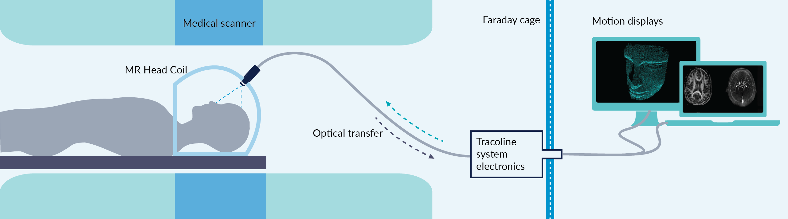
Patient motion was tracked on 136 patients scanned at a Siemens mMR Biograph hybrid PET/MRI scanner using an MRI compatible version, TCL2, of the markerless motion tracker, Tracoline3-4 (Figure 1).
Image series (pre- and post-contrast 3D T1 MPRAGE scans) included in the study (n = 172) were from patients with dementia (n = 73) and both pediatric patients (n = 53) and adults (n = 46) with brain tumors. These patients were included in different studies where the tracking equipment was used for quality insurance and general evaluation of patient motion. All data used in this study was anonymized.
The image quality was scored by an experienced radiologist as being not usable in clinical practice (positive class) or usable (negative class).
Baseline of motion was detected by applying a lowpass finite impulse response (FIR) filter to the motion data. The analyzed motion parameters were: 1) number of peaks with sudden movement >1 mm compared to baseline, 2) maximal motion after baseline subtraction, 3) percentage of time with motion less than 0.1 mm, 4-5) standard deviation of motion curve both with and without baseline subtracted.
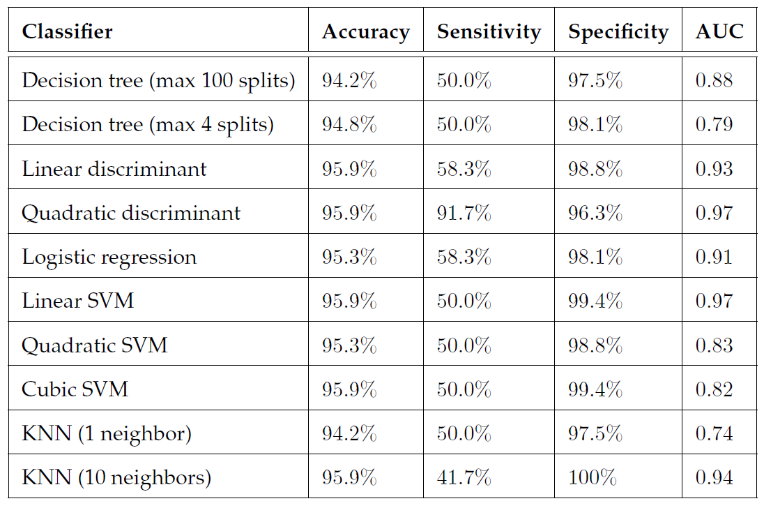
Based on these five parameters and the scoring of image quality, different classification algorithms were trained (decision trees, discriminant analysis, logistic regression, support vector machine and nearest neighbor) to determine which yielded the best performance. All classification algorithms were trained using 10-fold cross-validation.
Results
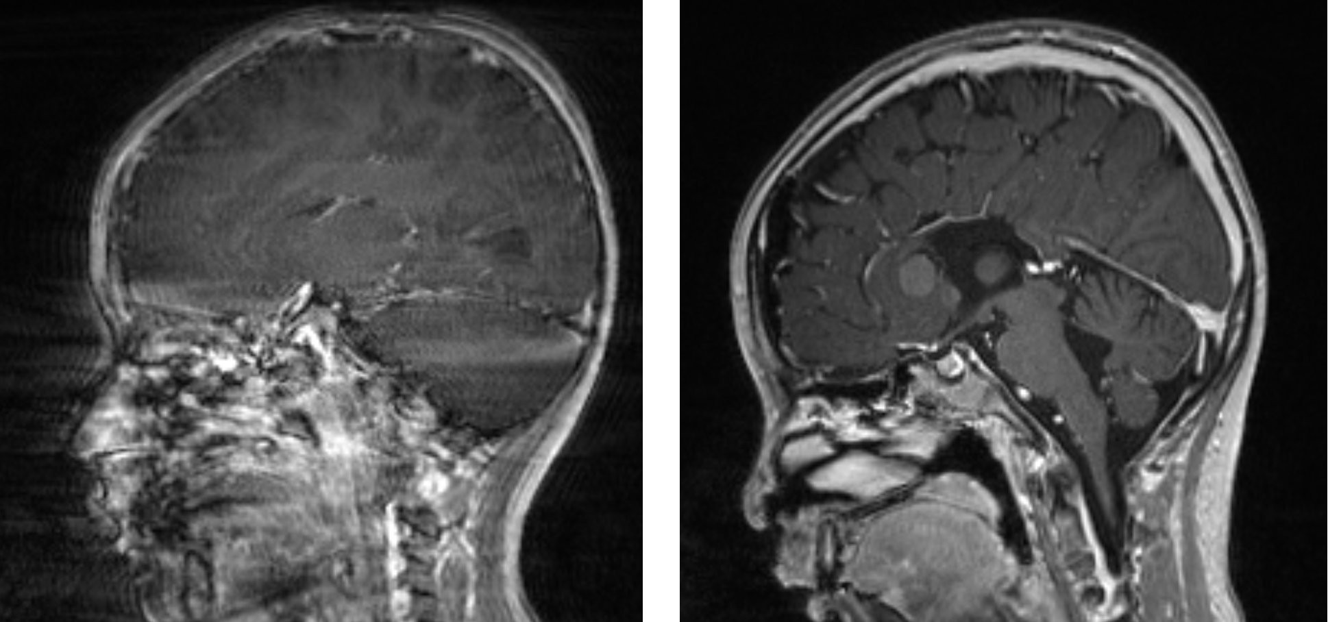
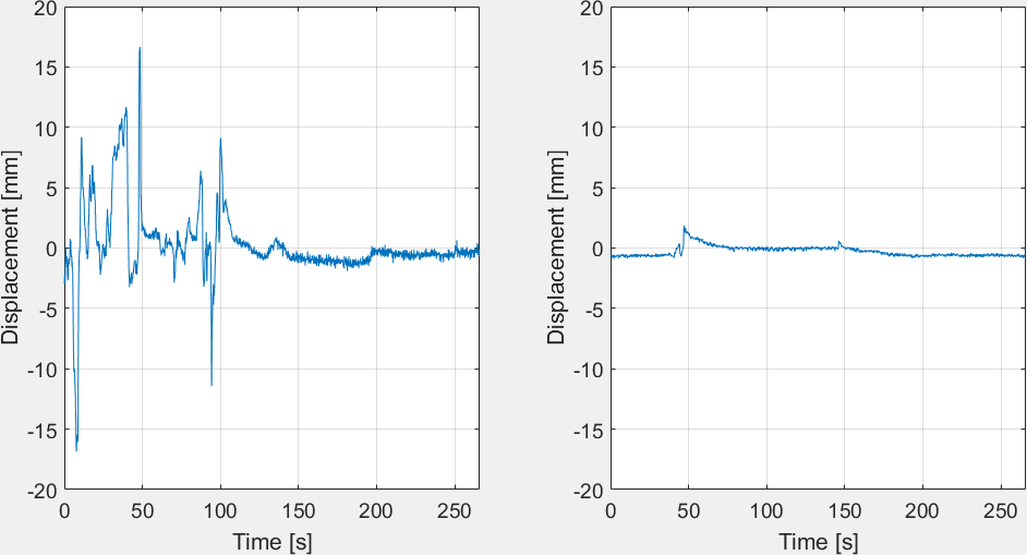
Examples of the influence of patient motion on image quality are provided in Fig. 2 with corresponding motion curves seen in Fig. 3. It was deemed that 12 sequences belonged to the positive class and 160 belonged to the negative class.
Quadratic discriminant analysis yielded the best performance with a classification accuracy of 95.9%, a sensitivity of 91.7%, specificity of 96.3% and AUC of 0.97. This means that the algorithm was able to correctly predict if a repeated scan was required in 11 of 12 cases. Performance of all trained classifiers is visualized in Fig. 4.
Discussion
Prediction of image quality is feasible using the markerless tracker with a high sensitivity and specificity. The algorithm presented here can easily be adopted to other image sequences, and possibly in the future be able to predict image quality during acquisition as all motion parameters can be estimated prospectively.
The dataset used in this work was highly imbalanced, with a limited number of negative cases compared to positive. One could have decided to exclude a subset of images from the positive class, but this would have further reduced the size of the dataset and was therefore not done.
The image scoring performed by the medical expert is to a high degree subjective and dependent on application. This lead to many borderline cases, where it is not clear if a repetition of recording was needed or not.
Conclusion
The objective with this work was to demonstrate the feasibility of using the markerless motion tracker, TCL2, to predict image quality in a routinely recorded MRI sequence. Based on 172 image series, scoring by an experienced radiologist and quadratic discriminant analysis, an accuracy of 95.9% was obtained with sensitivity of 91.7% and specificity of 96.3%. The technique could be adopted to other MRI sequences, and used to develop a tool for predicting image quality during image acquisition which could reduce costs and increase clinical throughput.Acknowledgements
This work was supported by Novo Nordisk Foundation. TracInnovations is thanked for their help in using and setting up tracking equipment.References
1. Andre, J. B., Bresnahan, B. W., Mossa-Basha, M., et al. Toward quantifying the prevalence, severity, and cost associated with patient motion during clinical MR examinations. Journal of the American College of Radiology. 2015;12(7):689-695.
2. Afacan, O., Erem, B., Roby, D. P, et al. Evaluation of motion and its effect on brain magnetic resonance image quality in children. Pediatric Radiology. 2016:1-8.
3. Olesen, O. V., Paulsen, R. R., Højgaard, L., et al. Motion Tracking for Medical Imaging: A Nonvisible Structured Light Tracking Approach. IEEE Transactions on Medical Imaging. 2012;31(1):79-87.
4. Olesen, O. V., Wilm, J., der Kouwe, A. V., et al. An MRI Compatible Surface Scanner. ISMRM 2014 pp. 1303.
Figures