0254
Characteristic Changes of Volume and Shape of Subcortical Structures in Obsessive-Compulsive Disorder1Radiology Department, Huaxi MR Research Center (HMRRC),West China Hospital of Sichuan University, Chengdu, People's Republic of China
Synopsis
We analysis subcortical nucleus volume and shape alterations in a relatively large sample of drug-naive adult patients with Obsessive-Compulsive disorder by using an automatically segmentation and vertex-based shape analysis protocol. Our findings indicated that (i)Gender effects were found in OCD, male patients seems are affected in reward system whereas females might have more disrupted stratum-pallidum-thalamus pathway which might be related to chronic stress. (ii)Shape analysis may provide more anatomical change information from a different perspective. Future structural research should consider shape and volume alterations together when explore brain changes.
Purpose
Obsessive-Compulsive disorder (OCD) is a common psychiatric disorder with obsessions and compulsions as the main symptom profile. It has been suggested to associate with dysfunction in cortico-striato-thalamo-cortical(CSTC) circuit[1]. Basal ganglia and thalamus were important hubs in this circuit and easily affected by drugs, however, few studies had investigated the morphological changes of those structures in drug naive patients. Herein, in current study, we aim to explore the volume and shape of subcortical structures in a relatively large sample of drug-naive OCD patients.Method
83 DSM-IV criteria diagnosed OCD patients and 93 age and sex matched healthy control were recruited and informed consents were obtained from all subjects. High resolution T1 weighted images were obtained using a volumetric 3-dimensional Spoiled Gradient Recall (SPGR) sequence (TR/TE = 8.5/3.4ms; flip angle = 12o; 156 axial slices with thickness = 1mm, field of view = 24×24cm2 and data matrix = 512×512) via a GE 3.0 T scanner.
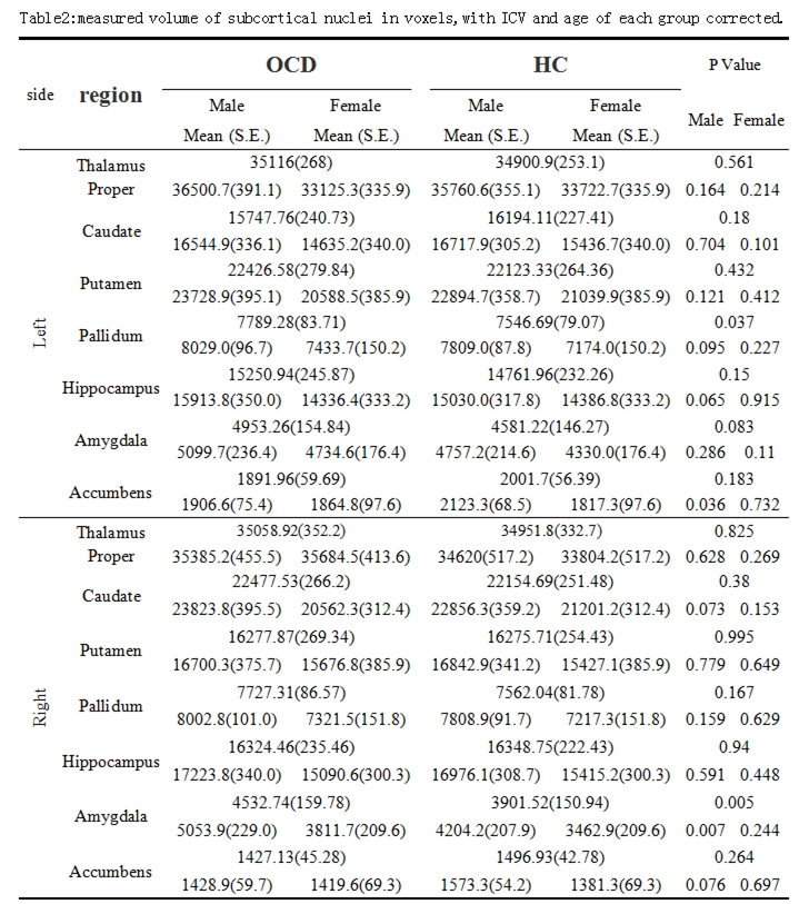
The structural data was automatically segmented using FSL software, volume of 7 subcortical nucleus are extracted as well as intracranial volume (ICV). Volume differences between two groups were compared using a general linear model (GLM) with age and intracranial volume as covariates. Diagnosis, hemisphere were entered as fixed factors. Correlations between volumetric measurements with illness duration, scores of YBOCS (Yale-Brown Obsessive Compulsive Scale), HAMA (Hamilton Anxiety Scale), and HAMD (Hamilton Depression Scale) were examined by using Pearson’s correlation under SPSS 17. Gender effect were further analysis by performing comparison between male and female patients separately. Shape analysis of subcortical nucleus was conducted with FIRST vertex analysis tool to yield a localized measure of shape differences between groups. Statistical of shape differences between two groups were also performed using a general linear model (GLM).
Result
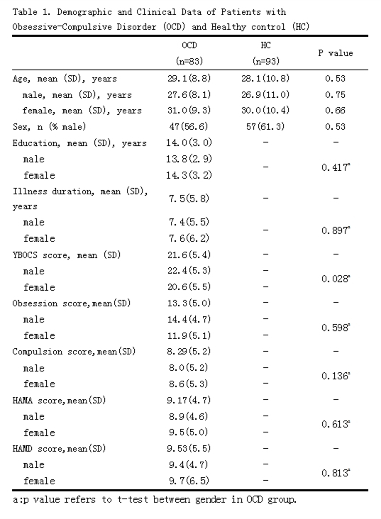
Demographic information for all subjects was shown in table1.
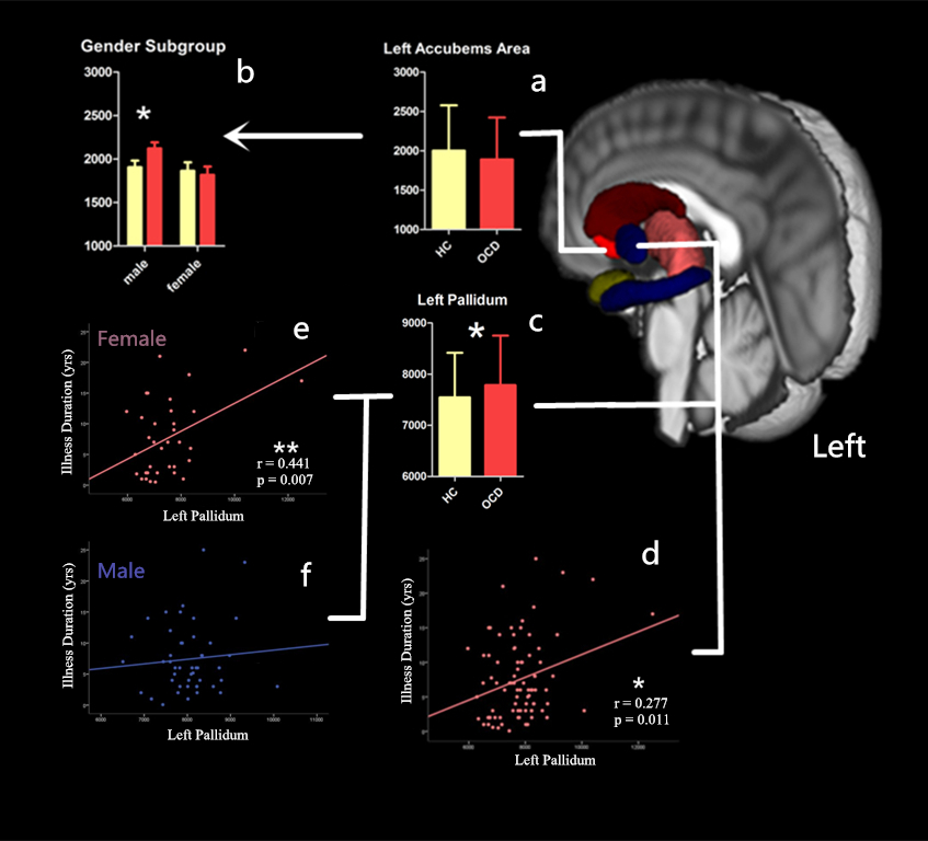
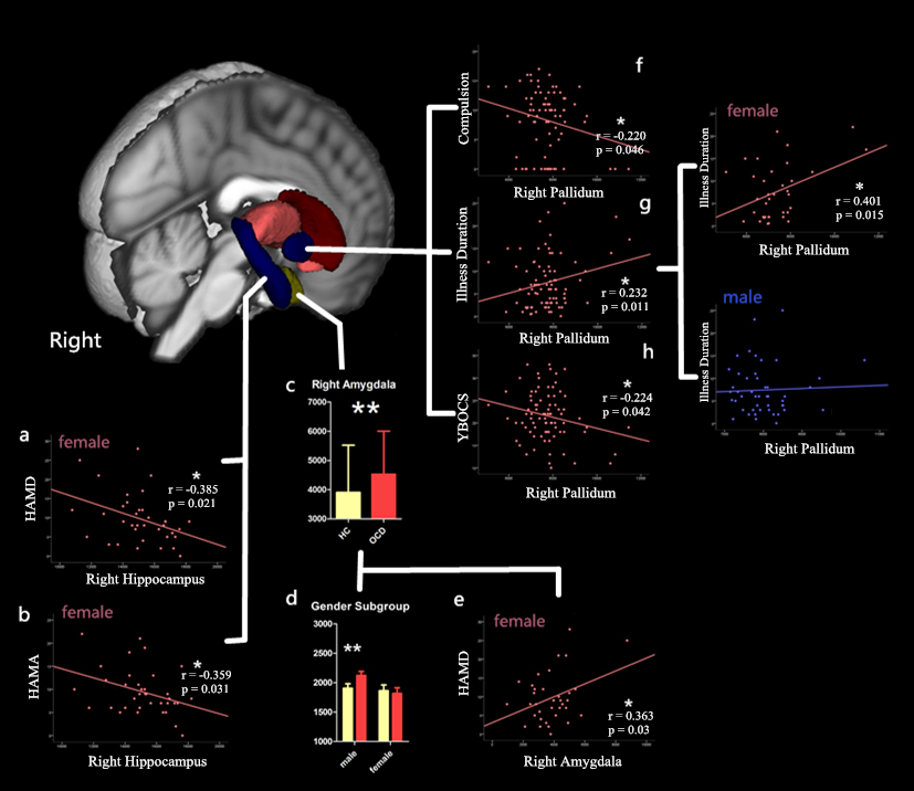
Comparing to healthy controls, OCD patients showed significantly enlarged pallidum (left side, p=0.037) and amygdala (right side,p=0.005)(table 2, figure 1 & 2). Male patients showed enlarged right amygdala(p=0.007) and shrinked left accumbens(p=0.036). Females, however, did not show significant difference comparing with HC.
Pallidum is significantly correlated with illness duration bilaterally, both in gender-combined analysis (left: r=0.277, p=0.011; right: r=0.232, p=0.035) and in female patients (left: r=0.441,p=0.007; right:r=0.401,p=0.015). Right pallidum is also correlated with compulsion (r=-0.220, p=0.046) and YBOCS scales (r=-0.224, p=0.042) when gender was combined. In female patients, HAMD score was found to be correlated with right hippocampus (r=-0.385,p=0.021) and amygdala (r=0.363,p=0.03) and HAMA was found to be correlated with right hippocampus(r=-0.359, p=0.031). No significant correlation was found in male patients.
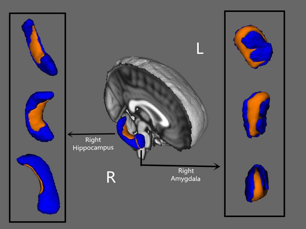
Vertex based shape analysis demonstrated altered morphology in right amygdala and right hippocampus. In hippocampus, deformation mainly happened in the ventromedial subregion. In Amygdala, it basically happens in dorsolateral area, as illustrated in Figure 3.
Disscussion & Conclusion
In current study, we found different pattern of subcortical abnormalities in male and female OCD patients. Accumbens is a critical nucleus in reward system, which is been affected mainly in male patients. Whereas, female patients seems suffer more from disrupted striatum-pallidum-thalamus integrity considering the fact that pallidum enlarged significantly and correlated with illness duration[2]. Stress seems another factor more prominent in female OCD as both HAMA and HAMD correlated with hippocampal volume. These findings highlight the potential importance of gender effects in OCD and suggest that further research on sex difference in OCD may be useful.
Shape analysis provide more information about morphometry from three-dimensional aspect. Even without significant volume alteration, there might be distortion of shape which implicate there might be dysfunction at histological and celluar level. Therefore, combining shape analysis with routine volumetric analysis might be worthwhile in the future structural study.
Acknowledgements
This study was supported by the National Natural Science Foundation (Grant No. 81671669 and 81171488)References
[1]Mohammed R.et al.(2012) Trends in Cognitive Sciences, Jan 6(1): 43-51,
[2]L.Menzies, et al.(2008) Neurosci Biobehav Rev,32:525-549
Figures