0235
Accelerating multi-contrast imaging in neuro-exam with sharable information1Electrical Engineering, Stanford University, Stanford, CA, United States, 2Radiology, Stanford University, Stanford, CA, United States
Synopsis
Neurological disorders results in great clinical challenges and high societal burdens. Currently multi-contrast MRI exams are frequently used for diagnosis because of the various tissue contrasts provides complementary diagnosis information to distinguish normal tissue from pathology. However, the cost of acquiring these multiple sequences is extensive scanning time, which significantly increases both the diagnosis cost and patients’ discomfort. Here we proposed a new approach to accelerate multi-contrast imaging by using Parallel Imaging, Compressed Sensing and sharable information. We validated the new approach with experiments on both patients and healthy subjects. We demonstrate that we can reduce the multi-contrast MRI scanning time significantly while preserving the diagnostic information.
Clinical Question
How can we quantify the anatomical and functional neurological pathology from multi-contrast imaging within a short time?Impact
Neurological disorder results in great clinical challenges and high societal burdens. Millions of people die due to neurological disorders every year [1]. Cerebrovascular disease, for example, is the 3rd most common cause of death in the USA. Mental illness has high cost both to individuals, families and society. MRI plays a critical role in diagnosis and clinical decision-making because of the myriad tissue contrast to distinguish normal tissue and pathology. The proposed technique targets for this vast population of patients with neurological disorders to reduce the scan duration and therefore the cost of multi-contrast neuro-imaging while preserving the diagnostic information.Approach
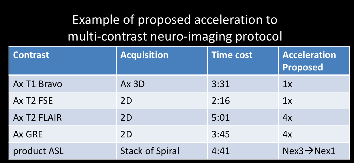
Currently, multiple sequences are performed consecutively for different contrasts. On example of study protocols are listed below. Multi-contrast neural imaging can provide fundamental diagnosis information from the complementary tissue contrasts. However acquiring multiple scans with different contrasts requires extensive scanning time which increase the cost of the diagnosis significantly.
Here we propose our technique to improve the reconstruction of undersampled multi-contrast imaging using the sharable information among different scans, an approach known as PROMISE [2]. We can use this information to increase acceleration factors and shorten the entire protocol. The proposed protocol is listed as following. First we acquire FSPGR T1-weighted and T2-weighted fast spin-echo (FSE) sequences which are relatively fast and provide good references for the sharable information , in structure and sensitivity information for improving Compressed Sensing (CS) and Parallel Imaging (PI) respectively [2,3]. Using these prior scans, we are able to greatly accelerate later sequences. Examples of the following accelerated scans include both anatomical scans (e.g. GRE, FLAIR) and functional scans (e.g. ASL). All these scans are time consuming and the proposed technique enables further undersampling with high reduction factor. For each highly undersampled scan, we reconstructed them separately [2] by solving the optimization regularized by sharable information. Sequential and separate reconstruction is used instead of reconstructing all the scans together [4] is due to the clinical need for reconstruction right after each scan.
The optimization objective function is as following, in which k-space x is recovered from sample y with under-sampling operator D, sensitivity information is encoded with PI self-calibration kernel G, CS regularization is conducted using Sparsity transform such as \(\Psi\) here for Wavelet transform. R is for optional image registration operator and F is the Fourier Transform. Both PI kernel G and weightings in the Sparsity Transform are extracted from sharable information for multi-contrast scans.
$$\hat{x}=\min_x{E(x)},{E(x)}=\parallel{DX-y}\parallel_2^2+\lambda_{PI}{\parallel{G_{sharable}x-x}\parallel_2^2}+\lambda_{CS}{\parallel{W_{sharable}\Psi R({F}^{-1}x})\parallel_1}$$
Gains and losses
Gains: By accelerating the multi-contrast scanning, we can shorten the entire multi-contrast neural imaging protocol while preserving the diagnostic information. A shorter neuro-exam is critical for patients with severe acute diseases such as stroke and brain hemorrhage, in which time is brain. Shorter scan time is also valuable to protect hearing due to extended exposure to loud noises, including its use in fetal imaging. Shorter acquisition times also may result in less motion artifact which is especially a problem for patients who cannot hold still in the scanner, such as children. Additional information can also be gained when we use the rest of normal MR scan time to acquire additional types of contrast, such as MRA, MRV, SWI, ASL etc. As a result, MRI can reach larger numbers of people, providing an exam without radiation at lower cost and shorter time requirements.
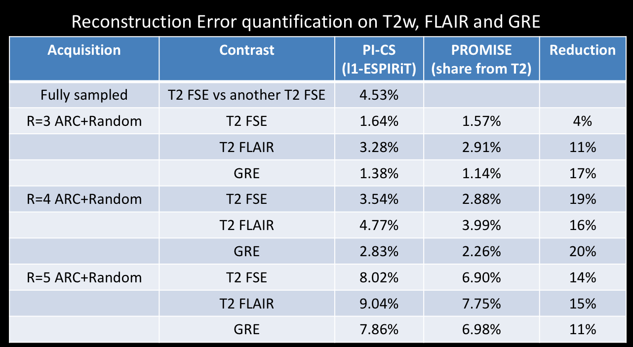
Losses: It is reasonable to expect that artifacts can occur when pushing the acceleration far. However, we can adjust the acceleration level based on our validation of diagnostic studies. Currently experiments have demonstrated that the method is robust for acceleration by a factor 4 for 2D imaging, much higher reduction factor possible with 3D imaging. Inter-scan motion is also a challenge for reconstruction using sharable information. Experiments shows the proposed technique is robust to motions on the order of several millimeters and image co-registration can further reduce the effects of larger motions.
Preliminary Data
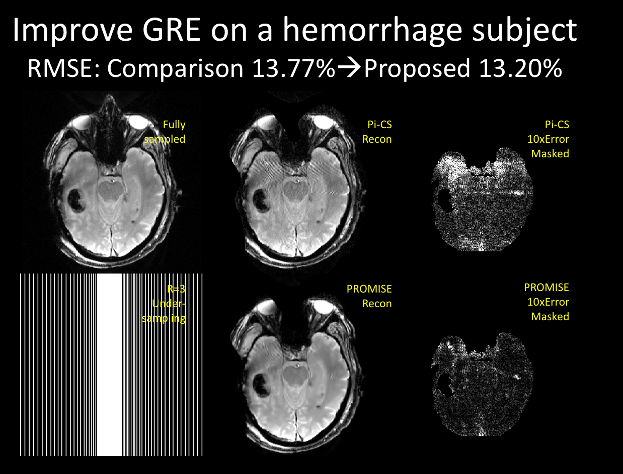
The diagnostic information to quantify the anatomical and functional pathology can be preserved perfectly and even potentially improved by using the proposed technique. We conducted various experiments on patients and healthy subjects to validate that the diagnosis quality is not jeopardized by significant reductions in scanning time. One example we have submitted in a patient with intraparenchymal hemorrhage shows that we can preserve good diagnostic information at a very high reduction factor. in a clinically relevant situation.Acknowledgements
Here we acknowledge GE Healthcare for supporting this work.References
1. http://www.who.int/mental_health/neurology/neurological_disorders_report_web.pdf
2. Gong, Enhao, et al. "PROMISE: Parallel-imaging and compressed-sensing reconstruction of multicontrast imaging using SharablE information." Magnetic resonance in medicine 73.2 (2015): 523-535.
3. Huang, Feng, and Enhao Gong. "Mri using spatially adaptive regularization for image reconstruction." U.S. Patent Application No. 14/916,200.
4. Chatnuntawech, Itthi, et al. "Vectorial Total Generalized Variation for Accelerated Multi-Channel Multi-Contrast MRI." Magnetic resonance imaging(2016).
Figures