0231
Targeted Rapid Knee MRI Exam using T2 Shuffling1Electrical Engineering and Computer Sciences, University of California, Berkeley, Berkeley, CA, United States, 2MR Applications and Workflow, GE Healthcare, Menlo Park, CA, United States, 3Radiology, Stanford University, CA, United States
Synopsis
We investigate the effectiveness of a targeted rapid pediatric knee MRI exam with total exam time of about 10 minutes. We aim to enable same-day MRI access, accommodating the abbreviated protocol between other scheduled patients. The protocol is based on T2 Shuffling, a four-dimensional acquisition that permits volumetric reconstruction of images with variable T2-contrast. Preliminary data for ten subjects referred for the targeted knee MRI exam is presented. Mean time from registration to exam completion averaged 48.5 minutes, with one outlier of 269 minutes due to technologist error in documentation.
Clinical Question
Can a rapid knee MRI protocol for pediatric knee pain enable near walk-in access to MRI?Impact
Pediatric knee pain is a common complaint in pediatric orthopedic clinics, with a variety of etiologies that require different clinical management. Typically, a parent must take time off work and a child time off school for the clinic visit. Though MRI often provides definitive diagnosis, cost is high, and scheduling requires parents and children to take another day off work and school. We aim to develop a rapid knee MRI protocol to enable same-day MRI access, accommodating the scan between two other patients.Approach
Although a rapid MRI may have high value, the realization of that value only occurs if the scan can be accommodated into a clinical schedule to yield incremental volume without expanding operating hours. Thus, this work presents the full integration of a fast imaging approach into a clinical workflow.
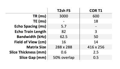
A four-dimensional fast spin-echo (FSE) acquisition that encodes the knee anatomy in volumetric fashion and also permits reconstruction of images with variable T2-contrast was implemented$$$^1$$$, termed T2 Shuffling (T2sh). The total protocol consisted of a localizer (1 minute), T2sh (7 minute), and a Coronal FSE T1 (3 minute). The scan parameters are summarized in Figure 1.
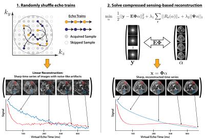
The overall methodology for the T2sh acquisition and reconstruction method is presented in Figure 2. The acquisition used a randomized view ordering to acquire phase encode samples during the spin-echo train. A compressed sensing-based reconstruction was implemented to resolve volumetric images at varying T2 contrasts.
Insurance plans were reviewed to subselect those plans with high likelihood waived authorization, rapid authorization, or retroactive insurance authorization. Patients with indications of anterior knee pain, or suspicion of osteochondral lesions, anterior cruciate ligament tears, or meniscal pathology covered by subselected insurance plans were eligible for rapid protocol knee MRI exams. A mechanism of communication between orthopedic clinic and an adjacent MRI scanner was established. Over a three month period, time from exam request to exam completion was recorded, along with time from patient registration to exam completion. Indications and clinical findings were also recorded.
Gains and Losses
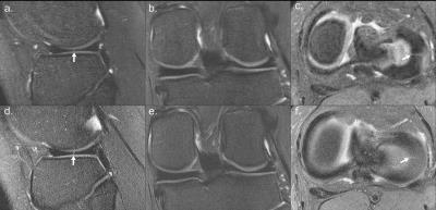
A prior study with 30 patients investigating a single sequence protocol using T2sh, compared against a conventional lengthy protocol, indicated that missing clinically relevant pathology is unlikely$$$^2$$$. A representative comparison between T2sh and conventional 2D MRI from the is presented in Figure 3. As additional patients can now be accommodated into the MRI schedule, the cost of the MRI is simply professional costs of the radiologist and minimal costs associated with reporting infrastructure. The magnet, technologists, and receptionists are already present. A significant additional benefit is same-day scheduling, which reduces lost productivity of parents and children at their work and school.Preliminary Data
Ten subjects had targeted knee MRI orders, with eight exams completed on the same day as requested. Of these eight subjects, mean time from order to completion was 138.6 minutes (minimum of 15 minutes, maximum of 310 minutes). Time from registration to exam completion averaged 48.5 minutes, with one outlier of 269 minutes due to technologist error in documentation. Omitting this outlier, mean time was 26.4 minutes. Clinical indications and MRI findings are shown in Table 1.Acknowledgements
We thank the following funding sources: National Institutes of Health (NIH) grants R01EB009690, R01EB019241, P41RR09784; Sloan Research Fellowship; Bakar Fellowship; GE Healthcare (research support)References
1. Tamir JI, Uecker M, Chen W, Lai P, Alley MT, Vasanawala SS, Lustig M. T2 Shuffling: Sharp, Multicontrast, Volumetric Fast Spin-echo Imaging. Magnetic resonance in medicine (2016). doi:10.1002/mrm.26102. [Epub ahead of print].
2. Bao S, Tamir JI, Young JL, Tariq U, Uecker M, Lai P, Chen W, Lustig M, Vasanawala SS. Fast comprehensive single-sequence four-dimensional pediatric knee MRI with T2 shuffling. Journal of magnetic imaging (2016). doi: 10.1002/jmri.25508. [Epub ahead of print].
Figures


