0191
Volumetric T2-weighted and FLAIR Imaging of Spine with Uniform Fat Suppression in a Single Acquisition1Radiology, UT Southwestern Medical Center, Dallas, TX, United States, 2Bioengineering, UT Dallas, Dallas, TX, United States, 3Advanced Imaging Research Center, UT Southwestern Medical Center, Dallas, TX, United States
Synopsis
2D T2-weighted turbo spin-echo (T2w-TSE), fluid attenuated inversion recovery (FLAIR) with and without fat suppression are widely used in the clinical brain and spine protocols to improve diagnosis. However, FLAIR suffers from low SNR and long scan times. In this work, we developed a dual-acquisition 3D TSE sequence combined with dual-echo Dixon based approach to generate T2-weighted and FLAIR images of the spine with and without fat suppression in a single acquisition using the similar acquisition time as 2D FLAIR. Uniform fat/water separation was achieved using a shared-field-map and complex subtraction was used to generate FLAIR-like images without artifacts.
Introduction
T2-weighted imaging and fluid attenuated inversion recovery with fat saturation (FLAIR-FS) are highly beneficial to improve lesion conspicuity in the brain and spine1,2. While FLAIR is routinely used in the brain, it often suffers from poor suppression of fluid and fat in spine due to increased B0 inhomogeneities. Additionally, the acquisition of both T2-weighted and FLAIR images further increases the total scan times increasing vulnerability to patient motion. Alternatively, the dual-acquisition based TSE sequence can provide robust fluid suppression by subtracting the longer echo time (TE) image from the shorter TE image, providing both a T2-weighted and a FLAIR image from the same acquisition2,3. However, extending this to spine imaging is challenging due to poor fat suppression. Thus, the purpose of this work was to develop a dual-acquisition 3D TSE sequence combined with dual-echo Dixon based approach for uniform fat suppression to generate T2-weighted and FLAIR images of the spine with and without fat suppression in a single acquisition.Theory
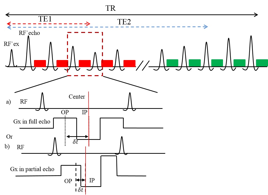
The proposed dual-acquisition 3D TSE sequence with dual-echo Dixon generates 4 sets of echoes, including two gradient echoes (in-phase, IP; out-of-phase, OP) at both shorter and longer TEs (TE1/TE2) in a single repetition (Fig.1). The four signals can be modeled as: $$S_1=(W_{TE1}+c_1F_{TE1})e^{i\phi_1}~\\S_2=(W_{TE1}+c_2F_{TE1})e^{i\phi_2}~\\S_3=(W_{TE2}+c_1F_{TE2})e^{i\phi_3}~\\S_4=(W_{TE2}+c_2F_{TE2})e^{i\phi_4}$$with$$c_n=\sum_mw_me^{i(2\pi\Delta{f_m}+iR_m)\delta{t_n}}$$where the index m indicates spectral peaks of fat with off-resonance frequency $$$\Delta{f_m}$$$ and transverse relaxation rates $$$R_m$$$. $$$\delta{t_n}$$$ is the echo time shift with respect to Hahn echo. $$$W$$$ and $$$F$$$ are considered as complex, $$$W'=We^{i\psi_1};~F'=Fe^{i\psi_2}$$$. With this approach, the two echoes at the shorter TE can undergo standard Dixon reconstruction using the estimated $$$ \Delta{\Phi_{TE1}}=e^{i(\phi_2-\phi_1)} $$$. This generates $$$W_{TE1}$$$ and $$$F_{TE1}$$$ as4:$$\left(\begin{array}{c}W_{TE1}'\\F_{TE1}'\end{array}\right)=\frac{1}{c_2-c_1}\begin{bmatrix}c_2&c_1\\-1&1\end{bmatrix}\left(\begin{array}{c}S_1\\S_2\Delta\Phi_{TE1}^*\end{array}\right)$$Since the majority of the tissue signals decay at longer TE (TE2), the signal to noise ratio (SNR) will be poor for proper fat/water separation at TE2. However, the B0 field should remain the same between TE1 and TE2 and hence, $$$\Delta{\Phi_{TE2}}=e^{i(\phi_4-\phi_3)}$$$ is identical to $$$\Delta{\Phi_{TE1}}$$$. Using this shared B0 field map between shorter TE and longer TE, $$$W_{TE2}'$$$ and $$$F_{TE2}'$$$ can be calculated as:$$\left(\begin{array}{c}W_{TE2}'\\F_{TE2}'\end{array}\right)=\frac{1}{c_2-c_1}\begin{bmatrix}c_2&c_1\\-1&1\end{bmatrix}\left(\begin{array}{c}S_3\\S_4\Delta\Phi_{TE1}^*\end{array}\right)$$Using these two reconstructed water-only images, the fat suppressed FLAIR-like images can be calculated as:$$I=Real[(W_{TE1}'-W_{TE2}')e^{-i\psi_1}]$$with$$\psi_1=\angle{W_{TE1}'}$$.
Methods
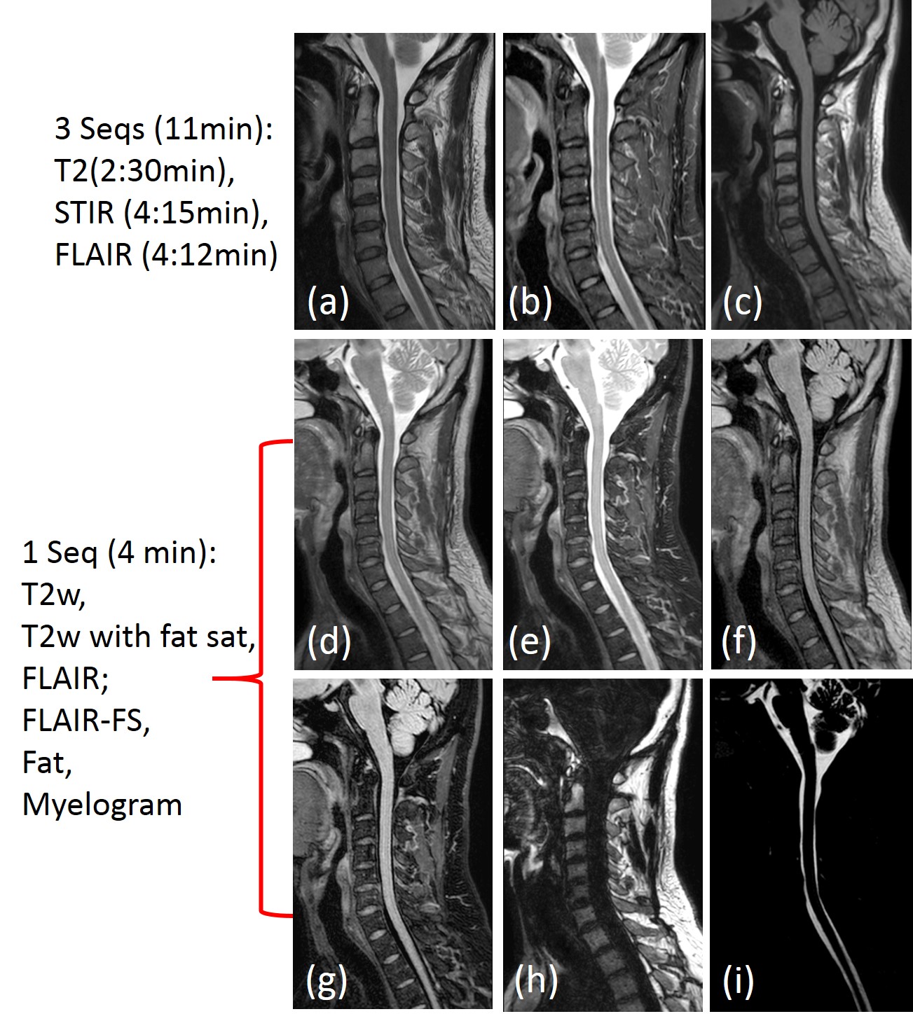
The dual-acquisition 3D TSE sequence with dual-echo Dixon was implemented on a 3T Ingenia scanner (Philips Healthcare, Best, The Netherlands) and is shown in Figure 1. The acquisition and reconstruction methods were evaluated and compared against standard 2D FLAIR and 2D STIR of the spine in 5 healthy volunteers with IRB approval and written informed consent. The typical imaging parameters of the proposed sequence included: sagittal and/or axial orientation; FOV = 360×360×199 mm; Resolution = 1×1×1 mm; SENSE = 2; equivalent TE1/TE2 = 72/304 ms, $$$\delta{t}$$$= 1.1 ms, partial readout of 0.8, total scan time = 4:05 minutes. The multi-slice 2D T2w, STIR and FLAIR images were acquired using the same parameters except: TR = 2500/6000ms (STIR/FLAIR); TE = 35/120ms (STIR/FLAIR); TI = 240/2000ms (STIR/FLAIR) and a slice thickness of 3mm. The total scan time for the 2D T2-weighted (2:30 min), 2D T2-STIR (4:15 min) and 2D T2-FLAIR (4:12 min) sequences were about 11 mins.Results
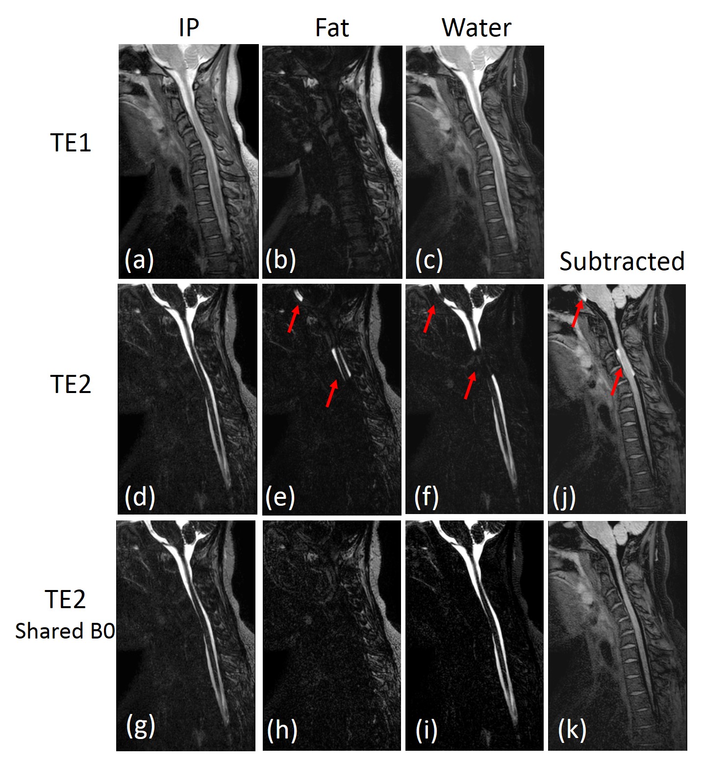
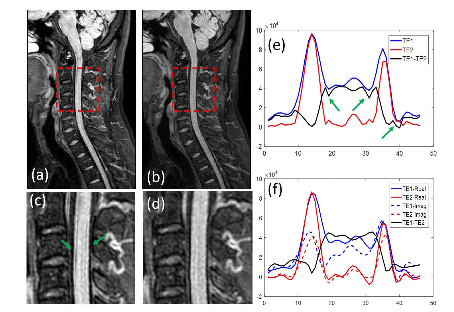
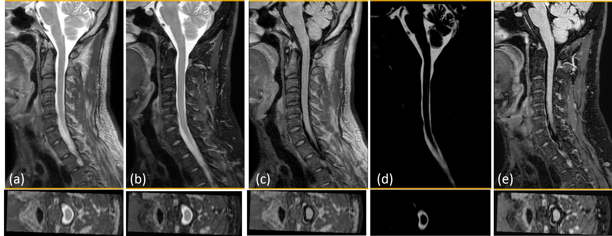
IP images at shorter TE (a) and longer TE (b) are shown in Figure 2. Shorter TE (TE1) images with higher SNR provided robust fat/water separation (b, c), while longer TE (TE2) images had fat/water swapping (e, f) and resulted in subtraction artifacts (j). Using a shared-field-map between TE1 and TE2 images, robust fat/water separation (h, i) in longer TE (TE2) image was successfully achieved and the subtraction artifacts were eliminated (k). Figure 3 compared magnitude (a, c) and complex subtraction (b, d) between the TE1 and TE2 water-only images. Magnitude subtraction results in subtraction errors (c, e) due to magnetization modulation and loss of phase information. Using the shared-field-map reconstruction and phase preservation, these artifacts can be eliminated (d, f). Multi-contrast 3D images (T2-weighted, T2-weighted with fat saturation, FLAIR, FLAIR-like with fat suppression, Fat-only and Myelogram etc., Fig. 4.d-i) can be generated using proposed sequence in 4:05 min, while standard multi-slice T2-weighted, STIR and FLAIR sequences required 11 mins in total. Figure 5 shows representative reformatted multi-contrast images from a single acquisition.Conclusion
The proposed dual-acquisition 3D TSE with dual-echo Dixon using shared-field-map between shorter and longer TE images generates multi-contrast high resolution volumetric images of the spine in clinically feasible scan times.Acknowledgements
No acknowledgement found.References
[1] Chagla GH, Busse RF, Sydnor R. et al. Three-dimensional fluid attenuated inversion recovery imaging with isotropic resolution and nonselective adiabatic inversion provides improved three-dimensional visualization and cerebrospinal fluid suppression compared to two-dimensional flair at 3 tesla. Investigative radiology. 2008; 43(8):547-551.
[2] Park J, Park S, Kim EY, et al. Phase-sensitive, dual-acquisition, single-slab, 3D, turbo-spin-echo pulse sequence for simultaneous T2-weighted and fluid-attenuated whole-brain imaging. Magnetic resonance in medicine. 2010; 63(5):1422-1430.
[3] Essig M, Deimling M, Hawighorst H, et al. Assessment of cerebral gliomas by a new dark fluid sequence, high intensity REduction (HIRE): a preliminary study. Journal of Magnetic Resonance Imaging. 2000; 11(5): 506-517.
[4]Eggers H, Brendel B, Duijndam A, et al. Dual-echo Dixon imaging with flexible choice of echo times. Magnetic resonance in medicine. 2011: 65(1): 96-107.
Figures