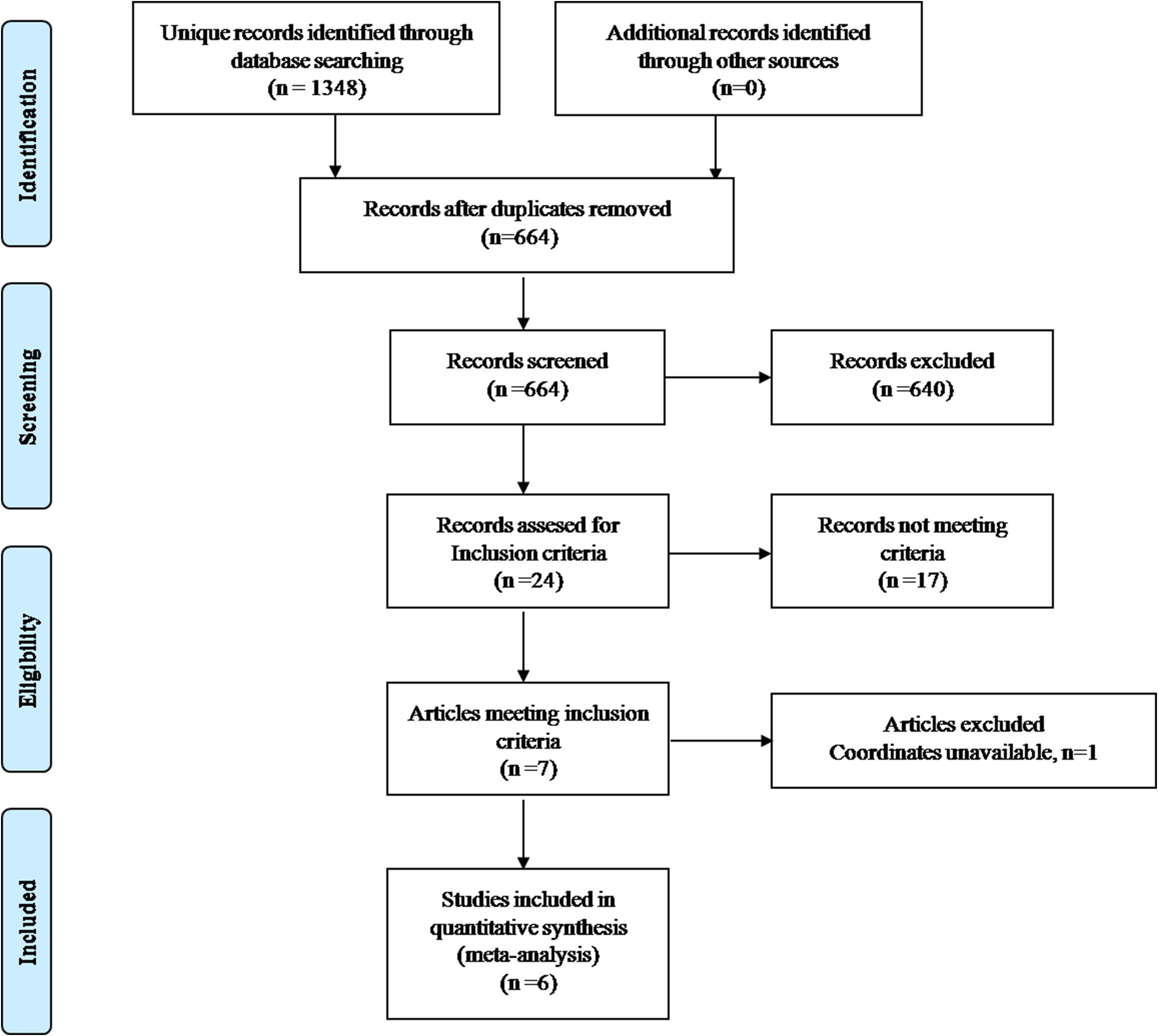
0190
Altered grey matter volume in patients with type 2 diabetes mellitus1Department of Radiology, Union Hospital of Tongji Medical College, Huazhong University of Science and Technology, Wuhan, People's Republic of China, 2Department of Radiology, Henan Provincial People's Hospital & the People's Hospital of Zhengzhou University, Zhengzhou, People's Republic of China
Synopsis
Our meta-analysis indicates that patients with T2DM have significantly and robustly reduced grey matter, mainly in the cortical-striatal-limbic networks. The meta-regression results suggest that T2DM patients with longer illness duration may have smaller grey matter volume in the right MTG. Our finding supports the notion that T2DM could lead to subtle diabetic brain structural changes, which may be correlated with cognitive impairment inT2DM patients.
PURPOSE
Previous studies of voxel-based morphometry (VBM) have found that patients with type 2 diabetes mellitus (T2DM) exhibit grey matter alterations, but these findings are inconsistent and have not been quantitatively reviewed. Therefore, we conducted a quantitative meta-analysis of VBM studies of patients with T2DM.
METHODS
The anisotropic effect size version of Seed-based D Mapping (AES-SDM) (http://www.sdmproject.com) method was applied to quantitatively estimate the regional grey matter abnormalities in T2DM patients. AES-SDM is a new coordinate-based meta-analytic technique that has been applied to many diseases, including depression, posttraumatic stress disorder, migraine and dementia. The method has been shown to be superior in some respects to the earlier methods, for example, activation likelihood estimation and multilevel kernel density analysis. For example, it can combine both positive and negative differences in the same map, thereby preventing a particular voxel from appearing to be significant in opposite directions. Additionally, AES-SDM enables several complementary analyses, such as jack-knife, subgroup and meta-regression analyses, that can be used to assess the robustness and heterogeneity of the results. First, we performed a pooled meta-analysis of the included studies. Then, a subgroup analysis, including data sets of patients with T2DM but without mild cognitive impairment, was performed. We also used meta-regression to explore the effects of some demographics and clinical characteristics.RESULTS
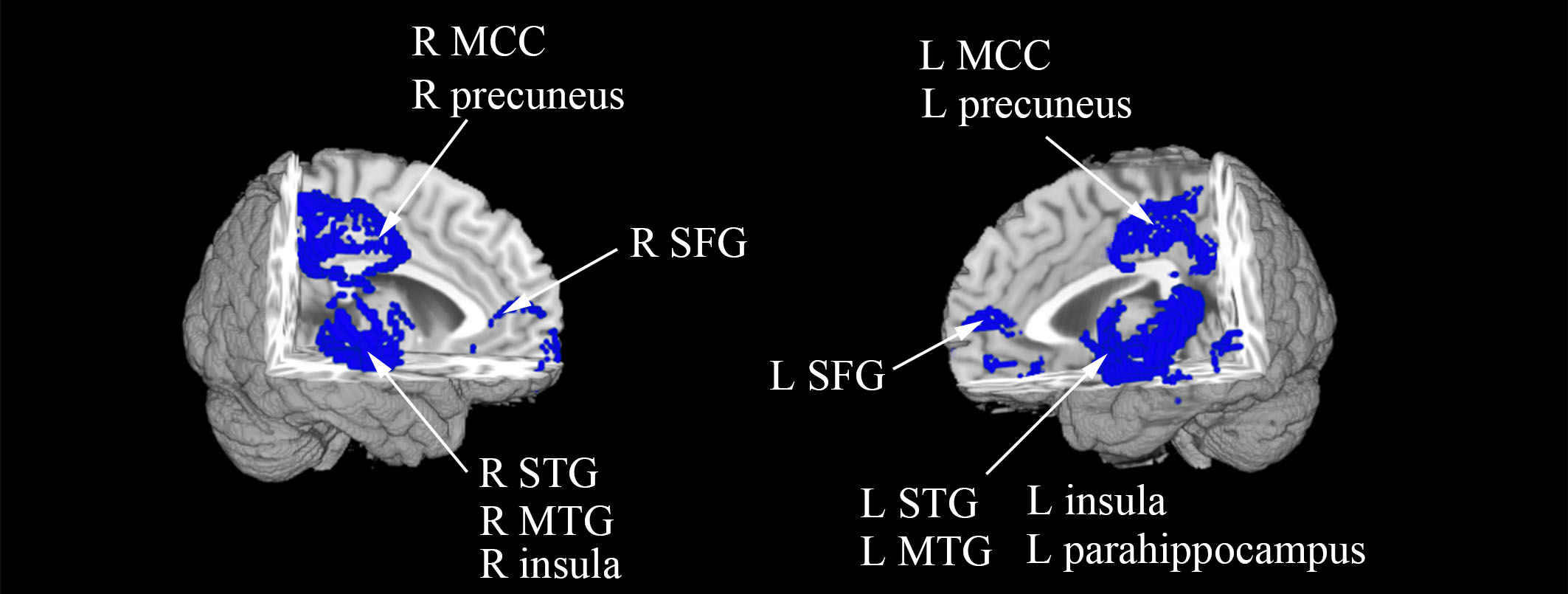
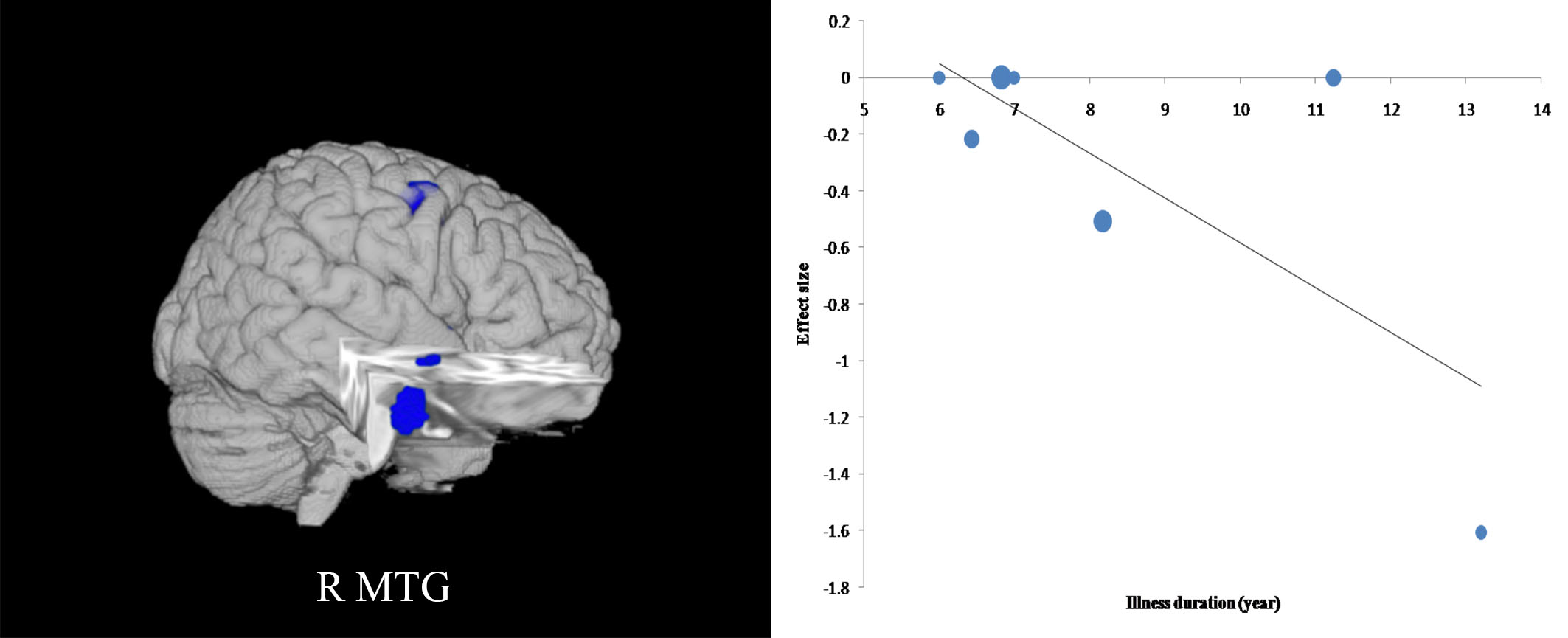
Six studies, with 7 datasets comprising 490 participants with T2DM and 508 non-T2DM controls, were included in the pooled meta-analysis. And 6 data sets that compared 462 patients without mild cognitive impairment and 479 healthy controls were analyzed in the sub-group meta-analysis. The pooled and subgroup meta-analyses found that T2DM patients showed robustly reduced grey matter in the bilateral superior temporal gyrus, middle temporal gyrus, medial superior frontal gyrus, insula, median cingulate cortex, precuneus cortex and the left lentiform nucleus extending into the parahippocampus. The meta-regression also found that illness duration was negatively associated with grey matter in the right middle temporal gyrus.DISCUSSION
To our knowledge, this study is the first meta-analysis of VBM studies in patients with T2DM that examines how age and clinical characteristics affect the grey matter volumes. Both the pooled and subgroup meta-analyses identified reduced grey matter volume in the cortical-striatal-limbic networks, but brain regions with increased grey matter volume were not detected. A previous study also observed cortex-limbic networks in patients with Alzheimer disease1. The results of our study may aid in the understanding of the underlying neurodegenerative process in T2DM. Additionally, the meta-regression analysis revealed that illness duration was negatively associated with grey matter volume in the right MTG, which indicated that T2DM patients with longer illness duration tended to have a smaller right MTG.CONCLUSION
The present meta-analysis is the first study to use SDM to identify grey matter reductions in the cortical-striatal-limbic networks in T2DM patients, thereby implicating this finding in the pathophysiology of cognitive impairment in T2DM patients. The meta-regression results may also reveal an effect of illness duration on brain grey matter alteration in T2DM patients. Our finding supports the notion that T2DM could lead to subtle diabetic brain structural changes, which may be correlated with cognitive impairment inT2DM patients. Longitudinal studies that investigate the dynamic brain structure changes of T2DM patients and the relationship between these alterations and cognition will help us better understand these results.Acknowledgements
This study was supported by the National Natural Science Foundation of China (grants 81271565, 31470047 and 81271534).References
1.Thompson PM, Hayashi KM, de Zubicaray G, et al. Dynamics of gray matter loss in Alzheimer's disease. J Neurosci. 2003;23(3):994-1005.Figures