0180
Combination of integrated dynamic shimming and readout-segmented echo planar imaging for diffusion-weighted MRI of the head and neck region at 3 Tesla.1Department of Radiology, University Hospital Tuebingen, Tuebingen, Germany, 2Siemens Healthineers, Erlangen, Germany, 3Department of Experimental Radiology, University Hospital Tuebingen, Tuebingen, Germany
Synopsis
The purpose of this study was to evaluate possible improvements in EPI-based DWI of the head/neck at 3 Tesla using a combination of readout-segmented EPI and dynamic shimming. We assessed ADC quantification in an anthropomorphic phantom and evaluated the presence of geometric distortions, signal losses, ghosting artifacts, and overall image quality in both, phantom and in-vivo data from 10 volunteers. We found that combining integrated
Purpose
Diffusion-weighted Imaging (DWI) of the head and neck region using echo planar imaging at higher static field strengths poses a challenge due to artifacts stemming from static field inhomogeneities of the head and neck region. Especially at 3 Tesla, strong geometric distortions, signal loss and failure of fat saturation commonly occur when using standard, single shot EPI (sEPI). Recently, the introduction of readout-segmented EPI (rsEPI) and EPI with integrated dynamic shimming has been shown to improve overall image quality of DWI in the head and neck region. A combination of these techniques is technically feasible and promises further improvements. The purpose of this study was to implement and evaluate the performance of combined integrated shimming and readout-segmented EPI (irsEPI) for head and neck DWI MRI at 3 Tesla.Methods
The present study consisted of a phantom experiment and of volunteer measurements.
All MR acquisitions were performed on a clinical 3 Tesla MR scanner (Magnetom Skyra, 3 T, Siemens Healthineers, Erlangen, Germany). The following sequence parameters were used for both, the phantom as well as the volunteer study: matrix size, 128; FoV [mm] 260; slice thickness [mm], 5; flip angle [°], 90; TR [ms], 5600. TE [ms] and pixel bandwidth [Hz/px] were 67/67/56/56 and 1325/1325/770/770 for sEPI/iEPI/rsEPI/irsEPI respectively.
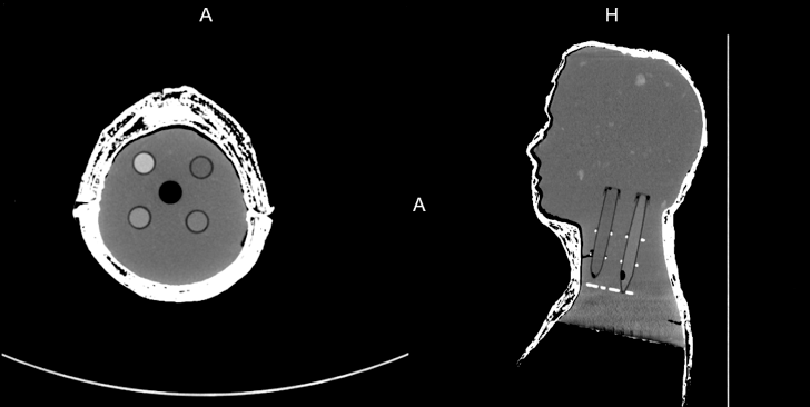
For the phantom experiments, an anthropomorphic phantom of the head and neck region was constructed by modeling a cast mold and filling it with a 3% agar solution. In addition, 5 plastic tubes were placed within the phantom containing air and sucrose solutions of different concentrations yielding different diffusivities. The phantom was examined using the DWI sequences described above (Figure 1).
For volunteer studies, 10 healthy volunteers (5 male, 5 females) were examined using the DWI sequences described above.
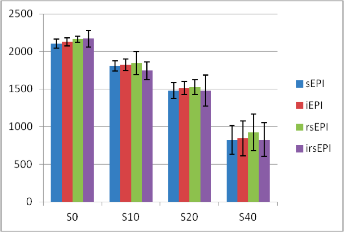
ADC values within the phantom compartments were measured and compared between the different sequences.
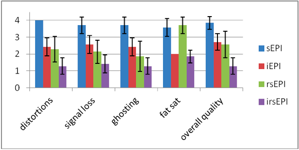
For phantom and volunteer measurements, the presence of geometric distortions, signal losses, ghosting artifacts as well as overall image quality were visually assessed on a 4-point scale by two radiologists in consensus. On the 4-point scale, 1 being the best possible outcome of the assessed category while 4 being the worst. In addition, failure of fat saturation was assessed in volunteer data.
Results
Quantification of ADC within the phantom compartments was comparable using the different sequence technologies without significant variations (Figure 2A).
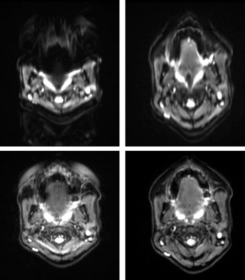
The combination of integrated shimming and readout-segmented EPI yielded significantly better overall image quality compared to sEPI, iEPI and rsEPI in phantom data as well as volunteer measurements (Figures 2B). Markedly reduced geometric distortions, signal loss and better fat saturation was observed using irsEPI (Figure 3).
Using irsEPI significantly improves image quality and reduces artifacts caused by magnetic field inhomogeneities in EPI based DWI of the head and neck region at 3 Tesla compared to alternative techniques. The clinical utility of this novel technique has to be further evaluated in patient studies.
Conclusion
Using irsEPI significantly improves image quality and reduces artifacts caused by magnetic field inhomogeneities in EPI based DWI of the head and neck region at 3 Tesla compared to alternative techniques. The clinical utility of this novel technique has to be further evaluated in patient studies.Acknowledgements
No acknowledgement found.References
1. Gatidis S, Graf H, Weiß J, Stemmer A, Kiefer B, Nikolaou K, Notohamiprodjo M, Martirosian P. Diffusion-weighted echo planar MR imaging of the neck at 3 T using integrated shimming: comparison of MR sequence techniques for reducing artifacts caused by magnetic-field inhomogeneities. MAGMA. 2016 Aug 8.
2. Zhang H, Xue H, Alto S, Hui L, Kannengiesser S, Berthold K, Jin Z. Integrated Shimming Improves Lesion Detection in Whole-Body Diffusion-Weighted Examinations of Patients With Plasma Disorder at 3 T. Invest Radiol. 2016 May;51(5):297-305.
3. Byeon J, Kim JY, Cho AH. Readout-segmented echo-planar imaging in diffusion-weighted MR imaging of acute infarction of the brainstem and posterior fossa: comparison of single-shot echo-planar diffusion-weighted sequences. Clin Imaging. 2015 Sep-Oct;39(5):765-9.
Figures