0175
Fast high-resolution diffusion MRI using gSlider-SMS, interlaced subsampling, and SNR-enhancing joint reconstruction1Electrical Engineering, University of Southern California, Los Angeles, CA, United States, 2A. A. Martinos Center for Biomedical Imaging, Department of Radiology, Massachusetts General Hospital, Charlestown, MA, United States
Synopsis
We describe a new approach that enables in vivo whole brain diffusion MRI with simultaneously high spatial resolution (660 µm isotropic voxels) and high angular diffusion encoding resolution (64 orientations at b=1500 s/mm2 and 4 b=0 s/mm2 images) in only 15 minutes. This is achieved by combining the gSlider-SMS acquisition strategy with constrained image reconstruction techniques that enable denoising (exploiting the fact that the diffusion images are smooth with correlated edge locations) and interlaced data subsampling (achieved by exploiting the same correlated edge constraints used for denoising, as well as through the use of q-space smoothness constraints).
Purpose
Recent progress has shown that in vivo whole-brain diffusion MRI can be acquired with simultaneously high spatial resolution (660 µm isotropic voxels) and high angular diffusion encoding resolution (64 orientations at b=1500 s/mm2 and 7 b=0 s/mm2 images) in as short as 25 minutes on the 3T CONNECTOM system1. This was achieved by combining the generalized SLIce Dithered Enhanced Resolution Simultaneous MultiSlice (gSlider-SMS) technique2 for SNR-efficient data acquisition with the SNR-enhancing joint reconstruction (SER)3,4 approach. Other recent work has shown that it possible to accelerate multi-encoding diffusion techniques like reversed-gradient field inhomogeneity correction5, gSlider-SMS6, and super-resolution reconstruction7,8 using the concept of “interlaced subsampling.” In this approach, each point in q-space is acquired with only a portion of its nominal set of spatial encodings to save time in data acquisition. While this would normally lead to poor image quality, it has been shown that high-quality image reconstruction is still possible by incorporating spatial and q-space smoothness constraints into the reconstruction5-8.
In this work, we show that gSlider-SMS, SER, and interlaced subsampling can all be combined together to provide substantial additional acceleration, and demonstrate in vivo human brain images with matching spatial coverage, spatial resolution, and diffusion encoding resolution to that described previously1, but reducing the acquisition time from 25 minutes to 15 minutes.
Methods
The proposed approach was tested by retrospectively undersampling the same 25 minutes of gSlider-SMS data described previously1. The conventional gSlider-SMS technique acquires simultaneous multislice diffusion data with slices that are 5x thicker than the nominal resolution, and uses 5 different RF encodings (that apply different phase modulations) to resolve the thinner slices that contribute signal to the thick slice. To simulate interlaced subsampling, we randomly discarded 2 out of 5 RF encodings for each of the 64 diffusion weighted images. We also discarded 3 out of the 7 images acquired without diffusion weighting (b=0 s/mm2), though retained the full set of encodings for the remaining unweighted images. This corresponds to keeping only 213 (58%) of the original 369 image volumes.
For simplicity, thick slices were first reconstructed using standard parallel imaging and simultaneous multislice reconstruction1. Subsequently, thin slices were estimated by solving the optimization problem: $$\hat{\mathbf{p}} = \arg\min_{\mathbf{p}} \|\mathbf{b} - \mathbf{E} \mathbf{p}\|_2^2 + \lambda_1 J(\mathbf{p}) + \lambda_2 R(\mathbf{p}).$$ The first two terms of this equation correspond to a data consistency constraint ($$$\mathbf{b}$$$ is the vector of acquired data measurements for all voxels, all diffusion encodings, and all RF encodings; $$$\mathbf{E}$$$ is the matrix representing the gSlider mapping from thin slices to subsampled phase modulated thick slices, and also includes the effects of shot-to-shot phase variations induced by the diffusion encoding; and $$$\mathbf{p}$$$ is the vector of high-resolution images to be reconstructed) and a spatial-smoothness regularization constraint for SER3,4 ($$$J(\cdot)$$$ is constructed based on a shared compound Markov Random Field edge model3, that encourages that image edges should be sparse, with correlation between the edge locations of different images). These two terms are very similar to what was used in previous work that combined gSlider-SMS with SER1. The third term $$$R(\cdot)$$$ is a regularization penalty that encourages smoothness of the signal in q-space, following previous approaches for interlaced subsampling5-8. For simplicity and consistent with some of the previous interlaced subsampling work5, we choose $$$R(\cdot)$$$ as simply the squared $$$\ell_2$$$-norm of the Laplace-Beltrami smoothness operator applied to the spherical harmonic representation of the q-space signal variations of each voxel9. Variables $$$\lambda_1$$$ and $$$\lambda_2$$$ correspond to regularization parameters that were adjusted manually to achieve the desired trade-off between spatial resolution and signal-to-noise ratio.
Our proposed new approach was compared against traditional gSlider-SMS from 25 minutes of data acquisition (without SER and without interlaced subsampling) and gSlider-SMS from 25 minutes of data acquisition with SER (without interlaced subsampling). After images were reconstructed, we computed orientation distribution functions (ODFs) using the Funk-Radon and Cosine Transform10 (using BrainSuite software, http://brainsuite.org/) and also estimated diffusion tensors.
Results
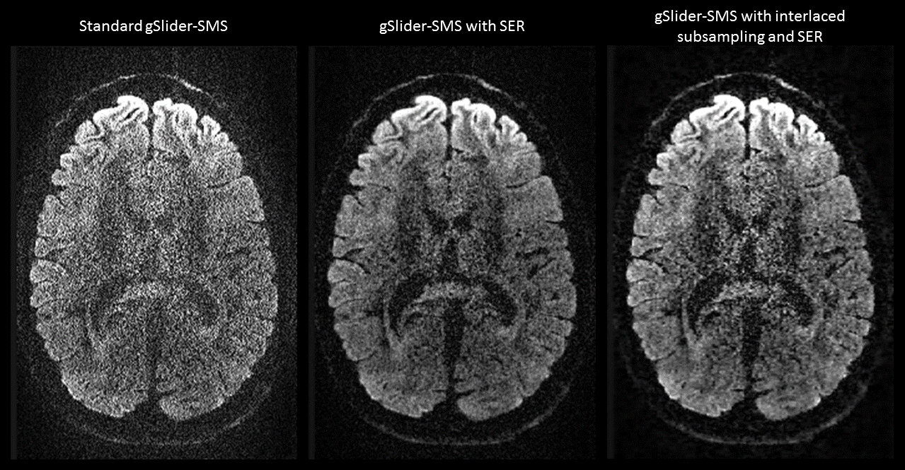
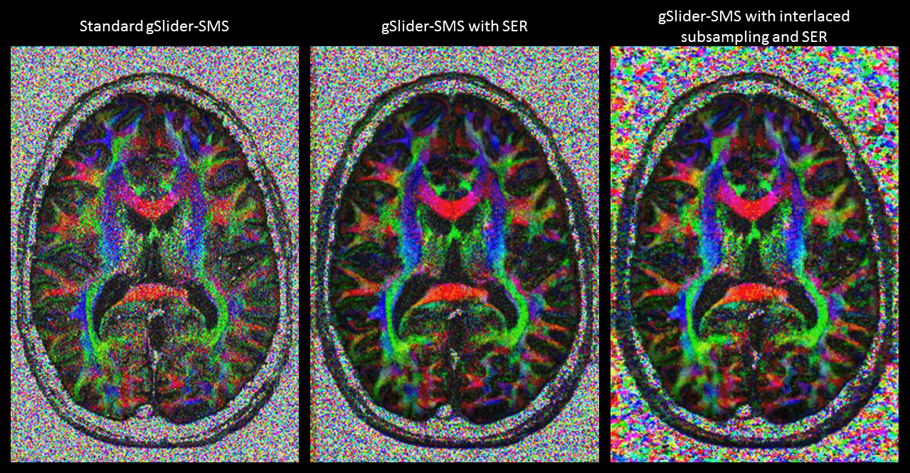
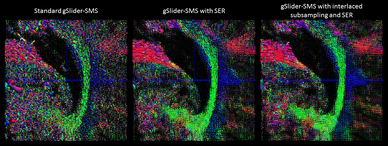
Figure 1 shows that our proposed 15-minute approach yields diffusion weighted images with similar quality to those obtained using gSlider-SMS with SER from 25 minutes of data, which are substantially better than the images obtained using gSlider-SMS from 25 minutes of data without SER reconstruction. Figures 2 and 3 show that this observation is also true for estimated diffusion tensors and ODFs.Discussion and Conclusions
This work proposed a new approach to fast diffusion MRI that enables state-of-the-art volume coverage, spatial resolution, and diffusion encoding resolution in only 15 minutes of data acquisition. We anticipate that this will substantially improve the practical utility of this approach for practical routine use in applications.Acknowledgements
This work was supported in part by NSF CAREER award CCF-1350563, and NIH research grants R21-EB022951, R01-NS089212, R01-NS074980, U01-MH093765, P41-EB015896, and R01-EB020613.References
1. Haldar JP, Fan Q, Setsompop K. “Whole-brain quantitative diffusion MRI at 660 µm resolution in 25 minutes using gSlider-SMS and SNR-enhancing joint reconstruction.” Proc. ISMRM 2016, p. 102.
2. Setsompop K, Stockmann J, Fan Q, Witzel T, Wald LL. “Generalized SLIce Dithered Enhanced Resolution Simultaneous MultiSlice (gSlider-SMS) to increase volume encoding, SNR and partition profile fidelity in high-resolution diffusion imaging.” Proc. ISMRM 2016, p. 607.
3. Haldar JP, Wedeen VJ, Nezamzadeh M, Dai G, Weiner MW, Schuff N, Liang Z-P. “Improved diffusion imaging through SNR-enhancing joint reconstruction.” Magn Reson Med 69:277-289, 2013.
4. Kim JH, Song S-K, Haldar JP. “Signal-to-noise ratio-enhancing joint reconstruction for improved diffusion imaging of mouse spinal cord white matter injury.” Magn Reson Med 75:1499-1514, 2016.
5. Bhushan C, Joshi AA, Leahy RM, Haldar JP. “Improved B0-distortion correction in diffusion MRI using interlaced q-space sampling and constrained reconstruction.” Magn Reson Med 72:128-1232, 2014.
6. Ning L, Setsompop K, Rathi Y. “A combined compressed sensing super-resolution diffusion and gSlider-SMS acquisition/reconstruction for rapid sub-millimeter whole-brain diffusion imaging.” Proc ISMRM 2016, p. 4212.
7. Ning L, Setsompop K, Michailovich O, Makris N, Shenton ME, Westin C-F, Rathi Y. “A joint compressed-sensing and super-resolution approach for very high-resolution diffusion imaging.” NeuroImage 125:386-400, 2016.
8. Van Steenkiste G, Jeurissen B, Veraart J, den Dekker AJ, Parizel PM, Poot DH, Sijbers J. “Super-resolution reconstruction of diffusion parameters from diffusion-weighted images with different slice orientations.” Magn Reson Med 75:181-195, 2016.
9. Descoteaux M, Angelino E, Fitzgibbons S, Deriche R. “Regularized, fast, and robust analytical Q-ball imaging. Magn Reson Med 58:497-510, 2007.
10. Haldar JP, Leahy RM. “Linear transforms for Fourier data on the sphere: Application to high angular resolution diffusion MRI of the brain.” NeuroImage 71:233-247, 2013.
11. Shattuck DW, Chiang MC, Barysheva M, McMahon KL, de Zubicaray GI, Meredith M, Wright MJ, Toga AW, Thompson PM. “Visualization tools for high angular resolution diffusion imaging.” Proc MICCAI 2008, pp. 298-305.
Figures