0172
Diffusion Weighted Imaging using a Dixon based Single Shot Turbo Spin Echo1Radiology, UT Southwestern Medical Center, Dallas, TX, United States, 2Philips Research, Hamburg, Germany, 3Advanced Imaging Research Center, UT Southwestern Medical Center, Dallas, TX, United States
Synopsis
Diffusion weighted imaging using single-shot turbo spin-echo (DWI-SShTSE) is increasingly used due to its robustness to geometric distortions, but often suffers from incomplete fat suppression at 3T using spectrally-selective fat suppression methods (SPIR/SPAIR etc.) in challenging areas with large field inhomogeneities. STIR can improve the fat suppression but at the expense of reduced SNR. In this work, we developed a multi-echo Dixon DWI-SShTSE sequence with shared field map between lower and higher b-values for uniform fat suppression without using image navigator and increasing scan times. We also demonstrated its robustness to the phase variations due to diffusion gradients.
Introduction
Diffusion weighted imaging using Single-shot turbo spin-echo (DW-SShTSE) is increasingly used due to its robustness to geometric distortions compared to DW-EPI in challenging areas with B0 inhomogeneity such as the spine and skull base imaging1,2. DW images are always acquired with fat suppression due to the inherent hyperintensity combined with low diffusion coefficient of fat that reduces the conspicuity of lesions and may affect the apparent diffusion coefficient (ADC) measurement of water. Fat suppression with DW-SShTSE is commonly achieved using spectrally selective inversion recovery (SPIR/SPAIR), but this approach suffers from incomplete fat suppression in challenging areas with large field inhomogeneities. STIR can offer more uniform fat suppression but at the expense of reduced signal to noise ratio (SNR). While DW-SShTSE can be combined with a Dixon based acquisition for uniform fat suppression3, the additional phase variations induced by DW gradients and reduced SNR impose challenges for fat/water separation particularly at higher b-values.
Thus, the purpose of this work was to develop a DW-SShTSE using a multi-echo Dixon based acquisition4 combined with shared field map between lower and higher b-values for uniform fat suppression without increasing the total scan time.
Theory
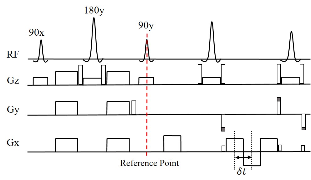
The phase insensitive diffusion weighting preparation5 was combined with a multi-echo Dixon SShTSE, where the In-Phase (IP) and Out-of-Phase (OP) images are acquired in the same repetition. After the reference point, the CPMG condition was imposed and the signal can be modeled as: $$S_{n,m}=(W_m+c_nF_m)e^{i(\phi_n+\psi_m)}~~~(1)$$ where $$$n,~m$$$ are the numbers of echo and b-value respectively. Water (W) and fat (F) are considered complex with $$$\phi_n$$$ corresponding to the field strength offset $$$\Delta{B_0}$$$ and echo times (TE). The additional phase induced by DW gradients ($$$\psi_m$$$) would be nonzero at b>0 and simultaneously affects the echoes acquired in the same repetition.
If IP and OP echoes are acquired at b=0, then $$$ \psi_0=0 $$$ and $$$\Delta{\Phi_0}=e^{i(\phi_2-\phi_1)}$$$. $$$W_0$$$ and $$$F_0$$$ at b=0 can be calculated using the conventional fat/water separation method6:$$\left(\begin{array}{c}W_0\\F_0\end{array}\right)=\frac{1}{c_2-c_1}\begin{bmatrix}c_2&c_1\\-1&1\end{bmatrix}\left(\begin{array}{c}S_{1,0}\\S_{2,0}\Delta\Phi_0^*\end{array}\right)~~~(2)$$ $$$c_1$$$ and $$$c_2$$$ are the complex vectors of fat with multiple spectral peaks at TE1 and TE2. If $$$\Delta{B_0}$$$ and TE are identical between different b values, $$$\Delta{\Phi_m}=e^{i(\phi_2-\phi_1)}=\Delta{\Phi_0}$$$. For b>0, eq.2 can be written as:$$\left(\begin{array}{c}W_m\\F_m\end{array}\right)=\frac{1}{c_2-c_1}\begin{bmatrix}c_2&c_1\\-1&1\end{bmatrix}\left(\begin{array}{c}S_{1,0}\\S_{2,0}\Delta\Phi_0^*\end{array}\right)e^{-i\psi_m}=\left(\begin{array}{c}W_m'\\F_m'\end{array}\right)e^{-i\psi_m}~~~(3)$$ $$$\left(\begin{array}{c}W_m'\\F_m'\end{array}\right)$$$ can be estimated using $$$S_{n,m}$$$ and shared field map $$$\Delta{\Phi_0}$$$. Although $$$\psi_m$$$ is unknown, it only modulates the phase and doesn’t affect the magnitude of the final reconstructed water and fat images at higher b-values, which are $$$|W_m|=|W_m'e^{-i\psi_m}|$$$ and $$$|F_m|=|F_m'e^{-i\psi_m}|$$$.
Methods
The sequence (Fig.1) was implemented on a 3T Ingenia scanner (Philips Healthcare, Best, The Netherlands). The proposed method was evaluated and compared against DW-EPI and DW-SShTSE with SPIR in the brain and spinal cord of 5 healthy volunteers with IRB approval and written informed consent. The typical imaging parameters of the proposed sequence included: sagittal and/or axial orientation; FOV = 220×220 mm; Resolution = 1.5×1.9 mm; Slice Thickness = 4mm; SENSE = 2; TE = 70 ms, $$$\delta{t}$$$ = 1.1 ms and 6 signal averages. The total scan time was approximately 3:00 min for 13 slices with 2 b-values (e.g. 0 and 500).Results
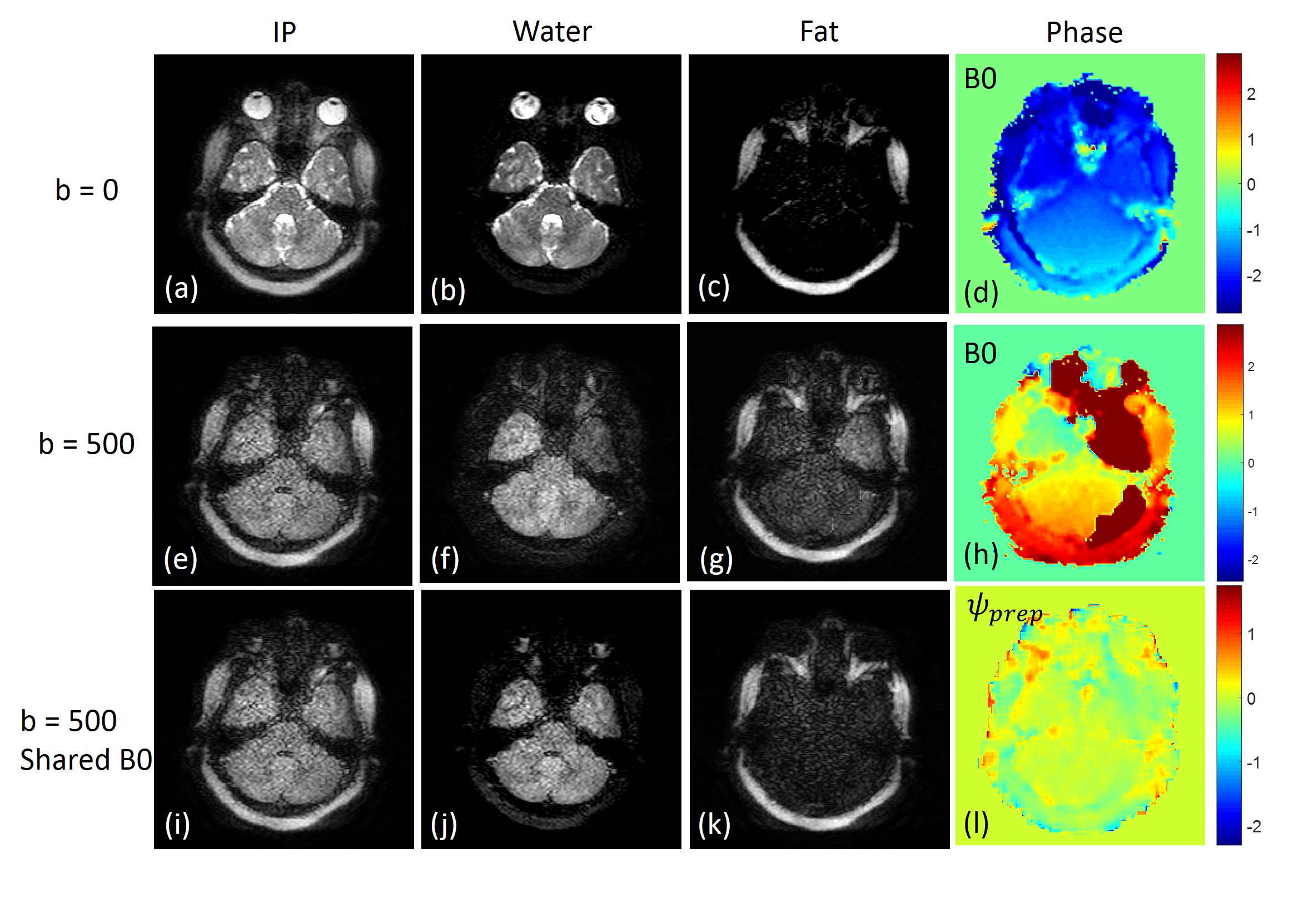
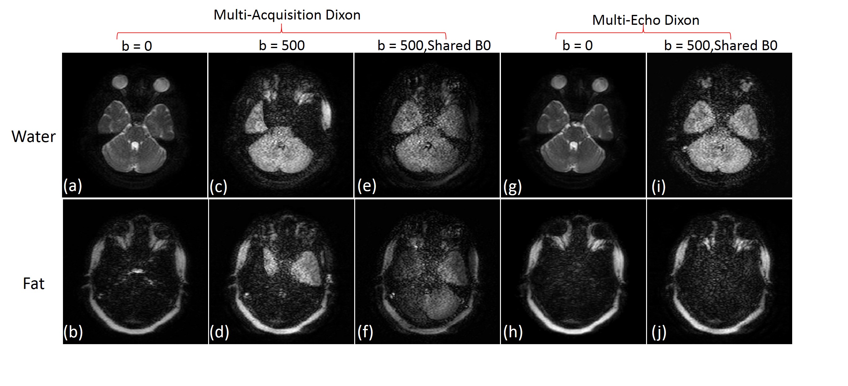
Figure 2 demonstrates the use of a shared field map between b=0 and b=500 for uniform fat/water separation. The native B0 from its own acquisition generates uniform fat/water separation at b=0 (fig.2, top row), but fails at b=500 due to low SNR (fig.2, middle row). Using the shared field map from b=0, uniform fat/water separation was achieved at b=500 (fig.2, bottom row). With proper fat/water separation, the additional phase modulation ($$$\psi_{prep}=e^{i\phi_m}$$$) due to the DW gradients can also be calculated (fig.2l). While the use of a shared field map achieves uniform fat/water separation with multi-echo Dixon acquisition, it fails in multi-acquisition due to different $$$\psi_m$$$ induced by DW gradients between different echoes acquired in separate repetitions (fig.3).
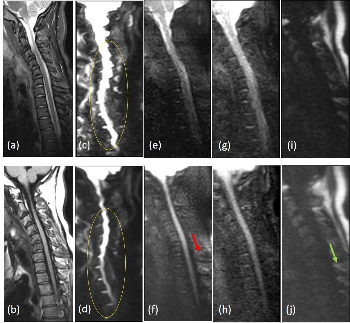
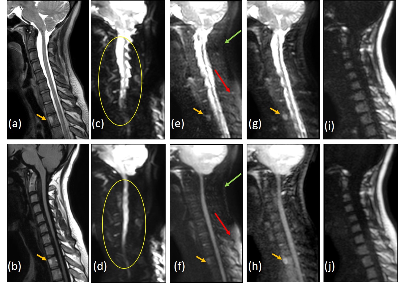
The shared field map approach using multi-echo Dixon also improved fat/water separation in spine imaging, which is more challenging due to much lower SNR and segmented appearance of the intervertebral spaces (fig.4). The proposed approach provides diffusion weighted images (figs.5g, 5h) with minimal to no image distortion compared to DW-EPI (figs.5c, 5d), and with uniform fat suppression compared to DW-SShTSE with SPIR (figs.5e, 5f).
Discussion
We have demonstrated multi-echo Dixon with a shared-B0-map that can improve fat/water separation in DW-SShTSE imaging without the need for additional image navigators. The resulting images are free of geometric distortion with uniform fat suppression, allowing the accurate measurement of water diffusion compared to DW-EPI and DW-SShTSE-SPIR even in locations with significant field inhomogeneities such as the cervical spine and skull base. Furthermore, this approach also allows the measurement of ADC in fat.Acknowledgements
No acknowledgement found.References
1, Stroman, PW, Wheeler-Kingshott C, Bacon M, et al. The current state-of-the-art of spinal cord imaging: methods. Neuroimage. 2014; 84: 1070-1081.
2, Foer DB, Vercruysse JP, Bernaerts A, et al. The value of single-shot turbo spin-echo diffusion-weighted MR imaging in the detection of middle ear cholesteatoma. Neuroradiology. 2007; 49(10): 841-848.
3, Hwang KP, et al, Diffusion weighted SSFSE with Dixon fat-water separation. Proc ISMRM 2008; 16.
4, Wang XZ, et al, Robust abdominal imaging with uniform fat suppression using Dixon based single shot turbo spin echo. Proc ISMRM 2016; 573.
5, Alsop DC. Phase insensitive preparation of single-shot RARE: Application to diffusion imaging in humans. Magnetic resonance in medicine. 1997; 38(4): 527-533.
6, Eggers H, Brendel B, Duijndam A, et al. Dual-echo Dixon imaging with flexible choice of echo times. Magnetic resonance in medicine. 2011; 65(1): 96-107.
Figures