0159
Topographic Mapping of Resting State fMRI Data1School of Physics, University of Nottingham, Nottingham, United Kingdom
Synopsis
Task-based fMRI can provide robust somatotopic mapping of digits of the hand. Resting state fMRI (rs-fMRI) provides the ability to parcellate brain areas based on their connectivity. Here, we use simultaneous multislice to acquire high spatial resolution fMRI resting state data with a short TR to determine whether we can topographically map connectivity within the sensorimotor cortex. Seed based locations of the index finger (Digit 2) and little finger (Digit 5) are defined from somatotopic travelling wave and finger tapping tasks, and used to demonstrate significant topographic mapping in rs-fMRI data.
Introduction
Task-based fMRI has provided the ability to form detailed maps of the somatotopic representation of fingertips in the human primary somatosensory cortex (SI)1,2. Resting state fMRI (rs-fMRI) studies3 have shown that it is possible to functionally parcellate brain areas based on their connectivity, with recent studies showing the mapping of motor cortex4 between hemispheres, and that higher functional correlations exist between the centrally located hand region, than between the foot and tongue5. Here, we use high spatial resolution fast simultaneous multislice (SMS)/multiband (MB) imaging at 7T to determine if there is significant topography in the connectivity of rs-fMRI data between Digit 2 (D2) and Digit 5 (D5).Methods
Data Acquisition
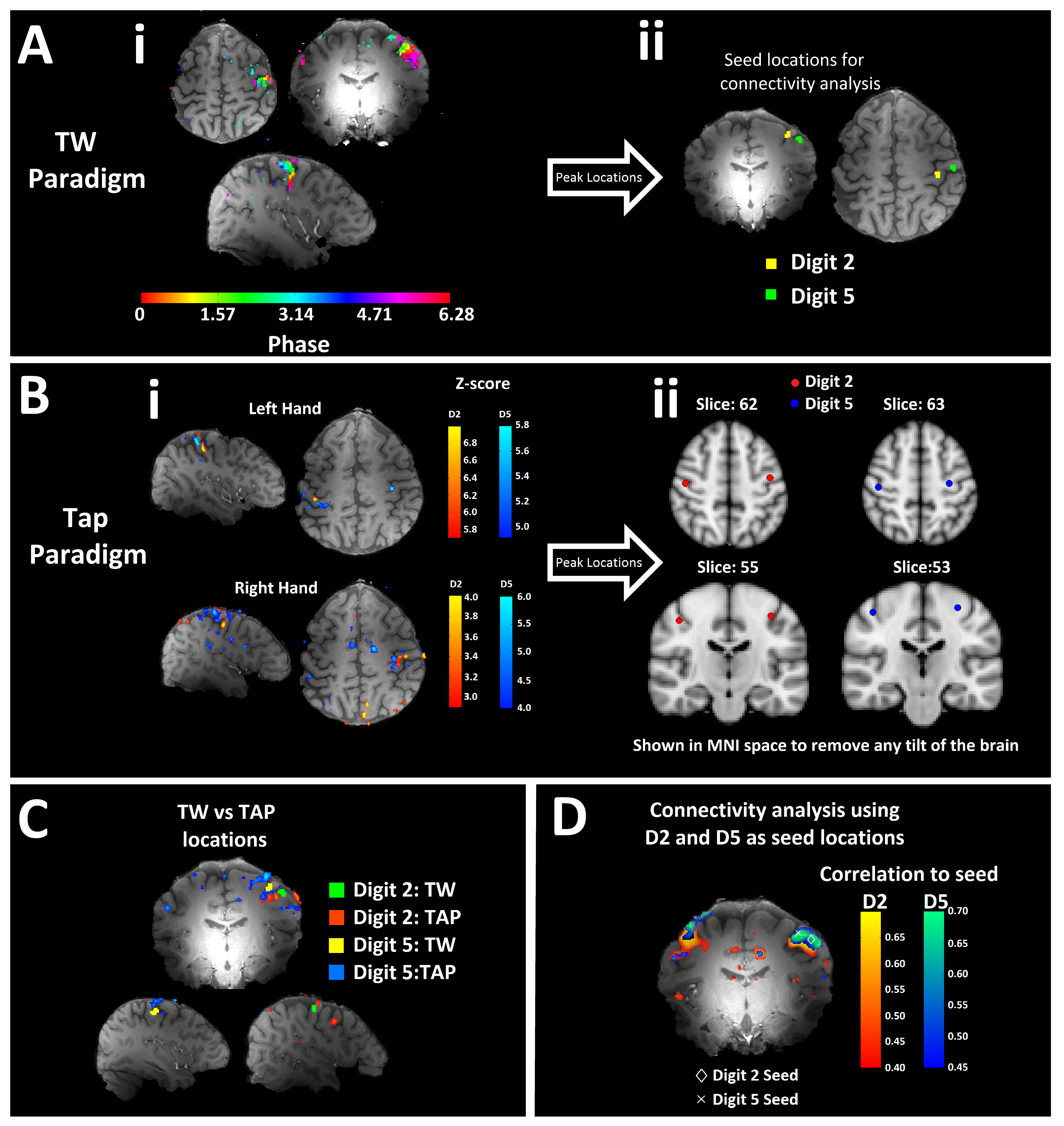
Seven subjects (2 female, age 24±2 years) participated in the study. fMRI data were collected using a 7T Philips Achieva scanner equipped with a 32-channel receiver coil (NOVA Medical). For the fast rs-fMRI scan, GE-EPI fMRI data (TR = 600 ms, MB-factor = 3, SENSE 1.5, 2 mm3 isotropic resolution, FOV = 128x60x192 mm, FA = 46°, TE = 25 ms) were acquired for 5 minutes whilst subjects had their eyes open. In addition, subjects participated in task-based fMRI measures to generate somatosensory and motor maps. Somatotopic maps were formed using a ‘travelling wave (TW)’ paradigm1 (TR= 2 s, TE= 25 ms, FA=75°) in which piezo-electric stimulators sequentially stimulated each digit of the hand for 4s of a 20 s cycle either in a forward (from the thumb to the little finger) or backward (from little finger to the thumb) ordering for 8 cycles6. Subjects also performed a task paradigm where they performed a D2 or D5 movement for 8 s followed by 20 s rest, repeated 3 times for each digit per hand.
Data Analysis
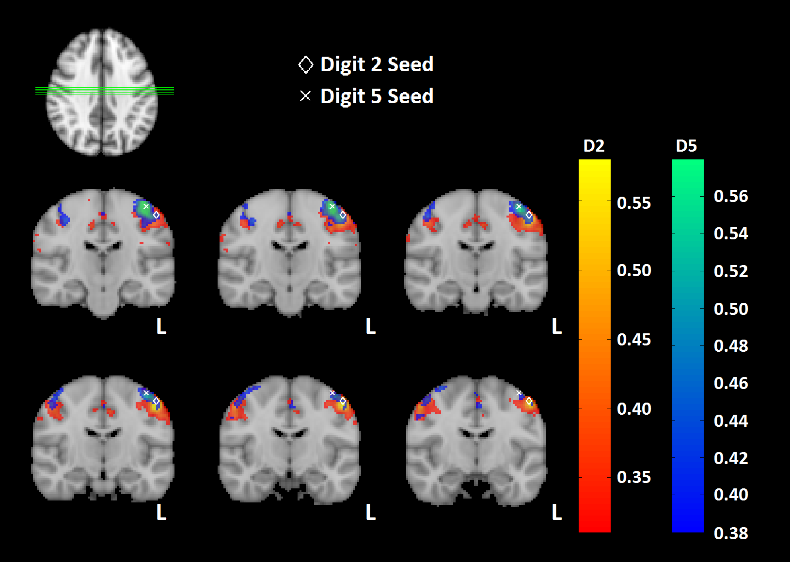
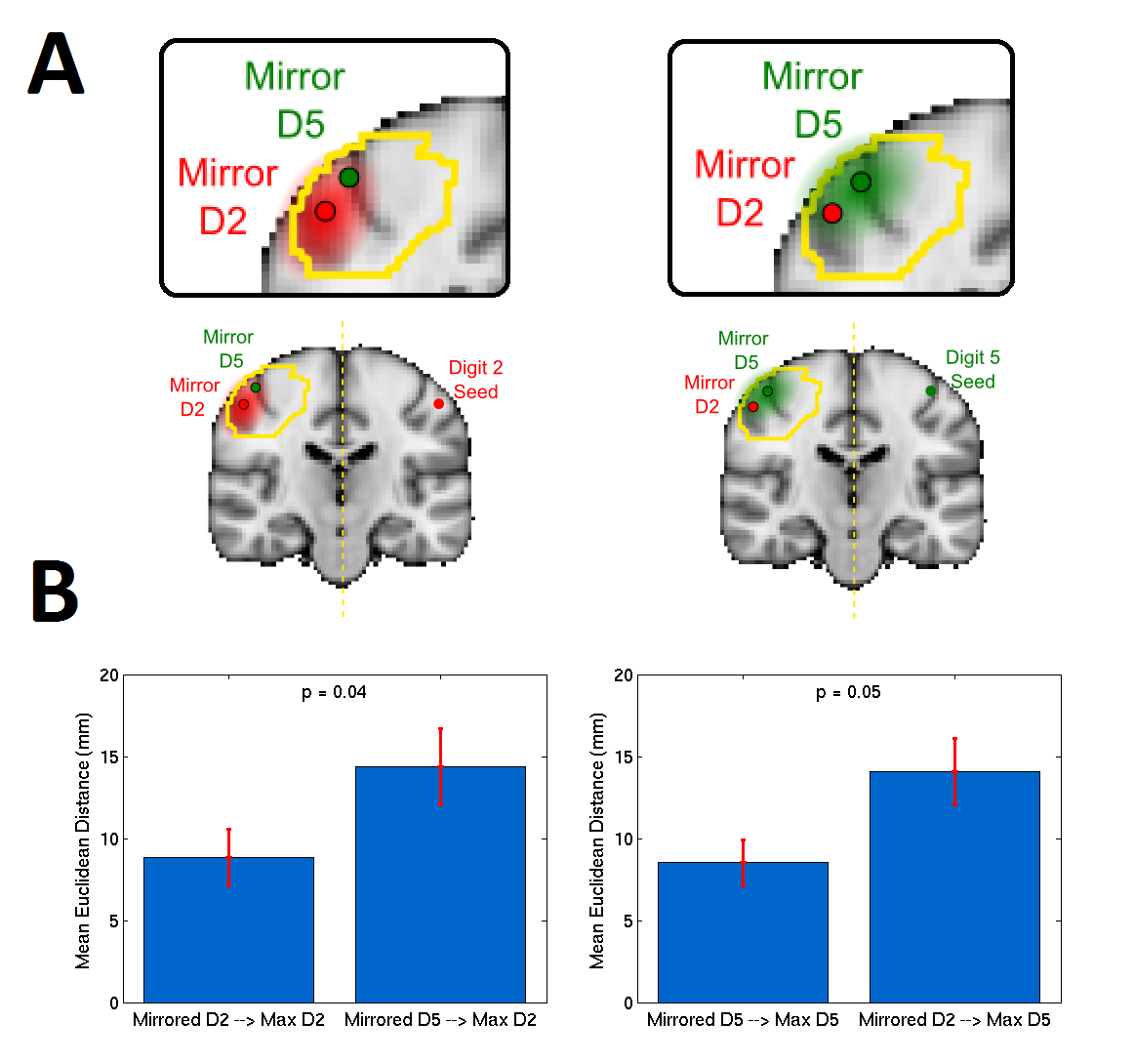
To determine the locations of D2 and D5, the TW fMRI data were analyzed using Fourier analysis in mrTools (http://www.cns.nyu.edu/heegerlab) to calculate the coherence and phase of the best-fitting 1/20 Hz sine wave at each voxel. These peak locations were confirmed using the motor fMRI data, which were analysed using a GLM. The peak locations of D2 and D5 were then used as seed locations in the rs-fMRI data. Connectivity maps were generated for each seed (i.e. D2 and D5). Significance was determined by first mirroring the seed location for each digit across the cortex. The mean Euclidean distance from the mirrored seed location to the maximum of the connectivity map in the contra-lateral motor cortex was found, and contrasted with the distance to the other digit’s mirrored seed (see Figure 3A).
Results
Fingertip somatotopic maps showed a clear orderly pattern of phase variation corresponding to the five digits in SI (Figure 1Ai), and the motor task clearly defined D2 and D5 (Figure 1Bi), as well as showing symmetry across the cortex (Figure 1Bii). This enabled a seed-based connectivity analysis to be done using seed locations representing both Digit 2 and Digit 5 (shown on an individual subject in Figure 1D, and on a group level in Figure 2), whereby a separation in the connectivity maps is seen. Figure 3 illustrates the significance of the spatial separation of the rs-fMRI connectivity maps. Given the symmetry seen in the motor maps, the seed locations were mirrored across the cortex (Figure 3A) in order to give a new region of interest, and the peak connectivity within a motor mask was found for each map, along with the Euclidean distance to the mirrored seed (Figure 3B). The mean Euclidean distance from the peak of the D2 connectivity map to the mirrored D2 location was significantly smaller than the distance to the mirrored D5 location, and a similar pattern was seen when comparing the D5 connectivity map to D5 location (p < 0.05 for both).Discussion
High spatial and temporal resolution MB data can be used to parcellate rs-fMRI data topographically, with group connectivity maps (Figure 2) showing clear separation between D2 and D5 in the opposite hemisphere to the seed location. There was close agreement between a given digit (D2 or D5) location as defined from task based fMRI and its rs-fMRI digit connectivity map.Conclusion
This work has demonstrated the possibility to form rs-fMRI digit-based connectivity maps of individual sensorimotor regions. Future studies will expand this study to assess functional connectivity between sensory and motor cortex of individual digits and to assess whether it is possible to differentiate fine-scale functional networks within sub-areas of SI.Acknowledgements
This work was funded by an MRC grant.References
[1] Sanchez-Panchuelo, RM, et al. "Mapping human somatosensory cortex in individual subjects with 7T functional MRI." Journal of neurophysiology 103.5 (2010): 2544-2556.
[2] Sanchez-Panchuelo RM, et al. “Within-digit functional parcellation of Brodmann areas of the human primary somatosensory cortex using functional magnetic resonance imaging at 7 Tesla.” J Neurosci. 32.45 (2012):15815-22.
[3] Biswal, B, et al. "Functional connectivity in the motor cortex of resting human brain using echo-planar mri." Magnetic resonance in medicine 34.4 (1995): 537-541.
[4] Van Den H, et al. "Specific somatotopic organization of functional connections of the primary motor network during resting state." Human brain mapping 31.4 (2010): 631-644.
[5] Long X, et al. “Functional connectivity-based parcellation of the human sensorimotor cortex.” Eur J Neurosci. 39(8) (2014): 1332-42.
[6] Sánchez-Panchuelo, RM, et al. "Regional structural differences across functionally parcellated Brodmann areas of human primary somatosensory cortex." Neuroimage 93 (2014): 221-230.
Figures