0143
Enhanced detection of cerebral arterial system with USPIO-enhanced MRI1Radiology, New York University School of Medicine, New York, NY, United States, 2Radiology, New York University School of Medicine, 3Wayne State University
Synopsis
Although small arterial system plays a key role in delivering oxygen and glucose to brain tissue, the in vivo detection is still challenging. Using a low dose of ultra-small-superparamagnetic-iron-oxide (USPIO) contrast agent, it will induce susceptibility contrast in the arterial blood, which enhances the small arteries visibility on susceptibility weighted imaging. This study has demonstrated its feasibility in human brain. Such technique has potential to unveil underlyingmicrovascular abnormalities in neurovascular diseases that cannot be done in vivo with any other conventional method in use today.
Introduction
The cerebrovascular system, in particular the arterial system at micro level, plays a key role in delivering oxygen and glucose to brain tissue. Small arterial abnormalities including both morphological (e.g., tortuosity & stenosis) and vascular density changes are implicated in a number of neurovascular and neurodegenerative disorder1. Although susceptibility weighted imaging (SWI) has dramatically improved the visibility of cerebral venous system due to the high concentration of paramagnetic deoxyhemoglobin in venous blood, it is limited for detection of arteries. At present, in vivo detection of the branching cerebral arteries and tiny arterioles where vasculogenic neuropathology often begins still remains challenging; such detection may be largely improved with ultra-small-superparamagnetic-iron-oxide (USPIO) enhanced MRI by inducing intravascular susceptibility into the arteries. Therefore, the purpose of this study is therefore to evaluate the potentiality of USPIO-enhanced MRI in improving the visibility of small arteries of human brains.Methods
This was an institutional board reviewed protocol using Ferumoxytol (or Feraheme 30mg/mL), which is an FDA approved USPIO for human study use) to enhance microvascular MR imaging. High resolution (in plane: 0.11x0.22mm2) pre- and post-Ferumoxytol (varied from 1mg to 4 mg/kg) images were acquired using a double echo 3D gradient echo (GRE) imaging at 7T in three volunteers. The dose of Ferumoxyltol is about a quarter of the dose used for treatment. Ferumoxytol is a blood pool (much larger than Gadolinium) contrast agent with a half-life about 15 hours. The imaging parameters include TR = 24-35ms, TE=8ms, 10ms, or 16ms, flip angle = 10°, and slice thickness = 0.8-1.25mm. The acquisition time is about 13-15 minutes per scan. The comparison was made using either single slice or minimal intensity projection (mIP) of original magnitude or the SWI data. Quantitative susceptibility mapping (QSM) was also generated using SWIM techniques 2. Susceptibility values of arterial and venous blood on QSM are used to separate arteries from veins on post-USPIO scans.Results
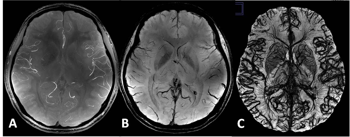
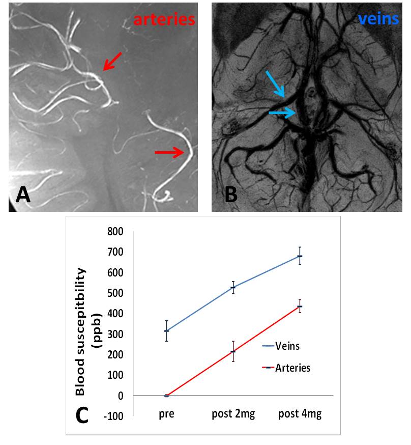
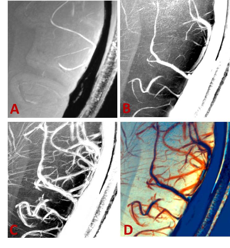
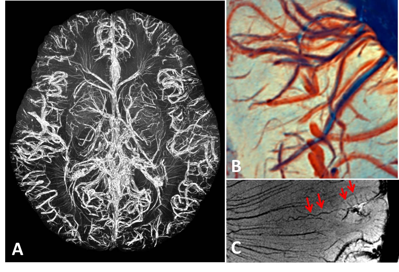
No side effects with injection of Ferumoxytol were reported from these volunteers. On pre-USPIO images, the shorter echo on 3D GRE is sensitive to in-flow effect that can be used for MR arteriogram (MRA) and longer echo is used to produce MR venogram (MRV). As shown in Figure 1, compared to pre-USPIO scans (Figure 1A & 1B), there is largely increased detectability of small vessels of both arteries and veins on post-USPIO scan (Figure 1C). Such increased detectability is more obvious on images with longer TE and higher dose ferumoxytol, in which susceptibility artifacts are also increased and challenging to QSM quantification. The susceptibility values of arterial and venous blood are notably different (Figure 2) and can be used to separate arteries from veins on post-USPIO scans. Using vascular tracking and the QSM susceptibility values of arteries and veins, the arterial and venous branches can be separated and color-coded (Figure 3D). The small vessel morphology and vascular density can also be evaluated based on the separated arterial and venous systems in both qualitative or quantitative ways. Figure 4 demonstrated a post-USPIO QSM intensity projected images of whole brain and selected region as well as a single magnitude image showing a tortuous artery that cannot be seen with conventional MRA.Discussion
USPIO-enhanced MRI at higher resolution showed an increased ability to detect not only the venous but also arterial system at a micro level due to the increased susceptibility and its blooming effect. Such technique may provide a direct visual inspection of the pathological source of microvascular abnormalities tied to different CNS diseases for clinical studies. Being able to detect and monitor the changes of small vessel changes earlier of the disease process will open the door to better understanding the etiology of the disease and to better treatment of the disease.Acknowledgements
This work was partly supported by R01 grant NS-076588 from National Institute of Health (NIH) and was performed under the rubric of the Center for Advanced Imaging Innovation and Research (CAI2R, www.cai2r.net), a NIBIB Biomedical Technology Resource Center (NIH P41 EB017183).References
[1] Brown WR, Thore CR: Review: cerebral microvascular pathology in ageing and neurodegeneration. Neuropathology and applied neurobiology 2011, 37:56-74.
[2] Haacke EM TJ, Neelavalli J, Cheng YC: Susceptibility mapping as a means to visualize veins and quantify oxygen saturation. Magn Reson Imaging 2010, 32:663-76.
Figures