0070
Ultrafast T2 mapping using echo-split GRASE acquisition and parametric POCSMUSE reconstruction1Department of Biomedical Engineering, University of Arizona, Tucson, AZ, United States, 2Brain Imaging and Analysis Center, Duke University Medical Center, Durham, NC, United States, 3Department of Diagnostic Radiology, The University of Hong Kong, Hong Kong, 4Department of Diagnostic Radiology, Keio University School of Medicine, Japan
Synopsis
Our novel ultrafast T2 mapping framework, which uniquely integrates echo-split GRASE acquisition and parametric POCSMUSE reconstruction, has the following major advantages. First, parametric T2 map and high-quality multi-contrast images can be derived from a single set of single-shot GRASE data, with inherently low susceptibility to motion artifacts. Second, contamination of stimulated and other high order echoes is minimized in the
Purpose
Conventional parametric T2 mapping procedures (e.g., based on FSE1) have two limitations. First, multi-TE data acquisition is time consuming and thus susceptible to motion artifacts. Second, T2 mapping based on CPMG scans is inaccurate due to the signal contamination of stimulated and other high-order echoes2.
Here we report a new ultrafast T2 mapping method that can address the above-mentioned limitations. First, the motion artifact is inherently eliminated in single-shot echo-split GRASE scan. Second, the stimulated and high-order echo contamination is largely reduced by echo-split GRASE sequence. Third, high-quality and accurate T2 map can be produced from a single set of echo-split GRASE data using a parametric multiplexed sensitivity encoding based on projection onto convex sets (parametric POCSMUSE) algorithm.
Methods
Acquisition with echo-split GRASE sequence
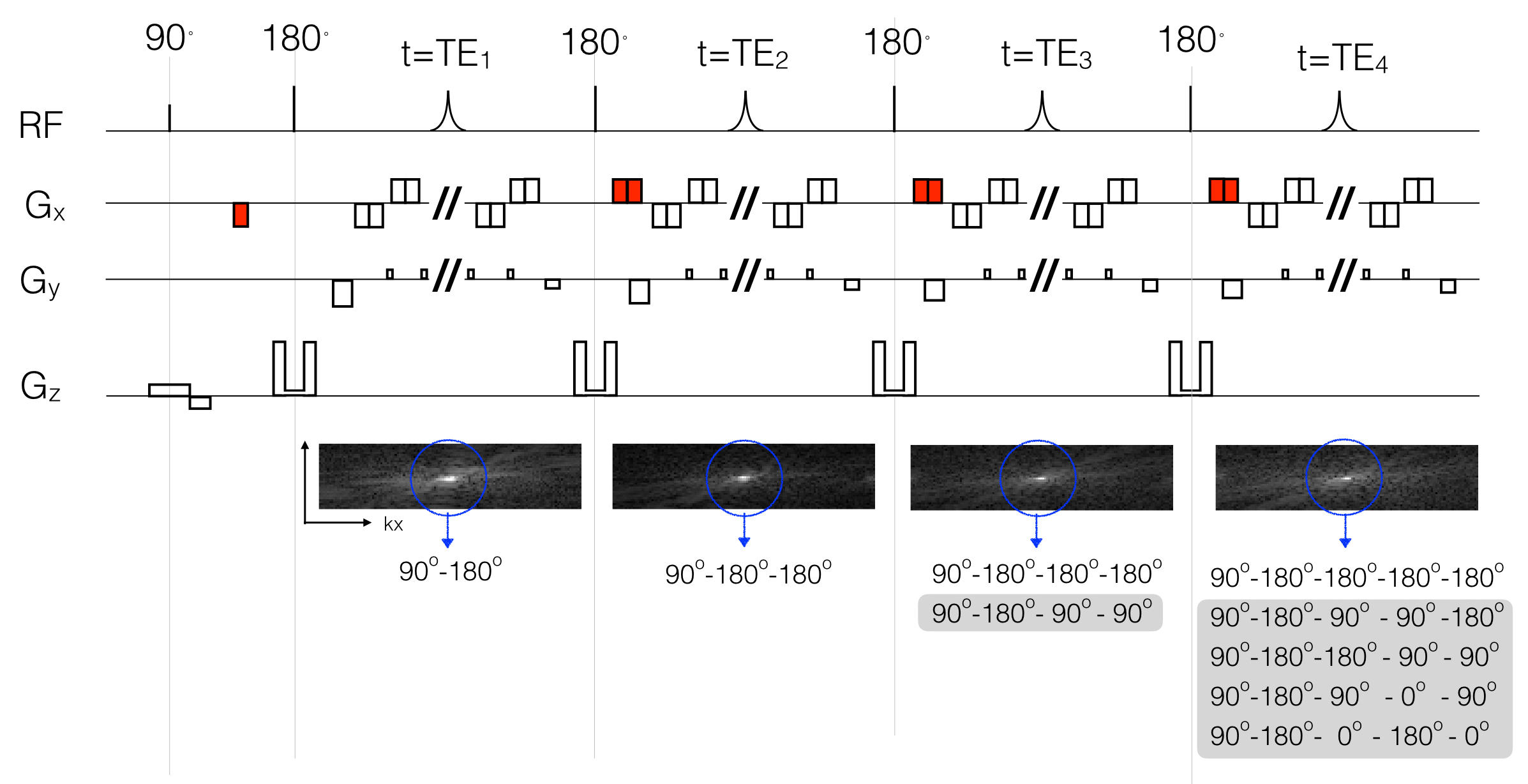
A novel single-shot echo-split GRASE sequence (Figure 1) is designed to acquire k-space data in a way that signals from multiple echo pathways are refocused at different kx locations. The signal intensities of different echo pathways across CPMG echoes can be analyzed with an extend phase graph (EPG) method3. As illustrated in Figure 1, 1) the primary echoes are always refocused at k-space center; 2) most high-order echoes are eliminated; 3) only five residual non-primary pathways occur in the third and fourth CPMG echoes. Instead of fitting the acquired data with exponential function, the signal changes from multiple pathways are thoroughly considered in our T2 mapping procedure as shown below.
Reconstruction with parametric POCSMUSE
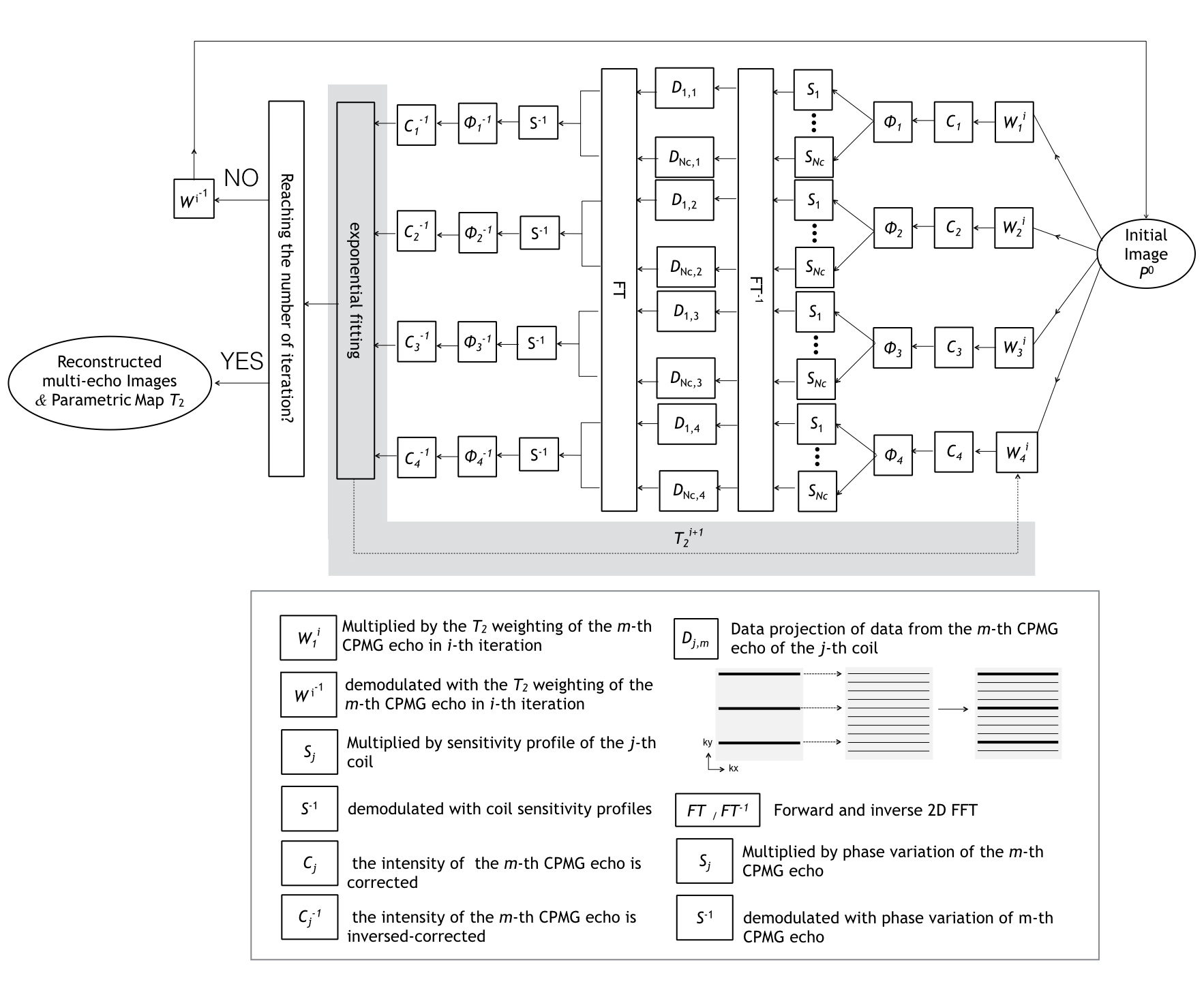
A parametric POCSMUSE reconstruction framework (extended from single-contrast POCSMUSE4) is developed to incorporate multiplexed parallel MRI and multi-echo-pathway signal modeling into a unified procedure. The multi-echo-pathway signals of four CPMG echoes are jointly assessed in an equation $$$Q=ETM_o$$$, where $$$E$$$ is the encoding matrix, $$$M_o$$$ are the primary echo signals at four TEs, and $$$T$$$ is the intensity correction based on EPG. With the prior knowledge of slice profiles and T1 ranges, this non-linear equation can be solved with iterative parametric POCSMUSE (see Figure 2). The reconstruct procedures are iterated until a set of multi-contrast images and a full-FOV T2 map are produced.
Our T2 mapping method was evaluated at 3 Tesla with four healthy volunteers. Firstly, single-shot echo-split GRASE data with four CPMG echoes (TE = 30, 60, 90 and 120 ms) were acquired and processed with parametric POCSMUSE to produce T2 maps. Secondly, four sets of fully-sampled 4-shot spin-echo EPI data were obtained with TE = 30, 60, 90 and 120 ms, respectively. These 4-shot EPI data, containing signals from only the primary echo pathway and with the same geometric distortion as GRASE data, were used as the T2 map reference. Thirdly, regular GRASE data with four CPMG echoes (TE = 30, 60, 90 and 120 ms) were acquired, processed with SENSE5, and exponentially fitted to produce T2 maps, as a comparison.
Results
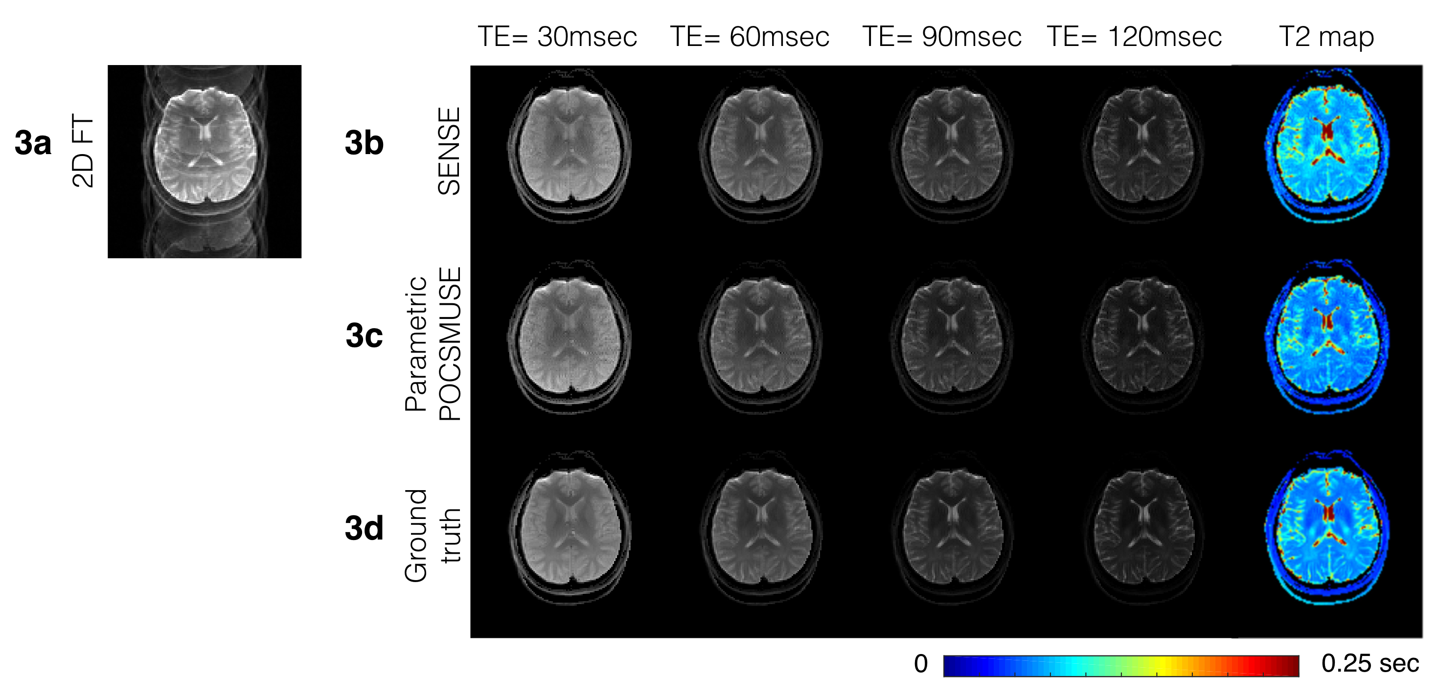
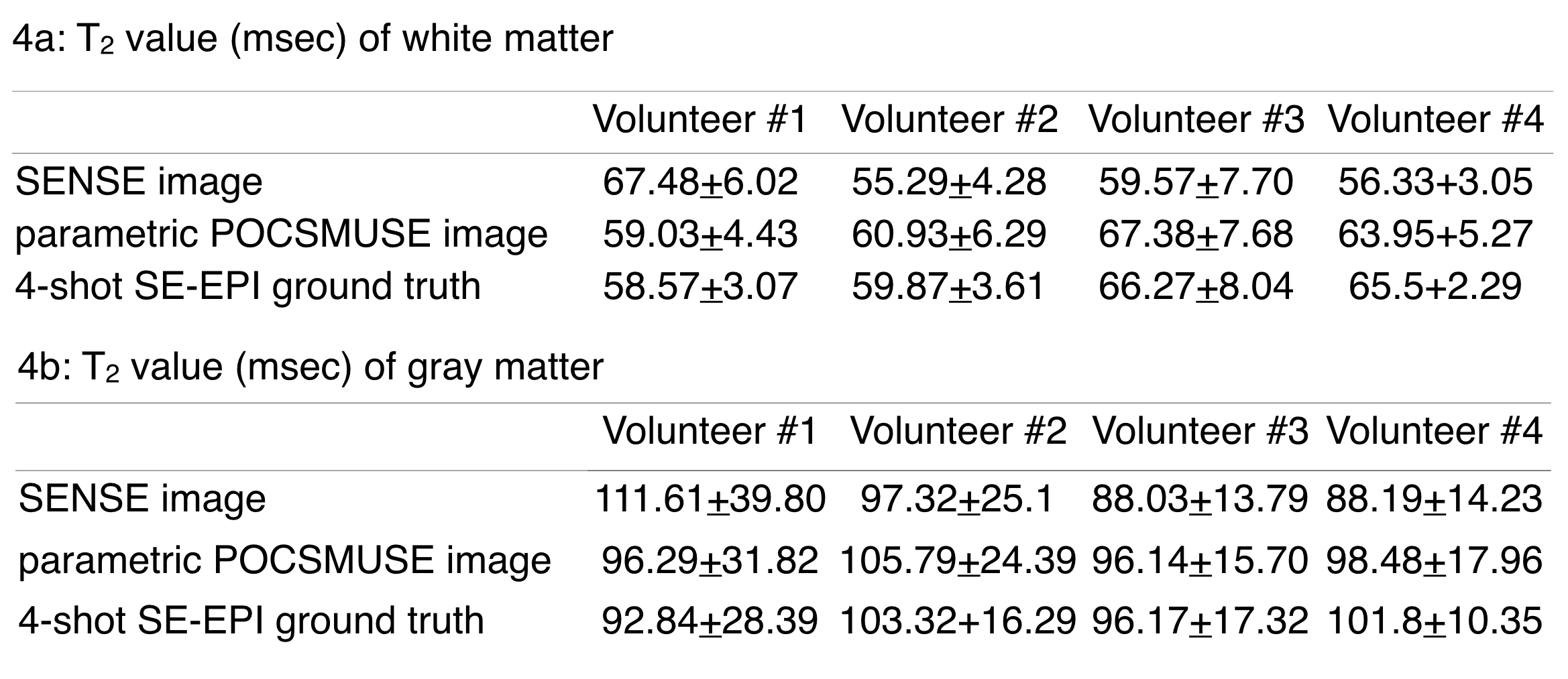
Figures 3 and 4 show the experimental results. Figure 3a demonstrates that 2D FT reconstruction of GRASE data is susceptible to aliasing artifact due to non-smooth T2 modulation in k- space. Figure 3b shows that although aliasing-free multi-contrast images can be produced with SENSE, the fitted T2 map significantly differs from the ground truth (Figure 3d). Figure 3c shows that high-quality and aliasing-free images can be produced with parametric POCSMUSE, and the T2 values measured from single-shot echo-split GRASE are very close to the ground truth. Figure 4 compares the T2 measures for all four volunteers, showing the superior performance of the parametric POCSMUSE, as compared with SENSE method.Discussion and conclusions:
Here we report an ultrafast T2 mapping method enabled by echo-split GRASE scan and parametric POCSMUSE reconstruction. The new method is based on single-shot acquisition and thus not vulnerable to motion artifacts. Signal contamination from stimulated and high-order echoes is reduced in the developed pulse sequence, and parametric T2 maps can be accurately measured with parametric POCSMUSE. Experimental results acquired from four healthy adult volunteers demonstrate the successful performance of this new technology.Acknowledgements
No acknowledgement found.References
1. Hennig, J MRM 1986; 3(6):823
2. Hennig, J JMR 1988; 78:397
3. Hennig, J Concepts in MR 1991; 3
4. Chu, ML MRM 2014;74:1336
5. Pruessmann K.P. MRM 1999; 42(5):952
Figures