0053
Alterations in Brain Functional Connectivity and Global Cerebral Blood Flow in Collegiate Football Athletes over a Single Football Season1Michigan State University, East Lansing, MI, United States, 2Penn State University, University Park, PA, United States, 3Philips Healthcare, 4Northwestern University, Chicago, IL, United States, 5Purdue University, West Lafayette, IN, United States
Synopsis
There has been growing concern over sports-related brain injuries and their long-term effects. However, the cumulative effect on the brain of sub-concussive hits is still poorly understood. Eighteen male collegiate student football athletes completed multi-modal MRI scans before and after a football season. We found significant changes of functional connectivity to the default-mode network, along with significant increase of cerebral blood flow both globally and at the postcentral gyrus. These changes point to the need for further investigation of the long-term development of brain networks in the presence of sub-concussive hits, and the potential relationship with brain vascular modification.
INTRODUCTION
There has been growing concern over sports-related brain injuries and their long-term effects (1). However, the cumulative effect on the brain of sub-concussive hits is still poorly understood. Collegiate student football athletes, who frequently encounter sub-concussive hits, are an ideal population to address this question.
METHODS
Eighteen male Penn State University football players (21.6 ± 1.28 years old) completed high-quality MRI scans before and after the football season, using a Siemens 3T Prisma MR scanner with a 32-channel head coil. High-resolution 3D T1-weighted, resting-state fMRI (rs-fMRI), arterial spin labeling (ASL) and DTI brain images were collected. In rs-fMRI, a 10-min EPI dataset (awake and eyes closed) was acquired at 72 axial slices with 35.8-ms TE, 2000-ms TR, 90° flip angle, 2mm × 2mm × 2mm voxel size, and 208mm × 208mm field of view (FOV). 3D pulsed ASL images were acquired with 40 axial slices, 15.62-ms TE, 4600-ms TR, 1.5mm × 1.5mm × 3mm voxel size, 192mm × 192mm FOV, 700-ms bolus duration, PICORE Q2TIPS, and 2× parallel acceleration. DTI were acquired at 72 axial slices with 2mm × 2mm × 2mm voxel size, 30 diffusion-weighted volumes with b = 1000 s/mm2, and 2× parallel acceleration. A BodiTrak system was used to count sub-concussive hits ≥ 80G and ≥ 25G during practices, but not during games.
AFNI (2) was used to analyze the rs-fMRI data. Pre-processing steps involved spike removal, motion correction, slice-timing correction, alignment to volumetric images, and spatial blurring of 4-mm full width at half maximum. Baseline, system-induced signal trends up to a 4th order polynomial nature were removed. The mean signal time courses in CSF and white matter were modeled as nuisance signals. A band-pass filter of 0.009 Hz – 0.08 Hz was also applied. Functional connectivity to the nodes of default-mode-network (DMN) (right and left isthmi of cingulate cortex (ICC) and the right and left hippocampi) were recently found to be altered after concussion (3,4), and thus were used as seed regions. Correlation was calculated between the time course in every voxel of the brain against the space-averaged time course from each seed region. In the ICBM 452 template, whole-brain within-group ANOVA was performed on the Fisher’s Z transformed values of the correlation coefficients to assess brain network modification.
The cerebral blood flow (CBF) map from ASL on each subject was aligned to the T1-weighted high-resolution volumetric images based on 12-paramter affine transformation in AFNI (2). The CBF mean value was extracted from each of the 70 cortical regions based on FreeSurfer segmentation (5). Two-tail paired t-tests on CBF were applied to the mean cortical CBF and these cortical regions to assess the difference between pre-season and post-season.
RESULTS
The players experienced cumulative totals of 4.6 ± 4 hits ≥ 80 G and 134 ± 87 hits ≥ 25G over 53 practices throughout the football season.
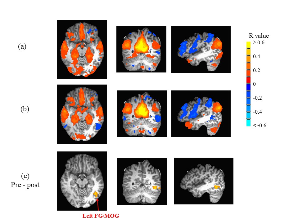
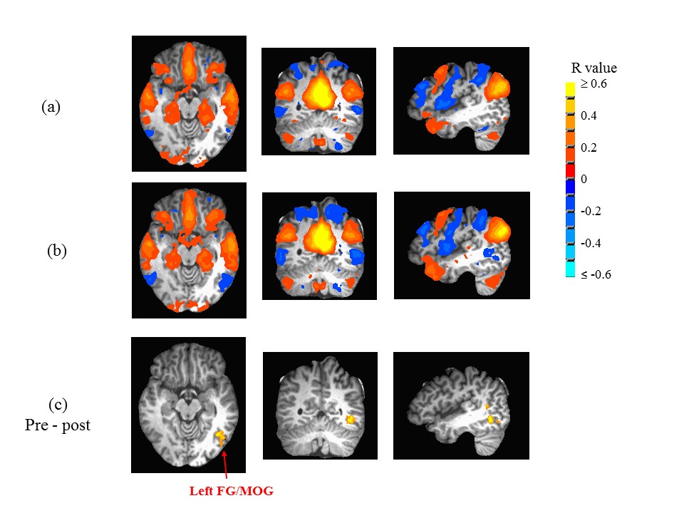
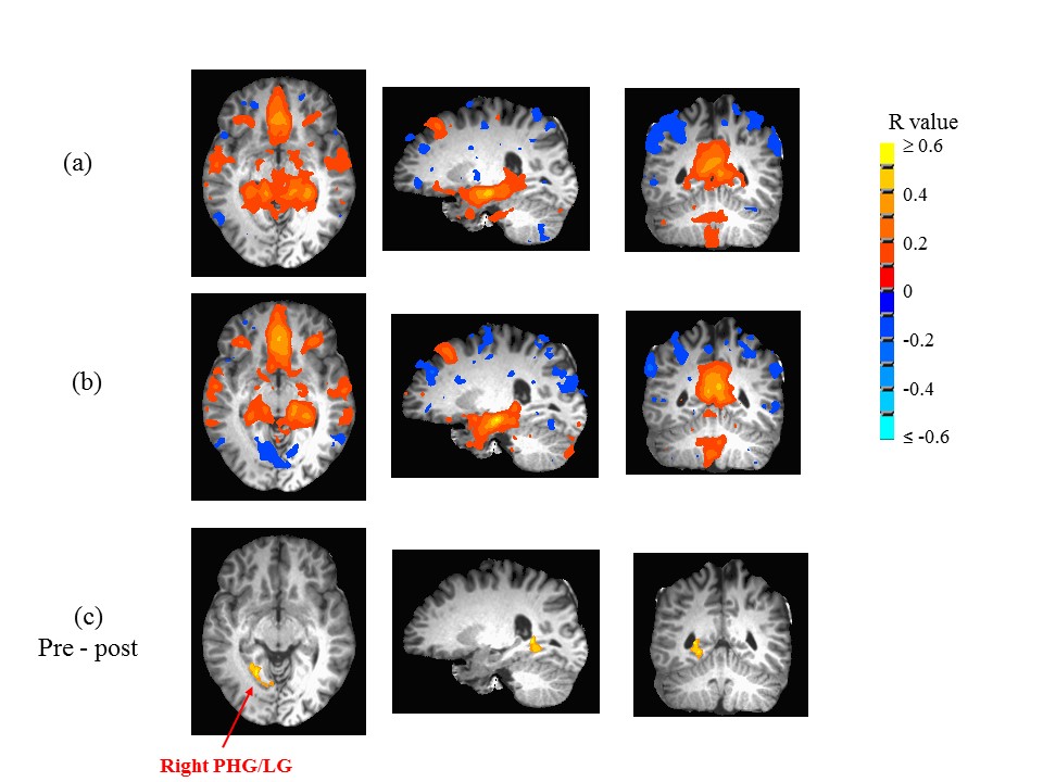
The whole-brain ANOVA revealed that the functional connections to the right ICC, left ICC and left hippocampus were significantly changed over a season. In both the right and left ICC, there was a significant change in the relation to the left fusiform gyrus/middle occipital gyrus, from a weak positive correlation at pre-season to an anti-correlation at post-season (Fig. 1 and Fig. 2). The left hippocampus was found significantly changed from weak positive correlation to anti-correlation with the right parahippocampal gyrus (Fig. 3).
A significant global increase of CBF was found from 65.7 ± 9.3 ml (blood)/100g (tissue)/min to 70.3 ± 9.0 ml (blood)/100g (tissue)/min over the season (p = 0.048). A significant increase of CBF was also observed in the right postcentral gyrus, from 48.5 ± 10.1 ml (blood)/100g (tissue)/min to 57.1 ± 9.7 ml (blood)/100g (tissue)/min (p = 0.0157 after Bonferroni correction).
No significant changes on the DTI metrics (fractional anisotropy (FA), and mean, axial and radial diffusivity values) were found on the overall FA skeleton or the 48 anatomical regions based on the ICBM-DTI-81 white-matter atlas (6) over the season.
DISCUSSION AND CONCLUSION
Over only a single football season, significant changes were found in functional connectivity to the DMN, while the structural connectivity remained stable as suggested by the DTI results. The changes in functional connectivity were accompanied by significant increase of CBF, both globally and at postcentral gyrus. These alterations suggest that sub-concussive hits, even when not clinically-diagnosed as “concussion”, can produce functional connectivity and CBF outcomes consistent with those observed in documented cases of brain damage. These changes emphasize the importance of further studies on how sub-concussive hits may affect the long-term development of brain networks and brain vascular modification.Acknowledgements
We want to thank Dr. Josef Pfeuffer at Siemens Healthcare, Erlangen for providing us with the advanced 3D ASL WIP package.References
1. McKee AC, Cantu RC, Nowinski CJ, Hedley-Whyte ET, Gavett BE, Budson AE, Santini VE, Lee HS, Kubilus CA, Stern RA. Chronic traumatic encephalopathy in athletes: progressive tauopathy after repetitive head injury. J Neuropathol Exp Neurol 2009;68(7):709-735.
2. Cox RW. AFNI: software for analysis and visualization of functional magnetic resonance neuroimages. Comput Biomed Res 1996;29(3):162-173.
3. Zhang K, Johnson B, Gay M, Horovitz SG, Hallett M, Sebastianelli W, Slobounov S. Default mode network in concussed individuals in response to the YMCA physical stress test. J Neurotrauma 2012;29(5):756-765.
4. Zhu DC, Covassin T, Nogle S, Doyle S, Russell D, Pearson RL, Monroe J, Liszewski CM, DeMarco JK, Kaufman DI. A potential biomarker in sports-related concussion: brain functional connectivity alteration of the default-mode network measured with longitudinal resting-state fMRI over thirty days. J Neurotrauma 2015;32(5):327-341.
5. Fischl B, Salat DH, Busa E, Albert M, Dieterich M, Haselgrove C, van der Kouwe A, Killiany R, Kennedy D, Klaveness S, Montillo A, Makris N, Rosen B, Dale AM. Whole brain segmentation: automated labeling of neuroanatomical structures in the human brain. Neuron 2002;33(3):341-355.
6. Mori S, Oishi K, Jiang H, Jiang L, Li X, Akhter K, Hua K, Faria AV, Mahmood A, Woods R, Toga AW, Pike GB, Neto PR, Evans A, Zhang J, Huang H, Miller MI, van Zijl P, Mazziotta J. Stereotaxic white matter atlas based on diffusion tensor imaging in an ICBM template. Neuroimage 2008;40(2):570-582.
Figures