0052
Diffusion Tensor Imaging Reveals Persistent Effects on White Matter Microstructure in High School Football Players with History of Sports-Related Concussion1Electrical and Computer Engineering, Purdue University, West Lafayette, IN, United States, 2Biomedical Engineering, Purdue University, 3Mechanical Engineering, Purdue University, 4Basic Medical Sciences, Purdue University
Synopsis
Diffusion Tensor Imaging has been considered a promising and sensitive imaging technology to detect subtle changes in white matter for people with mild traumatic brain injury. Although many studies have examined the immediate and near-term brain changes associated with sports-related concussions, the potential long-term consequences have been less-frequently investigated. In this study, a retrospective analysis was conducted on a subset of the Purdue Neurotrauma Group database to characterize the relationship between history of concussion and white matter diffusion properties.
INTRODUCTION
Diffusion Tensor Imaging (DTI) has been considered a promising and sensitive imaging technology to detect subtle changes in white matter (WM) for people with mild traumatic brain injury (mTBI) [1]. Although many studies have examined the immediate and near-term (6-12 months) brain changes associated with sports-related concussions, the potential long-term consequences have been less-frequently investigated. In this study, a retrospective analysis was conducted on a subset of the Purdue Neurotrauma Group database [2] to characterize the relationship between history of concussion and WM diffusion properties.METHOD
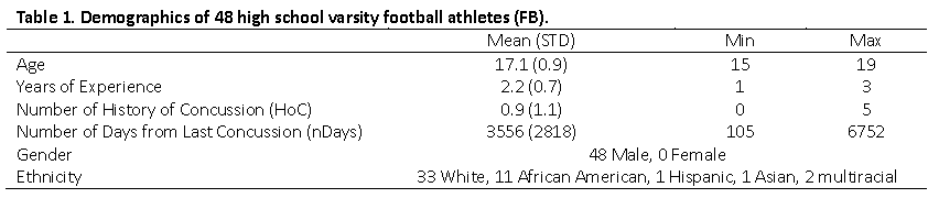
1) Participants: 48 male high school varsity football athletes (ages: 15-19; mean=17.1) underwent multiple MR imaging sessions as part of a longitudinal study including diffusion-weighted imaging (DWI). Data from a session taking place ~4.5 months after the end of their competition season, and ~4.5 months prior to the next season of participation, were selected to minimize recent exposure to sub-concussive events.
2) Data Acquisition: MR imaging was conducted using a 16ch Nova Medical brain array on a 3T GE Signa HDx. DWI were acquired using a spin-echo echo-planar imaging sequence (TR/TE=12,000/83.6 msec; matrix=96x96; FOV=24 cm; 46 axial slices; 2.5 mm isotropic resolution) with 30 diffusion encoding directions at b=1000 s/mm2 and one volume acquired at b=0 s/mm2.
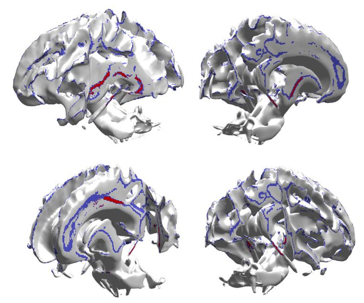
3) Processing: Image processing was performed based using FSL 5.0 toolbox and tract-based spatial statistics (TBSS) [3-4]. After correcting for motion and eddy current-induced distortion [5], DWI were segmented and eigenvalues and fractional anisotropy (FA) were estimated for each subject. After excluding images that failed visual quality inspection, data were nonlinearly co-registered to the FMRIB58-FA-template. The population mean FA image was thresholded (FA>0.2) to create a mean WM skeleton. The aligned FA image of each subject was projected onto the mean WM skeleton. The same nonlinear registration and skeleton projection were applied to diffusivity (MD) images.
4) Analysis: Average FA and MD for each subject were calculated for the 48 WM tracts defined in JHU-ICBM-DTI-81. Multiple regression analysis was conducted in the tracts to assess association between FA or MD and the history of diagnosed concussions (HoC; number of previous diagnosed injuries). Covariates included in the model were age (in years), ethnicity, years of high school football experience, and the number of days from the last concussion until the imaging session (nDays).
RESULTS
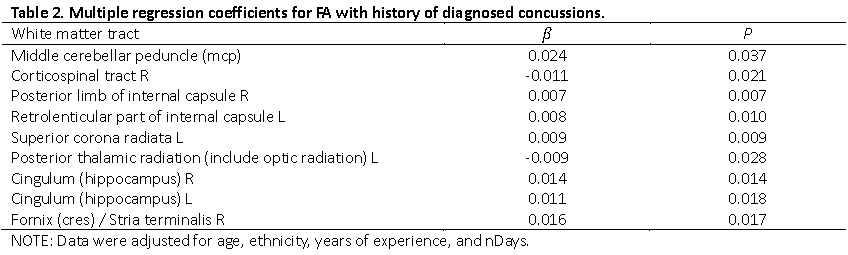
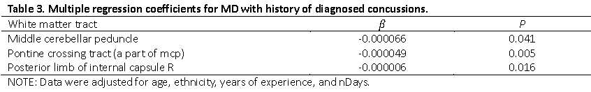
In multiple regression analyses, HoC was significantly associated with FA for nine WM tracts with seven exhibiting a positive correlation: the middle cerebellar peduncle ($$$\beta$$$=-0.024, p=0.037), right corticospinal tract ($$$\beta$$$=-0.011, p=0.021), right posterior limb of internal capsule ($$$\beta$$$=0.007, p=0.007), left retrolenticular part of internal capsule ($$$\beta$$$=0.008, p=0.010), left superior corona radiata ($$$\beta$$$=0.009, p=0.009), left posterior thalamic radiation including optic radiation ($$$\beta$$$=-0.009, p=0.028), right cingulum (hippocampus) ($$$\beta$$$=0.014, p=0.014), left cingulum (hippocampus) ($$$\beta$$$=0.011, p=0.018), and the right fornix (cres) / stria terminalis ($$$\beta$$$=0.016, p=0.017). There were three tracts found to exhibit significant association between HoC and MD, all exhibiting a negative relationship: the middle cerebellar peduncle ($$$\beta$$$=-0.000066, p=0.041), pontine crossing tract ($$$\beta$$$=-0.000044, p=0.012), and the right posterior limb of internal capsule ($$$\beta$$$=-0.000006, p=0.016).DISCUSSION
The analysis of DTI measurements suggests that sports-induced concussions may produce long-term local effects on axonal health in male football athletes. Statistically-significant associations were found between the number of previous concussion diagnoses and diffusion properties in multiple white matter tracts. The detected tracts are associated with sensory, somatosensory, optic, acoustic, and gustatory function as well as emotion, sleep, verbal memory, motor, equilibrium, and respiration. Exhibited changes for FA were higher values in 7 of 9 tracts, and lower MD in each of the 3 correlated tracts. Diagnosis of concussion is typically reported to produce reduced FA and elevated MD [6-8] (though controversies exist on the direction of change [9-10]). The higher FA and lower MD values observed here for subjects with greater numbers of previous concussions are hypothesized to be due to inflammation or swelling associated with axonal dilation [11] with no torn or broken axons, possibly suggesting a chronic state of physiologic response to injury.CONCLUSION
Findings suggest that sports-related concussions result in altered white matter health and that may persist for several years due either to extended repair mechanisms or chronic inflammation that may preclude complete recovery to pre-injury health.Acknowledgements
No acknowledgement found.References
1. Murugavel, Murali, et al. A longitudinal diffusion tensor imaging study assessing white matter fiber tracts after sports-related concussion. J Neurotraum 31.22. 2014; 1860-1871.
2. Talavage, T. M., et al. Functionally-detected cognitive impairment in high school football players without clinically-diagnosed concussion." Journal of Neurotrauma. 2014; 31.4: 327-338.
3. Smith, Stephen M., Mark Jenkinson, Heidi Johansen-Berg, et al. Tract-based spatial statistics: voxelwise analysis of multi-subject diffusion data. Neuroimage 31, no. 4. 2006; 1487-1505.
4. S.M. Smith, M. Jenkinson, M.W. Woolrich, et al. Advances in functional and structural MR image analysis and implementation as FSL. NeuroImage. 2004; 23(S1):208-219.
5. Andersson, J. L., & Sotiropoulos, S. N. An integrated approach to correction for off-resonance effects and subject movement in diffusion MR imaging. Neuroimage. 2016; 125, 1063-1078.
6. Inglese, Matilde, et al. Diffuse axonal injury in mild traumatic brain injury: a diffusion tensor imaging study. Journal of neurosurgery 103.2. 2005; 298-303.
7. Arfanakis, Konstantinos, et al. Diffusion tensor MR imaging in diffuse axonal injury. American Journal of Neuroradiology 23.5. 2002; 794-802.
8. Rugg-Gunn, F. J., et al. Diffusion imaging shows abnormalities after blunt head trauma when conventional magnetic resonance imaging is normal. Journal of Neurology, Neurosurgery & Psychiatry 70.4. 2001; 530-533.
9. Mayer AR, Ling J, Mannell MV, Gasparovic C, et al. A prospective diffusion tensor imaging study in mild traumatic brain injury. Neurology 2010; 74(8):643–50.
10. Bazarian J, Zhu T, Blyth B, Borrino A, Zhong J, Subject-specific changes in brain white matter on diffusion tensor imaging after sports-related concussion. Magn Reson Imaging. 2012; 30:171-180.
11. Povlishock J and Katz D. Update of neuropathology and neurological recovery after traumatic brain injury," J Head Trauma Rehab. 2004; 20 (1) 76-94.
Figures