0051
Long Term Changes in White Matter Following Sport-Related Concussion Measured by Diffusion Kurtosis Tensor Imaging: 6 months follow up1Department of Neurosurgery, Medical College of Wisconsin, Milwaukee, WI, United States, 2Department of Biophysics, Medical College of Wisconsin, Milwaukee, WI, United States, 3Department of Psychiatry, Medical College of Wisconsin, Milwaukee, WI, United States
Synopsis
We investigated chronic white matter changes in high school and collegiate football players with history of sport-related concussion using diffusion kurtosis tensor imaging. Results demonstrated that the symptoms normalized after one week but, mean diffusivity remained significantly low in concussed football players. These findings have implications for determination of recovery following concussion.
Introduction
Studies indicate that sport-related concussion
(SRC) results in rapid onset of specific physical changes and cognitive
deficits2, 6. However, SRC has historically been viewed as a minor injury without
long-term consequences because no abnormalities are seen in conventional MRI
and most athletes report resolution of symptoms within 7-10 days of injury.
While such symptom assessments are convenient, recent studies suggest that full
clinical recovery precedes physiological recovery. This means that injury can
persist even after symptoms have subsided6. However, the duration of these physiological alterations is unclear. Therefore,
understanding the trajectory of structural brain recovery is critical for
informing symptom management, return-to-play decisions, and prevention of
long-term deficits. Given that white matter (WM) tracts are especially
vulnerable to shear-strain injury caused by SRC1, 3, advanced
diffusion MRI techniques such as diffusion kurtosis tensor imaging (DKTI) show
great promise as biomarkers of brain injury and recovery following SRC. We have
previously reported widespread decreases in mean diffusivity and increased
axial kurtosis in the acute and sub-acute stages of injury (24 hours, 8 days)
in a group of concussed high school and collegiate athletes4. Here we investigated long-term chronic WM changes in our
sample using DKTI.Methods
Of the 26 concussed high school and college football players in the study (mean age = 17.4; SD = 1.7), 17 completed all time points (24 hours, 8 days and 6 months). 18 matched control athletes (mean age = 17.7; SD = 1.7) also completed identical protocols. The subjects underwent MRI imaging and assessment of symptoms (Sport Concussion Assessment Tool-3, SCAT3 and Standardized Assessment of Concussion, SAC), Balance Error Scoring System (BESS) and cognition at all three time points following SRC. For the study presented here, DKTI data at 6 months were analyzed using Tract-Based Spatial Statistics (TBSS)8. Additionally, Pearson correlations were used to investigate relationships between imaging findings and acute concussion severity measures (SCAT3). Data were acquired using GE MR750 3T MRI scanner equipped with a 32 channel head coil. DKTI was collected as part of multimodal imaging protocol using a single-shot SE-EPI sequence with 3mm isotropic voxels, four b=0 (reference images) and 60 diffusion-weighted images with b=1000s/mm2 and 2000s/mm2 (30 diffusion directions for each shell). Then, FA, MD, Dax, Drad (fractional anisotropy, mean, axial and radial diffusivity), MK, Kax and Krad (mean, axial and radial kurtosis) data were analyzed using TBSS workflow to compare groups. Diffusion measurements were also extracted from voxels with significant group differences and analyzed in relation to measures of concussion symptoms and cognition at 24 hours post-injury. Because of the longitudinal nature of this study with long follow-up time point, we performed test-retest reliability using concordance correlation coefficient (CCC)5 using repeat scans from control subjects. Average CCC was 0.91 for MD, 0.86 for Krad and 0.69 for Kax.Results
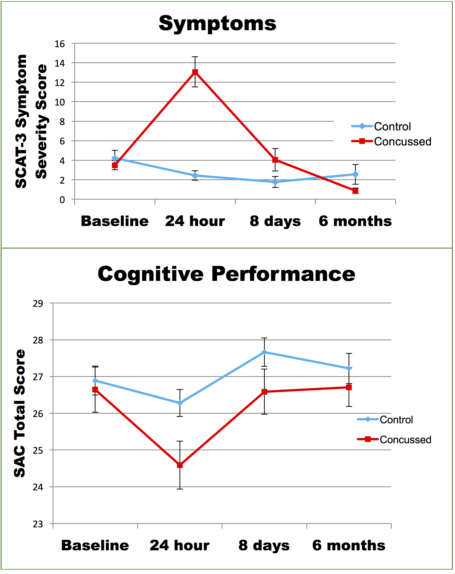
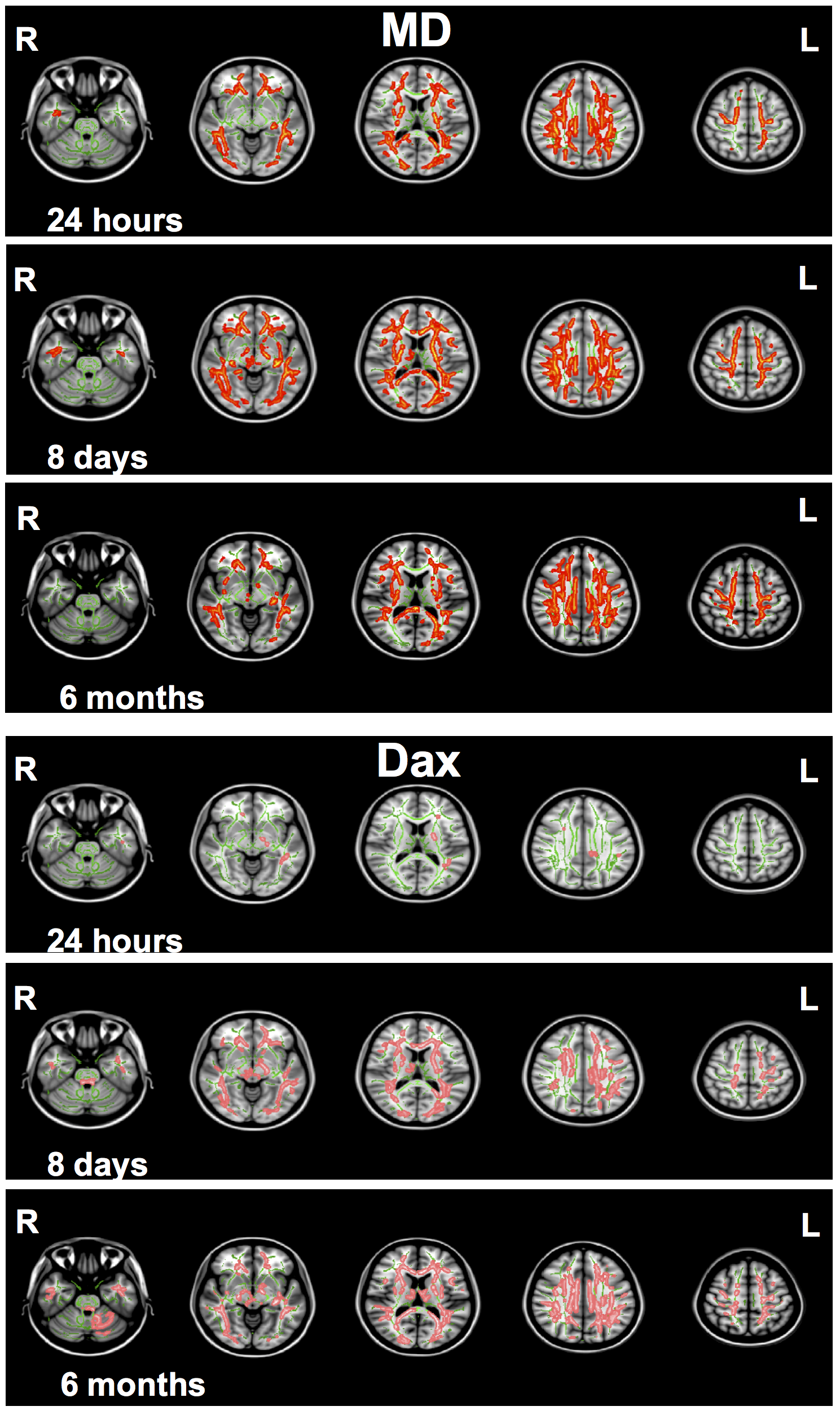
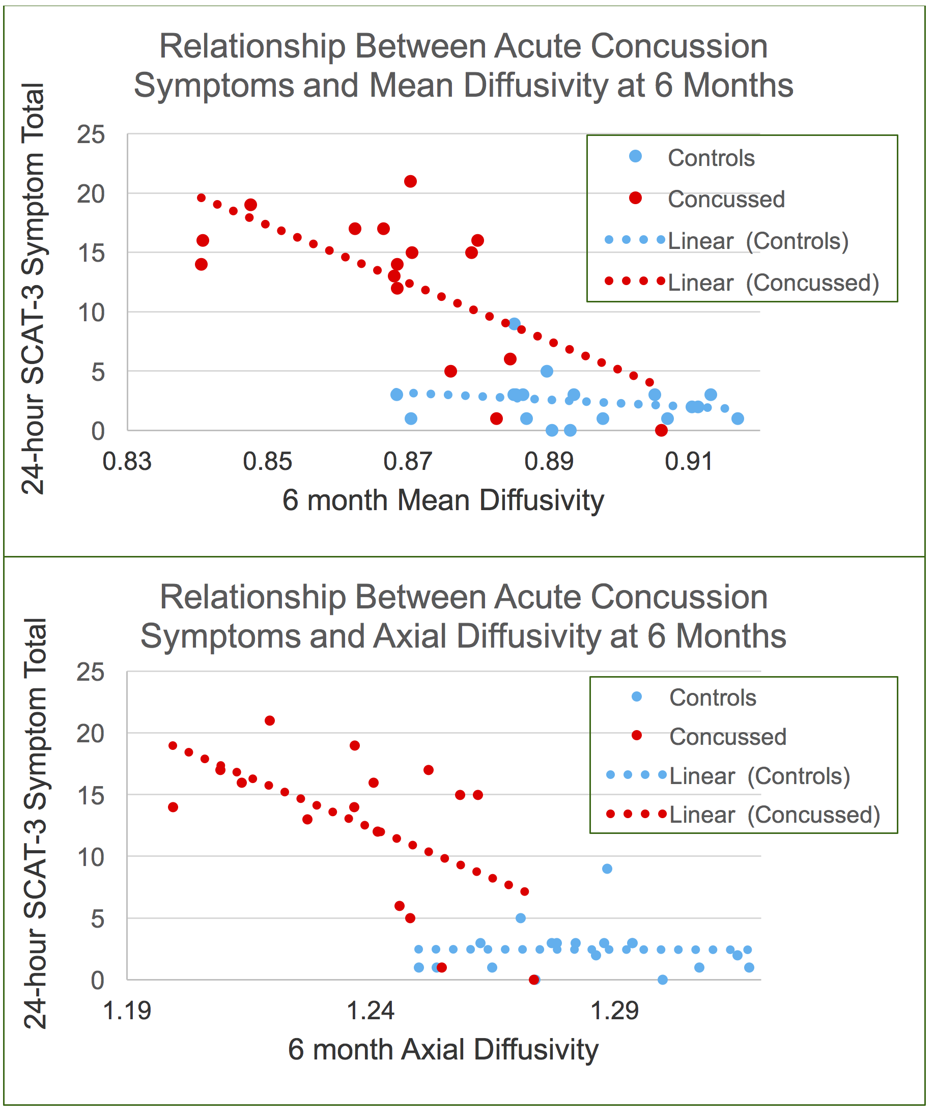
There were no group differences on measures of self-reported concussion symptoms, cognition, or balance at 6 months post-injury (Fig.1). However, the SRC group showed widespread decreased MD and Dax compared to controls at 6 months (Fig.2) (p<0.05, corrected for multiple comparisons). This was similar to acute (24 hours) and sub-acute (8 days) DKTI findings, which were included in Fig.2 for reference. Moreover, concussion symptoms (SCAT3 in concussed athletes) assessed at 24 hours after injury correlated negatively with MD and Dax at 6 months (Fig.3). There were no significant FA or diffusion kurtosis differences at 6 months.Discussion and Conclusion
These findings indicate that the concussed athletes showed significant WM alterations even after 6 months post-injury, despite normalization of clinical symptoms within a week after injury. Furthermore, Fig.3 illustrates that those with high injury severity had lingering effects measured by MD, while those with low injury severity returned to the levels of normal controls. Reduced diffusivity at 6 months post injury was associated with greater reported symptoms 24 hours post injury, suggesting a “dose-dependent” effect of SRC on WM based on the severity of the injury. These findings have implications for determination of recovery following SRC and concussion management. Further studies are needed to investigate the long-term impact of these WM changes.Acknowledgements
This project was supported by the National Center for Advancing Translational Sciences, National Institutes of Health (8UL1TR000055 and 1UL1-RR031973-01), the US Army Medical Research and Materiel Command (W81XWH-12-1-0004), and the NFL-GE Head Health Challenge I.References
1. Gentry, L.R., Imaging of closed head injury., Radiology, 191 (1994) 1-17.
2. Giza, C.C. and Hovda, D.A., The new neurometabolic cascade of concussion., Neurosurgery, 75 Suppl 4 (2014) S24-33.
3. Holbourn, A.H.S., Mechanics of head injuries, The Lancet, 242 (1943) 438-441.
4. Lancaster, M.A. et al., Acute white matter changes following sport-related concussion: A serial diffusion tensor and diffusion kurtosis tensor imaging study., Hum Brain Mapp, 37 (2016) 3821-3834.
5. Lin, L.I., A concordance correlation coefficient to evaluate reproducibility., Biometrics, 45 (1989) 255-268.
6. McCrea, M. et al., Acute effects and recovery after sport-related concussion: a neurocognitive and quantitative brain electrical activity study., J Head Trauma Rehabil, 25 (2010) 283-292.
7. McCrory, P. et al., Consensus statement on concussion in sport: the 4th International Conference on Concussion in Sport held in Zurich, November 2012., Br J Sports Med, 47 (2013) 250-258.
8. Smith, S.M. et al., Tract-based spatial statistics: voxelwise analysis of multi-subject diffusion data, Neuroimage, 31 (2006) 1487-1505.
Figures