0040
Optimized Single Pre/Post Contrast Protocol for MOLLI T1 Mapping with Inversion Group (IG) Fitting1Medical Imaging, University Health Network, Toronto, ON, Canada, 2Medical Imaging, University of Toronto, Toronto, ON, Canada
Synopsis
A major focus in cardiac research is the assessment of myocardial pathology using quantitative T1 mapping. A number of sequences are being investigated for this task. One candidate is MOLLI. It provides superior precision to other cardiac T1 mapping techniques. However, its precision is dependent on heart rate and range of T1 values present. Current attempts at optimizing precision are somewhat impractical, as they utilize separate MOLLI protocols for different heart rates, and for pre/post contrast imaging. This study identifies a single MOLLI protocol optimal for precision over a broad range of heart rates and pre/post contrast T1 values.
Introduction
A major focus in cardiac imaging research is the assessment of subtle diffuse myocardial pathology by means of quantitative T1 mapping. A number of cardiac T1 mapping sequences are currently being investigated for this task. One candidate is MOLLI [1]. It provides superior precision to other cardiac T1 mapping techniques. However, its precision is highly dependent on heart rate and the range of T1 values in the tissue. Therefore, its relative precision advantage is variable. To address this limitation, attempts have been made to optimize MOLLI precision. However, current optimizations are somewhat impractical, as they utilize separate MOLLI protocols for different heart rates, and for pre/post contrast imaging. The objective of this study is to identify a single MOLLI protocol that is optimal for precision over a broad range of heart rates and pre/post contrast T1 values.
Theory/Methods
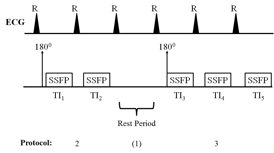
The basic MOLLI pulse sequence (Figure 1) consists of several 1800 inversion pulses. Following each 180, a series of SSFP images are acquired (=TI's). In conventional MOLLI, rest periods must be inserted prior to each 180 to allow full magnetization recovery. This is to avoid introducing bias into the subsequent T1 fits. However, we instead employ IG-fitting [2]. This technique provides accurate T1 maps for any combination of TI's and rest periods (including no rest period). This will provide maximum flexibility for the subsequent optimization (described next).
The precision of the fitted T1 is dependent on the distribution of TI's and rest periods. Our goal is to find the TI/rest period combination that is optimal. To perform the optimization, a MOLLI simulation is performed to calculate T1 precision over a range of pre- (100-600ms) and post-contrast (800-1200ms) myocardial T1 values, as well as a range of heart rates (50-80BPM). The simulation is performed for all possible TI/rest period combinations. For each combination, a cost metric is defined as the sum of precisions over all T1's and heart rates:
Cost Metric(TI, Rest Period) = Σi=HR ∑j=T1 σij(TI, Rest Period)
The optimal TI/rest period combination is defined the one which minimizes the cost metric.
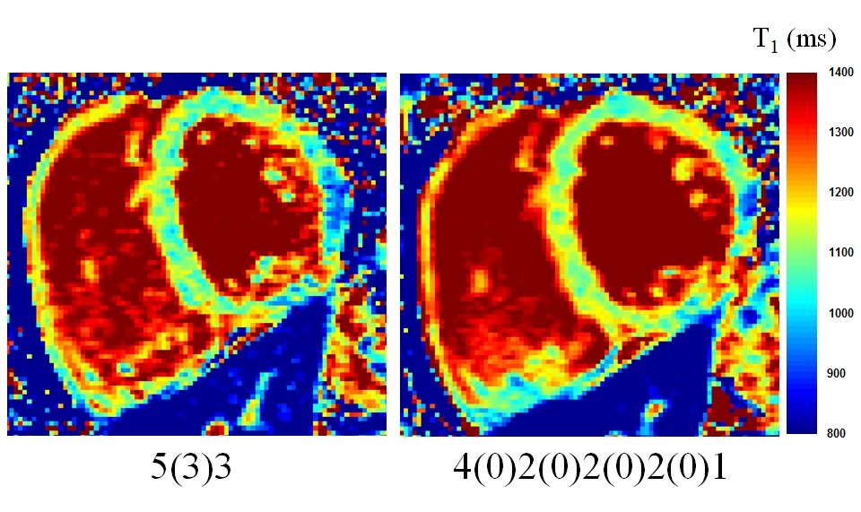
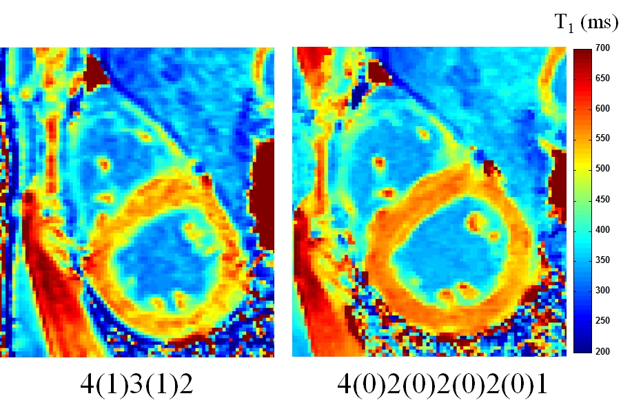
Standard MOLLI protocols acquire data over 11 heartbeats; specifically, a 5(3)3 protocol is used for pre-contrast imaging, and a 4(1)3(1)2 protocol for post-contrast (where non-bracketed numbers indicate TI's, and bracketed numbers indicate rest period heartbeats; see Figure 1). To provide a fair comparison with existing techniques, we therefore perform the optimization for an 11 heartbeat acquisition. The results of this calculation identified an optimal protocol of: 4(0)2(0)2(0)2(0)1. Note this protocol provides optimal precision over the range of pre- and post-contrast T1's and heart rates.
A total of 8 patients were imaged three separate times (total of 24 scans). Standard and optimized MOLLI protocols were acquired pre- and post-contrast. T1 precision was determined from the standard deviation over a myocardial ROI. To compare the precision of standard and optimized protocols, t-tests were performed. Statistical significance was set at the 95% confidence level.
Results
Figure 2 plots a pre-contrast example of images acquired with standard and optimized MOLLI protocols. In this case, the precision is 12% better in the optimized protocol image. Figure 3 plots a post-contrast example. In this case, the precision is 18% better in the optimized protocol image.
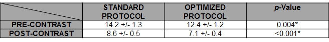
Table 1 lists the precision for standard and optimized protocols over all 24 scans. The precision of the optimized protocol is 12% (pre-contrast) and 17% (post-contrast) better than the standard protocol. In both cases, the difference is significant at the 95% confidence level. The range of heart rates was from 40BPM to 75BPM
Discussion/Conclusions
This study demonstrated that it is possible to employ a single MOLLI protocol that achieves better precision than standard MOLLI techniques. The improved precision was the result of the choice of TI/rest period combination. In particular, note that with standard MOLLI, rest periods are required between the 180's. Therefore, although the standard pre- and post-contrast protocols were acquired in 11 heart beats, only 8 (pre-contrast) or 9 (post-contrast) TI's were acquired. Since the optimized protocol employed the IG fitting [2], rest periods were not required. As a result, 11 data points were acquired in 11 heart beats. The optimized protocol also took into account different heart rates. As a result, the optimized protocol was able to achieve better precision over a wide range of T1's and heart rates. This improved precision may ultimately be beneficial in the assessment of subtle longitudinal changes in various myocardial pathologies.
Acknowledgements
Support from Siemens HealthcareReferences
[1] DR Messroghli et al., MRM 2004;52(1):141-146.
[2] MS Sussman et al., MRM 2016; 75(6): 2332-2340.
Figures