0008
Water diffusivity changes in the brain following exposure to low levels of focused ultrasound energy1Department of Diagnostic Radiology & Nuclear Medicine, University of Maryland School of Medicine, Baltimore, MD, United States
Synopsis
MR-guided Focused ultrasound (MRgFUS) for neuro-interventions is gaining popularity. However, the biological effects of low level exposure on the brain tissue are less understood. In this study, we used in-vivo MR diffusion kurtosis imaging (DKI) to measure water diffusion changes in vivo in rats at varying FUS exposure levels in the whole brain. Acoustic simulation and experimental data demonstrate more wide spread tissue diffusion changes and not just local changes with just a single exposure albeit for varied duration. Our results suggest that diffusion changes are mainly due to shearing forces exerted on the brain.
Introduction
MR-guided Focused ultrasound (MRgFUS) for neuro-interventions is gaining popularity not only for ablating tissues as in the case of the treatment of brain tumor, essential tremors and Parkinson’s disease, but also to study the effects of neuromodulation, and temporary opening of blood-brain barrier non-invasively. However, the biological effects of low level exposure on the brain tissue are less understood especially at locations that are removed from the focal spot. In this study, we used in-vivo MR diffusion kurtosis imaging (DKI) to measure brain water diffusion changes in vivo in rats at varying FUS exposure levels in the whole brain.Methods
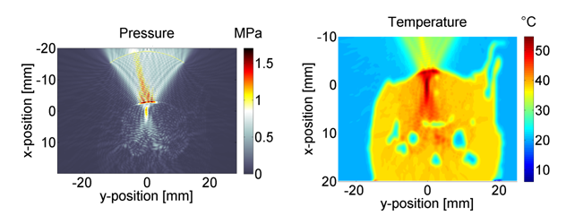
To systematically understand the brain structural and functional changes from focused ultrasound intervention, rats (n=8) were subject to various levels of MRgFUS exposure using an eight element array ultrasound transducer operating at 1.5 MHz (IGT, Paris France). Prior to commencing any MR imaging, CT scans of the rat brains were obtained to incorporate bone and tissue information to more accurately estimate the pressure distribution from FUS exposure as the acoustic wave passes through the skull. The pressure thus estimated can then be related to the changes in the observed diffusion in the brain tissue. Imaging was performed on a 7T Bruker MR scanner (Bruker Biospin, Billerica MA). Once the focal spot was determined, each animal was subject to exposure at a specific power level and duration. Temperature monitoring near the focal plane was performed during the exposure using a FLASH sequence for MR thermometry and to determine temperature distribution in the brain. Prior to the exposure and 2 hours after FUS exposure, MR T2-weighted and DKI were performed on the rats. For DKI especially, the number of diffusion directions was 30, and b value per direction was 1000 s/mm2, 1500 s/mm2, and 2000 s/mm2 respectively. Mean kurtosis (MK) and mean diffusion (MD) were measured across 7 axial slices centered over the acoustic focal plane to arrive at whole brain diffusion parameters. To better understand the acoustic pressure distribution in rat heads, a k-space time-domain method was employed to simulate the acoustic field during the exposure by solving the full Westervelt equation1.Results
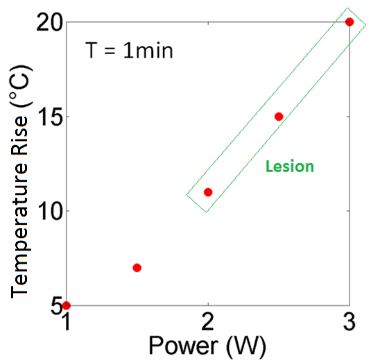
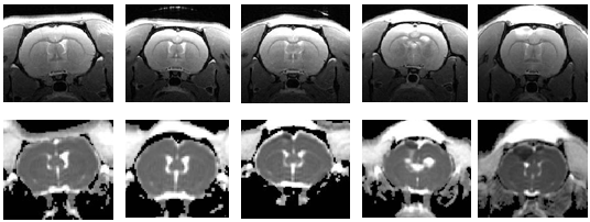
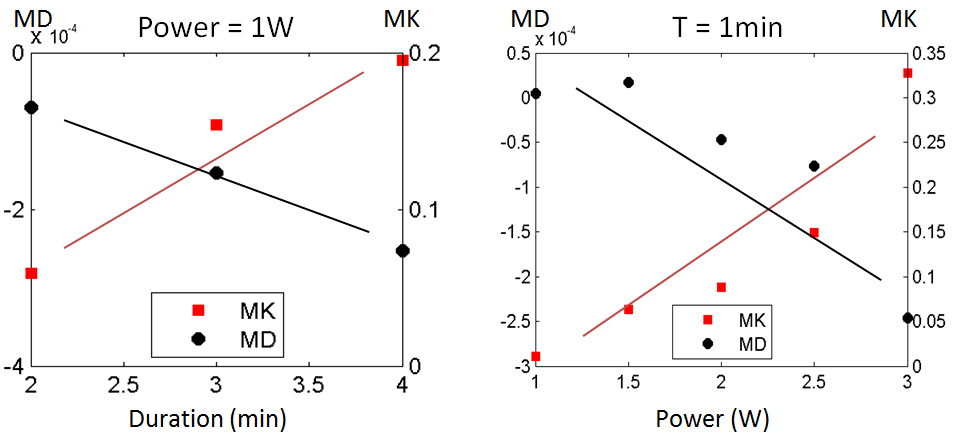
In all experiments, the focal spot located at same positon on cortex. Peak temperature was recorded near the focal plane during sonications ranging from 1-3 W for 1 min (Fig.1). Thermal lesions which were visible on T2-weighted images were observed when power was above 2 W for 1 min. The T2 weighted and diffusion kurtosis images before and after ultrasound exposure are shown in Fig.2 - the employed power from left to right is 1 W, 1.5 W, 2 W, 2.5 W and 3 W, respectively. Increased mean kurtosis (MK) and reduced mean diffusion (MD) were measured across 7 axial slices wiith increasing acoustic power and duration (Fig.3). Higher power has more of an effect on water diffusion properties compared to sonicating for a longer duration while delivering the same energy. This difference could be due to the ability of the in vivo tissue to dissipate of heat over time which may change the temperature gradient in the tissue. CT Hounsfield units for the rat skull were converted to bone related parameters2. Based on CT characterization, acoustic pressure simulation at 1 W on a rat brain is shown in Fig.4. It is found that the skull absorbs a significant amount of heat.Discussions
Our observation of decreased MD is consistent with the notion of decreased volume of the extracellular space or with increased uptake of water by the cells suggesting changes in cellular homeostasis. Increased MK suggests increased heterogeneity in the tissue microstructure suggesting increased astroglial reactivity associated with the rise in temperature. These results suggest that at even low exposure rates, where the pressures reaches ~400-500 kPa, water diffusion changes occur and can be detected by diffusion tensor imaging in the absence of any visible damage observed on T2-weighted image.Conclusions
Our results suggest that diffusion changes are mainly due to shearing forces exerted on the brain tissue that have an effect on the tissue environment far from the focal spot if only temporarily. Results also suggest that the whole brain may be affected even though the ultrasonic energy is focused to a spot and may have long term functional implications3 when multiple interventions are performed using this technique. We will continue to study the potential long term effects of such exposure or neuromodulation on cognition and behavior. Understanding the tissue level changes can also provide insights into effective means for clearing amyloid plaques4.Acknowledgements
No acknowledgement found.References
1. Hamilton M, Blackstock D. Nonlinear Acoustics. Academic Press; 1998.
2. Aubry J, Tanter M, Gerber J, et al. Optimal focusing by spatio-temporal inverse filter: part II. Experiments. J. Acoust. Soc. Am. 2001;110(48).
3. Zhuo J, Keledjian K, Xu S, et al. Changes in Diffusion Kurtosis Imaging and Magnetic Resonance Spectroscopy in a Direct Cranial Blast Traumatic Brain Injury (dc-bTBI) Model. PLoS one. 2015;10(8).
4. Jordan J, Ayala-Grosso Carlos, Markham K, et al. Antibodies Targeted to the Brain with Imaged-Guided Focused Ultrasound Reduces Amyloid-β Plaque Load in the TgCRND8 Mouse Model of Alzheimer’s Disease. PLsS one. 2010;5(5).
Figures