5065
3D-Printed whole-brain holder for multiple orientation magnetic susceptibility measurements and precise dissection1Donders Institute for Brain, Cognition and Behaviour, Radboud University, Nijmegen, Netherlands, 2Department of Anatomy, Donders Institute for Brain, Cognition and Behaviour, Radboud University Medical Center, Nijmegen, Netherlands, 3Wellcome Centre for Integrative Neuroimaging, FMRIB, University of Oxford, Oxford, United Kingdom, 4Faculty of Social Sciences, Radboud University, Nijmegen, Netherlands
Synopsis
In this study, we present a whole-brain holder for ex vivo experiments which allows rotating the sample inside a conventional head coil (while ensuring no deformation occurs) and provides guidance for precise correspondence between the MR data and excised tissue. We demonstrate some of these features with two experiments aimed at validating magnetic susceptibility measurements using MRI, where small 5mm cube samples located in different slices through the whole brain can be excised with great precision.
Introduction
Quantitative susceptibility mapping (QSM) is known to reflect iron concentration in deep grey matter (DGM)1,2. Conversely, in white matter (WM) the correlation of susceptibility to paramagnetic iron or diamagnetic myelin is more complicated because of the discrepancy between the existing QSM model and the WM micro-structural organization3-5. This mismatch is justified by magnetic susceptibility, $$$\chi$$$, in WM is anisotropic3,6 and the signal phase (used to compute $$$\chi$$$) in WM suffers from microstructure-related effects4.
Studying WM microstructure-related effects in the GRE signal requires acquisition with multiple sample orientations with respect to the static field, B0, and is more feasible with post-mortem samples. Furthermore, post-mortem studies allow the validation of MRI findings using, for example, histology. Yet, registration between the histology sample and the whole-brain data is never straightforward because of tissue deformation.
Here we show a setup that allows for ex-vivo multiple orientations experiments and precise dissection guidance. We demonstrate its usage in the study of the bulk magnetic susceptibility of excised samples from a whole post-mortem brain experiment.
Materials and Methods
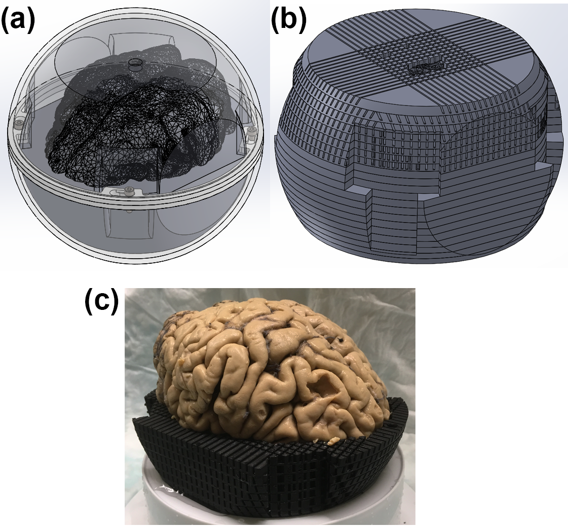
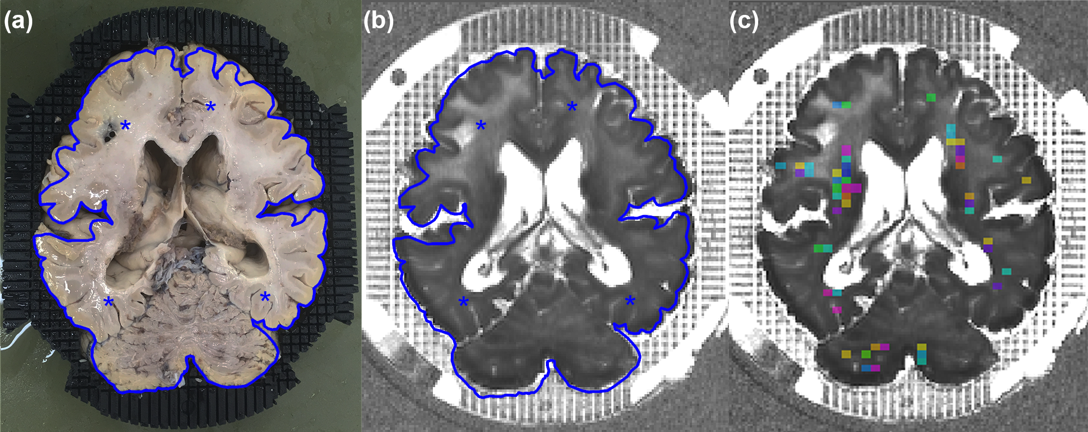
The setup consists of two parts: an outer-sphere and a custom 3D-printed holder (Fig.1). The holder consists of a stack of 20 5-mm thick, 3D-printed plates that remains fixed inside the sphere during rotation. The inner part has the exact shape of the brain specimen (obtained from a pilot scan) to make the specimen fit tightly, minimising tissue deformation when rotating or excising the sample. The outer part of the plates has a grid, providing landmarks in MRI images for dissection planning and guidance (Fig.2).
A formalin-fixed post-mortem brain specimen was scanned at 3T (Siemens, Erlangen, Germany) using a 64-channel head coil. Imaging protocol of whole-brain experiment consisted of:
- MP2RAGE with res=1mm isotropic, Tacq=5min;
- DWI spin-echo EPI, SMS=3, res=1.6 mm isotropic, TR/TE=3780/71.20ms, 2-shell (b=0/1250/2500s/mm2,17/120/120 measurements), NSA=16, Tacq=4.5hours;
- For 9 orientations with respect to B0, monopolar 3D ME-GRE was used with: TR/TE=39/2.15:3.05:35.7ms (12 echoes), res=1.8mm isotropic, flip angle=20°, Tacq=7min.
ROIs were defined in the 3D MRI image corresponding to dissectible regions (Fig.2c). The ROI selection criteria for our study was fibre direction homogeneity within the ROI. 16 samples (4 DGM and 12 WM) were excised and scanned inside an agarose gel-filled holder 7 days later. The imaging protocol of the dissected brain samples experiment consisted of:
- MP2RAGE as for the whole-brain experiment;
- DWI as for the whole-brain experiment, NSA=20, Tacq=5hours 40min;
- For 9 orientations with respect to B0, monopolar 3D GRE with 5 echoes, TR/TE=48/5.22:9.15:41.82ms, res=0.75mm isotropic, flip angle=11°, Tacq=14min.
All data were co-registered using a rigid-body transform to the GRE space using flirt7, allowing the calculation of the exact orientation and the main fibre direction of the samples in respect of B0. Good quality image coregistration was achieved in 5/16 excised samples and only these samples were analysed.
Field maps were computed8 and the background fields were removed using VSHARP9 for the whole-brain sample (mask obtained using FSL’s bet) and LBV10 for the excised samples. Whole-brain $$$\chi$$$ was computed from COSMOS10 and STI3,6 using the field maps acquired at 9 orientations. Excised samples $$$\chi$$$ was calculated using the field perturbation in the agarose gel surrounding the samples4, separated into an isotropic and anisotropic $$$\chi$$$ ($$$\chi_{isotropic}$$$ and $$$\chi_{anisotropic}$$$) with DTI results as the axonal orientation prior.
Susceptibility values estimated from the excised samples and their corresponding ROI averaged results of COSMOS and STI were compared.
Results
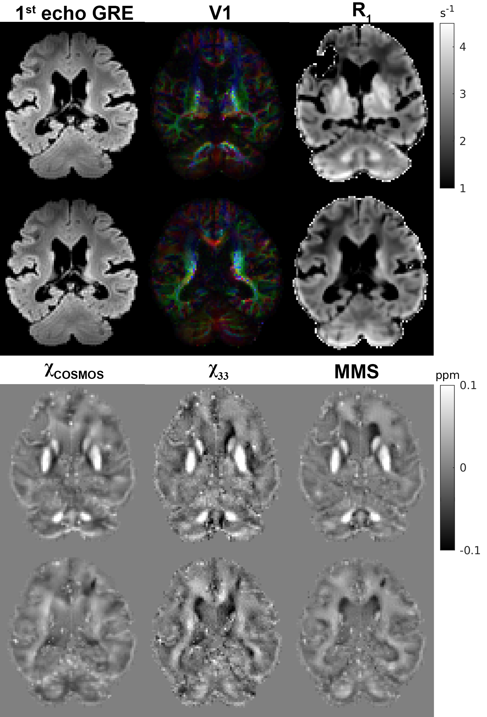
Fig.3 shows example data of the whole-brain experiment with the sample being scanned in multiple orientations allowing the calculations of high quality STI maps.
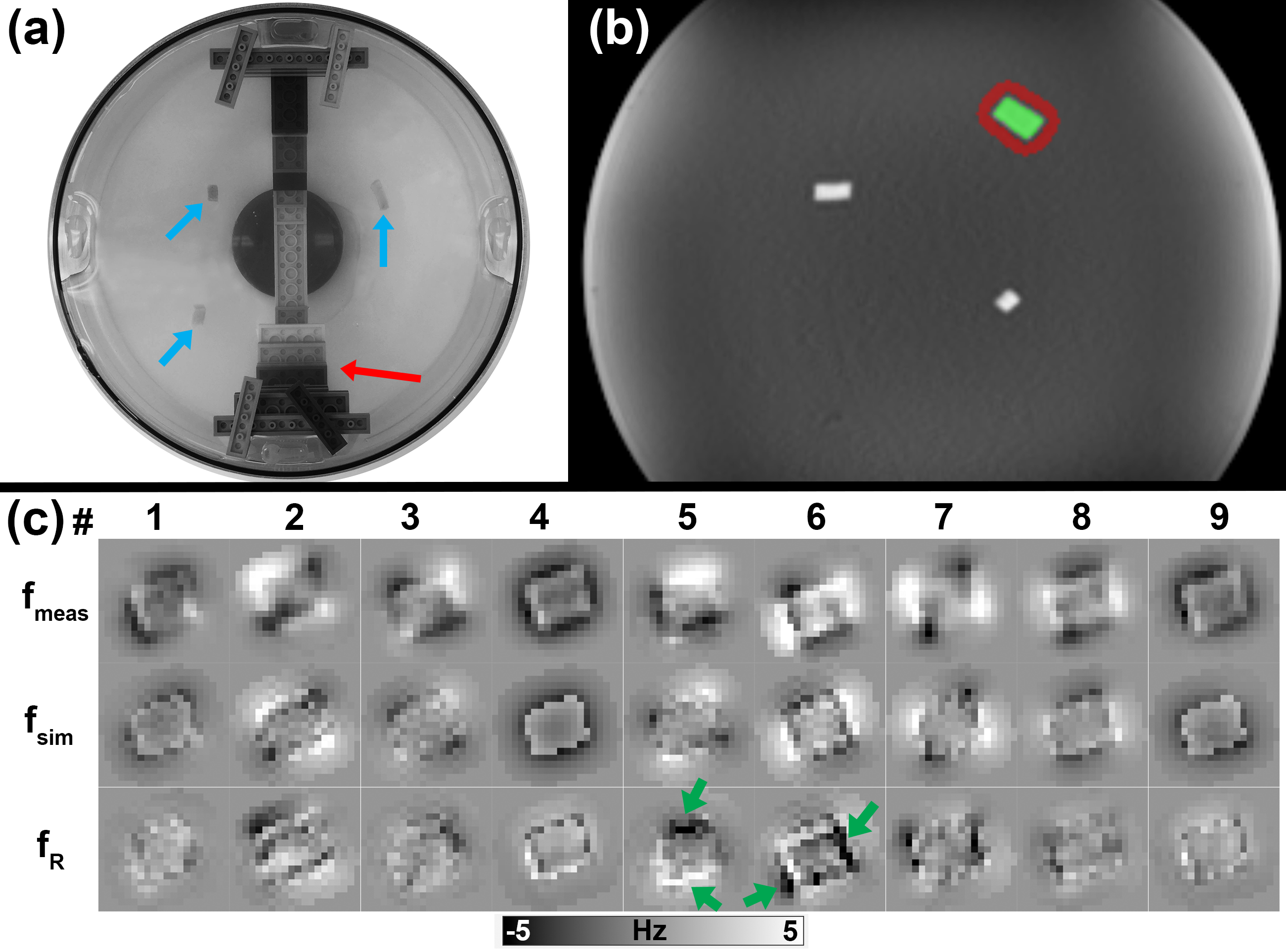
Fig.4c shows the measured ($$$f_{meas}$$$), simulated ($$$f_{sim}$$$) and residual frequency ($$$f_{R}=f_{meas}-f_{sim}$$$) from 1 excised sample. The residuals are clearly affected by the image registration accuracy, as small mismatches create large errors in the tissue/agarose interface. Image intensity variations caused by B1 field inhomogeneities and eddy current distortion hindered the accuracy of registration of different orientations MRI data (5/16 samples could be used).
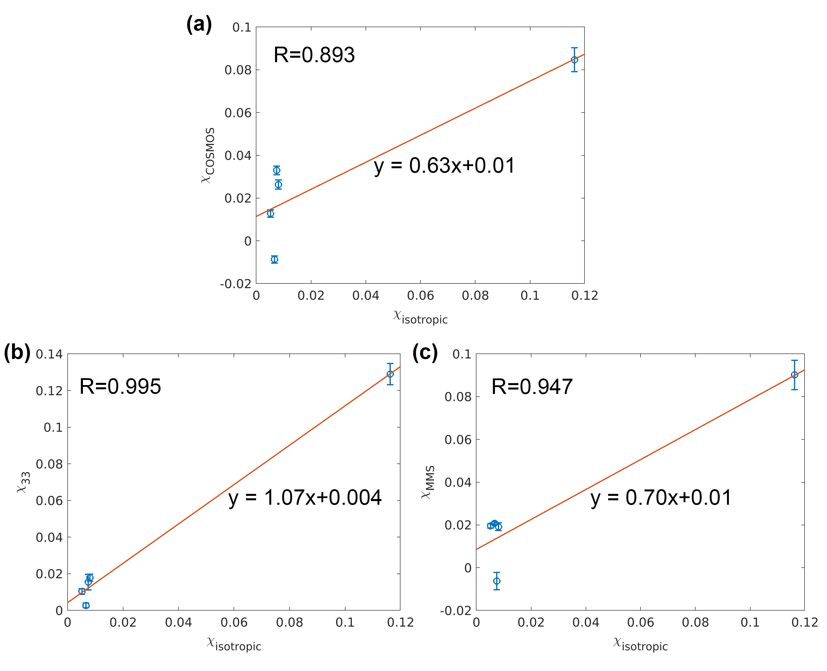
Fig.5 shows the regression plots of the excised tissue samples $$$\chi$$$ (obtained with the external field approach) versus the averaged susceptibility over the corresponding ROI. Good agreements in susceptibility were found, though the limited number of samples used and having only one DGM sample meant that the correlation is driven solely by this large $$$\chi$$$ outlier.
Conclusions
The holder setup provides an important guide to ensure the correspondence between MRI and the excised samples with a precision of ~1mm throughout the whole brain.
Future experiments will address some of the limitations of the current apparatus, including sample preparation, phantom building, ROI selection and acquisition strategy.
Acknowledgements
This work is part of the research programme with project number FOM-N-31/16PR1056/RadboudUniversity, which is financed by the Netherlands Organisation for Scientific Research (NWO). The authors would like to thank Man-Yan Lam for technical support in creating the essential 3D mesh files in AutoCAD.References
- Langkammer, C. et al.Quantitative susceptibility mapping (QSM) as a means to measure brain iron? A post mortem validation study. Neuroimage62,1593–1599 (2012).
- Sun, H. et al.Validation of quantitative susceptibility mapping with Perls' iron staining for subcortical gray matter. Neuroimage105,486–492 (2015).
- Li, X. et al.Mapping magnetic susceptibility anisotropies of white matter in vivo in the human brain at 7 T. Neuroimage62,314–330 (2012).
- Wharton, S. & Bowtell, R. Effects of white matter microstructure on phase and susceptibility maps. Magn Reson Med73,1258–1269 (2015).
- Yablonskiy, D. A. & Sukstanskii, A. L. Effects of biological tissue structural anisotropy and anisotropy of magnetic susceptibility on the gradient echo MRI signal phase: theoretical background. NMR Biomed30,e3655 (2017).
- Liu, C. Susceptibility tensor imaging. Magn Reson Med 63, 1471–1477 (2010).
- Jenkinson, M., Bannister, P., Brady, J. M. and Smith, S. M. Improved Optimisation for the Robust and Accurate Linear Registration and Motion Correction of Brain Images. NeuroImage, 17(2), 825-841, 2002.
- Robinson, S. D. et al.An illustrated comparison of processing methods for MR phase imaging and QSM: combining array coil signals and phase unwrapping. NMR Biomed30,e3601 (2017).
- Wu, B., Li, W., Guidon, A. & Liu, C. Whole brain susceptibility mapping using compressed sensing. Magn Reson Med67,137–147 (2012).
- Zhou, D., Liu, T., Spincemaille, P. & Wang, Y. Background field removal by solving the Laplacian boundary value problem. NMR Biomed27,312–319 (2014).
- Liu, T., Spincemaille, P., de Rochefort, L., Kressler, B. & Wang, Y. Calculation of susceptibility through multiple orientation sampling (COSMOS): A method for conditioning the inverse problem from measured magnetic field map to susceptibility source image in MRI. Magn Reson Med61,196–204 (2009).
Figures