5062
Susceptibility Contrast at Ultra-low Magnetic field with Superparamagnetic Nanoparticles1Institute of Medical Physics, The University of Sydney, Sydney, Australia, 2A. A. Martinos Center for Biomedical Imaging, Massachusetts General Hospital, Charlestown, MA, United States, 3ARC Centre of Excellence for Engineered Quantum Systems, The University of Sydney, Sydney, Australia, 4Department of Physics, Harvard University, Cambridge, MA, United States, 5Harvard Medical School, Boston, MA, United States
Synopsis
MRI scanners operating at ultra-low fields (ULF) promise to reduce the cost and expand the clinical accessibility of MRI. Here, we use an ULF (6.5 mT) MRI scanner and an efficient balanced steady-state free precession MRI protocol to image superparamagnetic iron oxide nanoparticles (SPIONS) in solution. We observe strong susceptibility effects due to the highly-magnetized state of SPIONs even at ULF. These susceptibility effects enable the most sensitive imaging of a contrast agent at ULF that we are aware of. These results will broaden the clinical applications of ULF MRI, and have implications for drug tracking and delivery in nanotheranostics.
Introduction
Electromagnet-based MRI scanners operating at ultra-low magnetic fields (ULF, < 10 mT) have recently demonstrated clinically-relevant imaging of the human brain.1 Due to low construction and operational costs, as well as ease of siting, ULF MRI scanners could become common screening tools particularly at remote hospitals and medical clinics.2
Clinical applications of ULF MRI are, however, limited by a lack of suitable contrast agents (CAs).1,3 Since relaxation-based CAs generally modulate signal intensity, their utility is limited by the inherently low signal-to-noise ratio (SNR) in the ULF MRI regime. One approach to improving the SNR in the ULF regime relies on sequences that can accommodate high-speed signal averaging, and in the case of balanced steady-state free precession (b-SSFP), the image intensity is weighted by the ratio T1/T2.1,4 This weighting, in combination with the convergence of T1 and T2 values at ULF,5 reduces the utility of relaxation-based contrast in ULF MRI. That said, the imaging of CAs via susceptibility effects remains unexplored at ULF. Indeed, susceptibility-weighted imaging has never been demonstrated in ULF MRI due to the negligible magnetizations induced in human tissues at low magnetic field.6
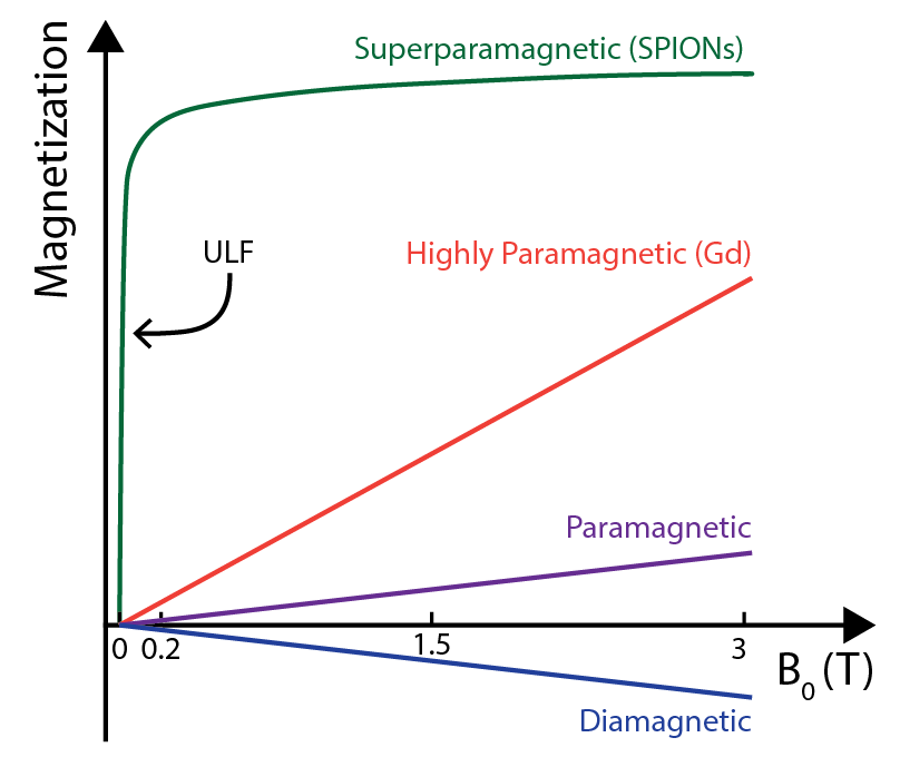
Here we describe our work with biocompatible superparamagnetic iron oxide nanoparticles (SPIONs) that demonstrate exceedingly high magnetizations in the ULF regime (see Figure 1).7 We exploit these magnetizations to perform sensitive, susceptibility-based CA imaging at ultra-low fields.
Methods
Imaging was performed at 6.5 mT (1H = 276 kHz) in an ULF MRI scanner1 using a probe that has been described previously.8 Data were acquired via a b-SSFP sequence with the following parameters: matrix size = 256 x 40 x 1, resolution = (0.8 x 0.9 x 30) mm3, TE/TR = 43/85 ms and NA = 10 or 40 (35 s or 2.3 min acquisition). The tip angle α was varied depending on the desired contrast (see results).9 Displayed images were zero-filled to (0.4 x 0.5) mm2 resolution.
PrecisionMRX SPIONs with a carboxylic acid coating were obtained from Imagion Biosystems. These SPIONs were chosen for their high magnetization of 35 A∙m2/kg Fe at 6.5 mT7 (approximately 3400 times higher than gadopentetic acid).10 The nanoparticles consist of a 25 nm magnetite core (Fe3O4) encapsulated in polymer (40 nm outer diameter). Magnevist (gadopentetic acid) from Bayer Schering Pharma was used as a gadolinium-based contrast agent. CAs were diluted in deionized water.
Results
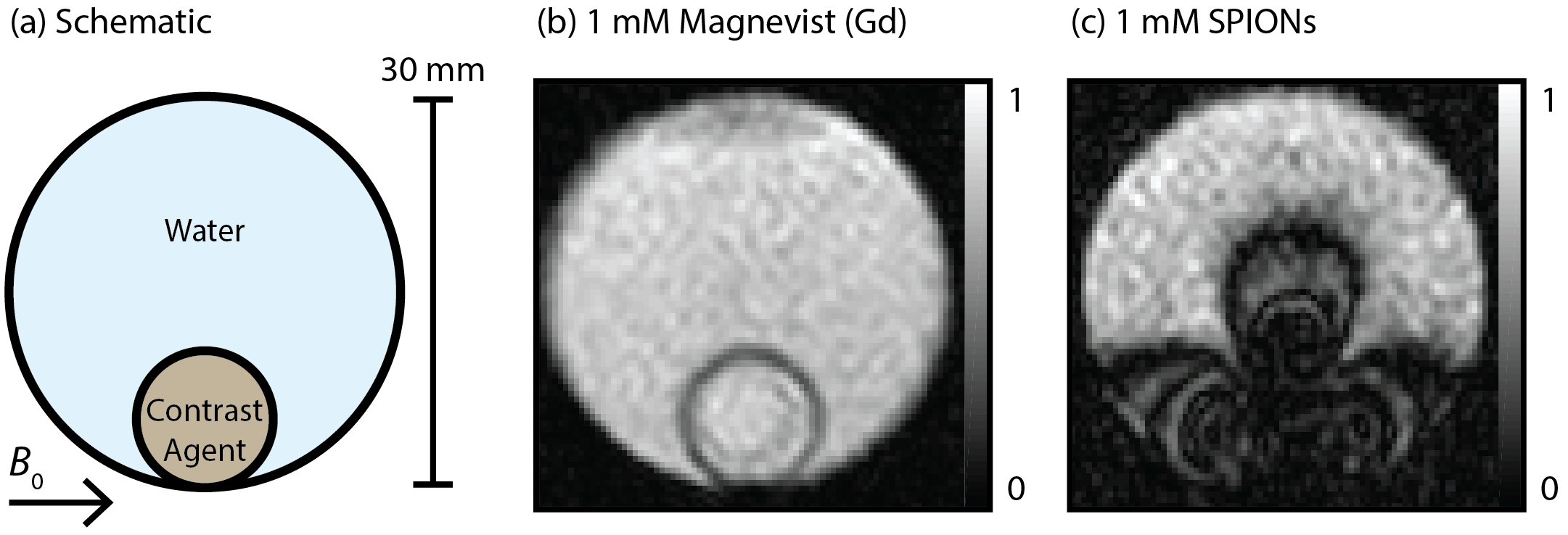
Images of phantoms containing water and CAs are shown in Fig. 2. Despite the significant concentration of gadolinium-based Magnevist, no apparent contrast is present in Fig. 2(b). Imaging with the same concentration of SPIONs (see Fig. 2(c)), results in clear contrast and an extensive dipolar susceptibility artifact covering a region much larger than the CA itself.
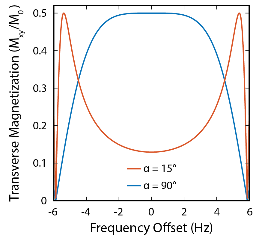
Varying the tip angle α used for b-SSFP acquisition modulates the MRI signal from susceptibility-shifted spins, providing a mechanism for generating positive contrast from SPIONs.9 For example, reducing α from 90° to 15° suppresses the MRI signal from on-resonant spins and boosts the MRI signal from off-resonant susceptibility-shifted spins, as shown in Fig. 3.
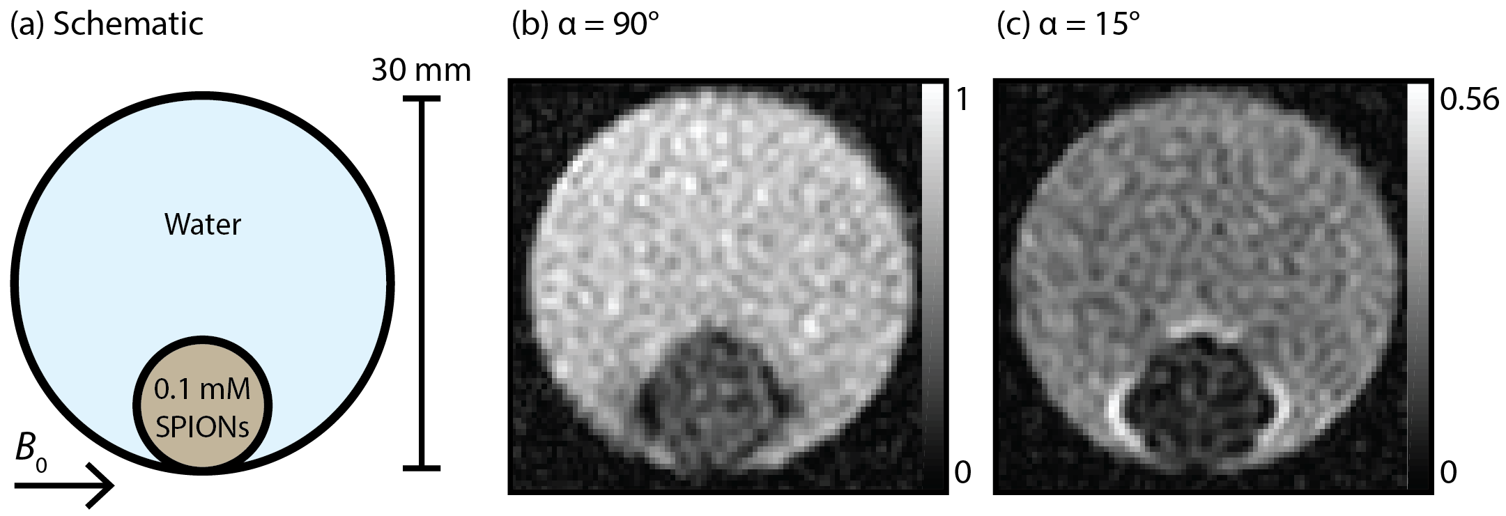
We demonstrate this positive-contrast effect in the images of 0.1 mM SPIONs shown in Figure 4. When a 90° tip angle is used (see Fig. 4(b)), water adjacent to the highly-magnetized SPIONs shows as dark due to a susceptibility-induced resonance-shift. Repeating the imaging with α = 15° (see Fig. 4(c)) shows these same water regions as bright. We note that the SPION solution remains dark in both images, presumably due to a large susceptibility-induced frequency-shift (greater than 1/2TR = 6Hz) and short T2 (47 ms).
Discussion
These results demonstrate ULF imaging of SPIONs at concentrations below those used clinically.11 This susceptibility-based approach, implemented with b-SSFP, enables high-resolution imaging over short acquisition times (more than 40 times faster than spin-echo T1-weighted imaging of CAs at ULF).12 The ability to switch between negative and positive contrast via tip angle will aid the unambiguous identification of CAs in clinical images.13 Identification may be further assisted by the suppression of background susceptibility artifacts at ULF – a challenge that has hindered the widespread use of SPIONs at high fields.14 Further, adjustments in TR could be used to minimize the size of susceptibility artifacts or to increase sensitivity to smaller resonance-shifts and hence enable imaging of even lower SPION concentrations.Conclusion
We have developed the most sensitive technique for imaging of CAs at ULF that we are aware of. This technique leverages the high magnetization of SPIONs at ULF to produce susceptibility contrast in an unexplored regime. Given the biocompatibility of SPIONs, these results will lead to new clinical applications of ULF MRI.Acknowledgements
This work was supported by The University of Sydney.References
1. M. Sarracanie et al., Sci. Rep. 5, 15177 (2015).
2. M. Espy et al., J. Magn. Reson., 229, 127-141 (2013).
3. D.E.J. Waddington et al., Nat. Commun., 8, 15118 (2017).
4. K. Scheffler et al., Eur. Radiol. 13, 2409-2418 (2003).
5. Bottomley et al., Med. Phys., 11, 425-448 (1984).
6. P. Magnelind et al., Diagn. Im. Europe, 11:49-51 (2016).
7. L. De Haro et al., Biomed. Eng.-Biomed. Tech., 60, 445-455 (2015).
8. D.E.J. Waddington et al., NMR Biomed., 31, e3896 (2018).
9. R. Dharmakumar et al., Phys. Med. Biol., 51, 4201 (2006).
10. P. Cantillon-Murphy et al., NMR Biomed., 22, 891-897 (2009).
11. T. Christen et al., Magn. Reson. Med., 70, 705-710 (2013).
12. X. Yin et al., Sci. Rep., 8, 11863 (2018).
13. T. Çukur et al., Magn. Reson. Med., 63, 427-437 (2010).
14. H. Wei et al. Proc. Natl. Acad. Sci., 114, 2325-2330 (2017).
Figures