5048
Does RF spoiling enhance human in-vivo brain MR Current Density Imaging (MRCDI)?1Danish Research Centre for Magnetic Resonance, Centre for Functional and Diagnostic Imaging and Research, Copenhagen University Hospital, Hvidovre, Denmark, 2Center for Magnetic Resonance, DTU Elektro, Technical University of Denmark, Kgs. Lyngby, Denmark, 3Department of Neurology, Copenhagen University Hospital, Bispebjerg, Denmark, 4High-Field Magnetic Resonance Center, Max-Planck-Institute for Biological Cybernetics, Tübingen, Germany, 5German Center for Neurodegenerative Diseases (DZNE), Bonn, Germany, 6Department of Biomedical Magnetic Resonance, University of Tübingen, Tübingen, Germany
Synopsis
MRCDI is an emerging modality for non-invasive measurement of weak currents in the human brain, which is important in several neuroscientific applications. It is based on current-induced field measurements and requires high sensitivity to the extrinsic field changes. Measurement sensitivity can be compromised by irrelevant field changes caused by physiological variation. Here, we compare the performance of the so far most sensitive MRCDI method based on steady-state free precession free induction decay (SSFP-FID) with its RF-spoiled counterpart fast low angle shot (FLASH). No significant sensitivity differences were observed in slices covering the upper part of the brain, but SSFP-FID had ~20% lower noise floors in lower slices. For the relevant acquisition parameters, FLASH exhibits no remarkable image quality enhancements in 2D.
Introduction
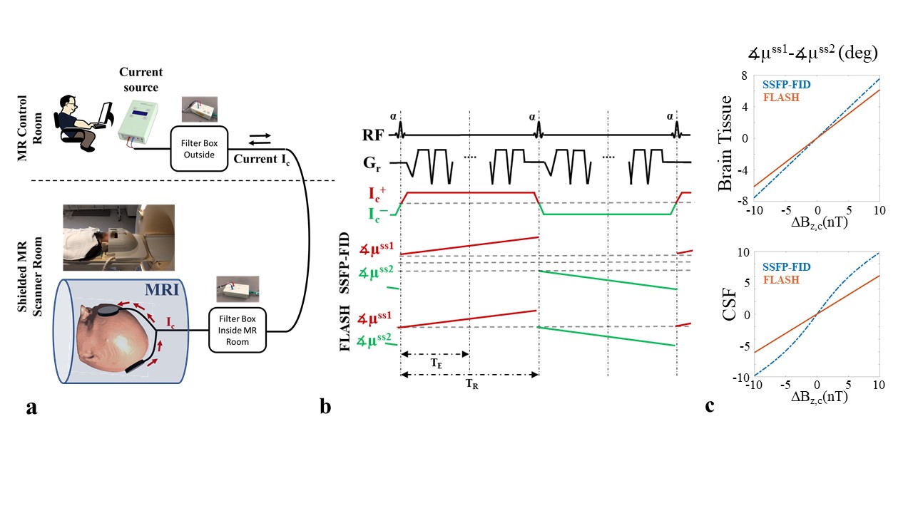
Exact knowledge of the spatial distributions of externally injected currents in the human brain is important for several neuroscientific applications. MRCDI is an emerging technique that combines MRI and weak current injection via electrodes to measure current flows in the brain. The component ∆Bz,c of the current-induced magnetic field parallel to the scanner field, modulates the phase of the MR signal. The signal phase can thus be used for ∆Bz,c calculations and current flow reconstructions (Fig. 1a). The quality of reconstructed current distributions directly depends on the sensitivity of the measured ∆Bz,c (1). Recently, reliable MRCDI measurements in the human brain with an unprecedented sensitivity of ~0.1nT have been reported (2). The used SSFP-FID method employs identical in-phase excitation pulses, and alternating currents to create two steady-states with opposite current-induced phases. As the method preserves FID coherence pathways, an additional phase difference is accumulated between two alternating steady-states. This mechanism might, however, increase the physiological noise. Here, we explore whether RF-spoiled FLASH (Fig. 1b,c), which is less sensitive to the extrinsically induced magnetic field, but which might also be more robust to physiological variation, can enhance the efficiency of human in-vivo MRCDI measurements.Methods
We simulated the phase sensitivity of SSFP-FID and FLASH to current-induced field changes for brain tissue and cerebrospinal fluid (CSF) based on Bloch equations and 3D rotation & relaxation matrices (3). In the simulations, the sequence parameters were tip angle α=30˚, repetition time TR=80 ms, echo time TE=40 ms, relaxation times T1=1.1 s and T2=100 ms for brain tissue, and T1=4.5 s and T2=1.5 s for CSF (Fig. 1c). We performed MRCDI measurements based on SSFP-FID and FLASH with multi-gradient-echo readouts in a spherical gel phantom (4) and in 3 healthy volunteers. The experiments were performed at 3T (MAGNETOM Prisma, SIEMENS Healthcare, Erlangen, Germany) with image matrix 112x90, voxel size 2x2x3 mm3, and α=30˚. In a first set of experiments, we sent alternating currents of Ic=+/-1 mA through a cable loop placed around the phantom. The current waveform was synchronized with the MR sequence. The current-induced fields ∆Bz,c were calculated from measured MR phase images (5). Four sets of experiments with identical total acquisition times of Ttot=3.6 mins were performed:
Exp 1: TR=60 ms, number of multi-gradient-echo readouts NGE=5, TE=[5.4, 13.6, 21.8, 30.0, 38.1] ms, and Nmeas=20 averages to increase the signal-to-noise-ratio.
Exp 2: TR=80 ms, NGE=5, TE = [7.5, 19.7, 31.9, 44.0, 56.1] ms, and Nmeas=15.
Exp 3: TR=100 ms, NGE=7, TE = [7.1, 18.7, 30.3, 41.8, 53.4, 64.9, 76.4] ms, and Nmeas=12.
Exp 4: TR=120 ms, NGE=7, TE = [8.3, 22.4, 36.5, 50.6, 64.7, 79.9, 93.2] ms, and Nmeas=10.
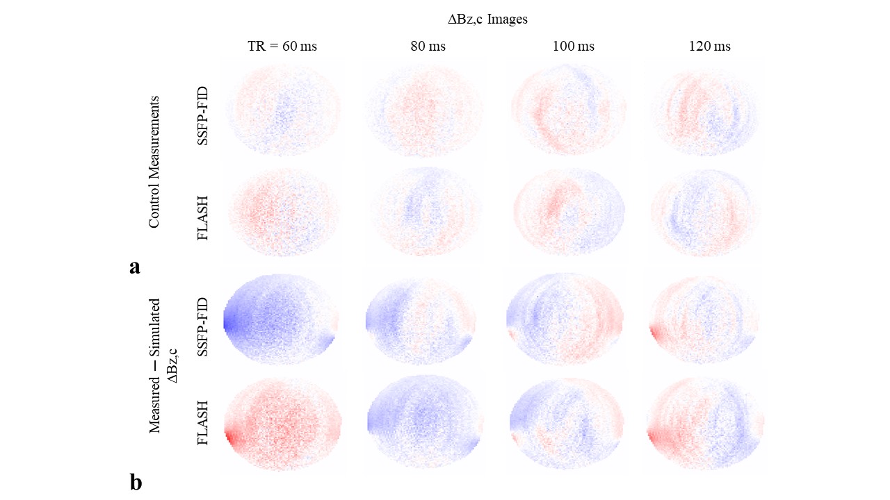
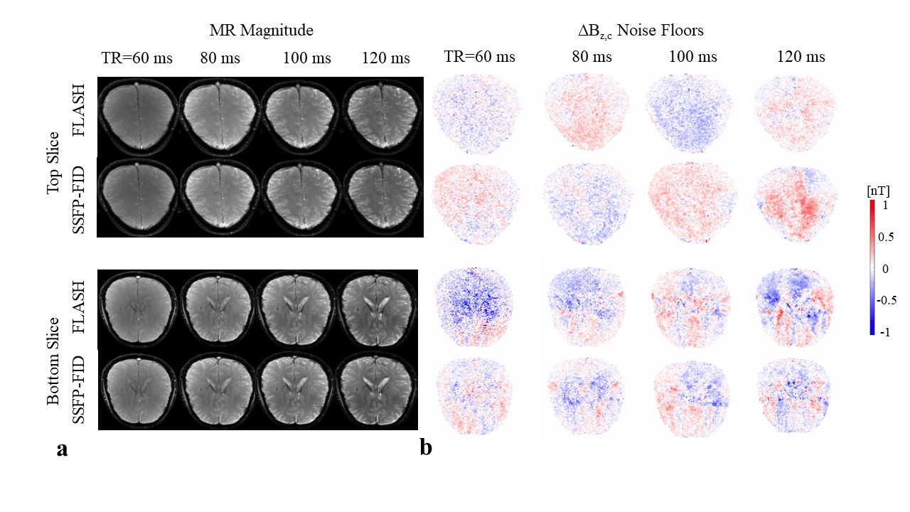
The experiments were performed with the lowest possible bandwidths and were repeated for SSFP-FID and FLASH. In order to image the cable paths, a T1-weighted PETRA scan was performed (number of slices Nsli=320, image matrix 320x320, voxel size (0.9mm)3, α=6˚, TR=3.61 ms, TE=0.07 ms, and inversion time TI=0.5 s), making the cables’s rubber coating clearly identifiable. The reconstructed cable paths were used to simulate the current-induced fields by the Biot-Savart law. The difference between the simulations and measurements were compared with current-free control experiments. In order to explore the noise floors of ∆Bz,c measurements in-vivo and the image qualities in the presence of physiological noise, Exp 1-4 were repeated without currents for two slices covering the top and lower part of the brain.
Results and Discussion
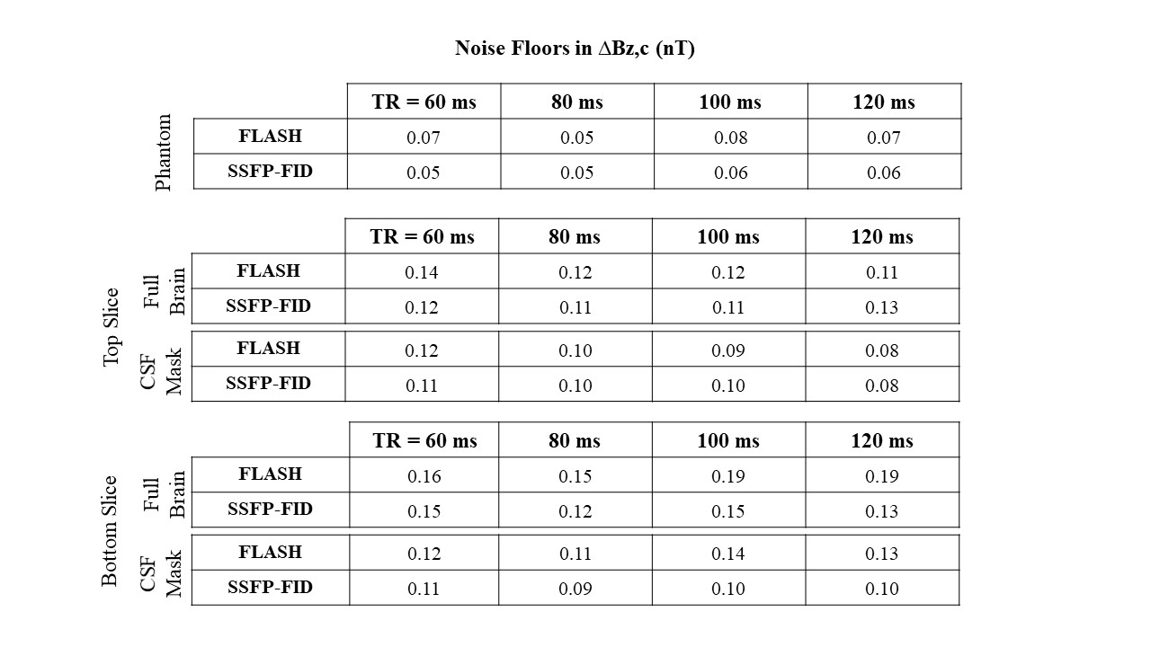
The simulations demonstrate that the phase sensitivity of SSFP-FID is 24% higher in the brain tissue and 2-fold higher in CSF (Fig. 1c). High correlations between ∆Bz,c simulations and phantom measurements (R2>0.98) validate both methods. The difference between measured and simulated current-induced fields are in the same range as the control experiments without currents (Fig. 2). For both the phantom and human data, no consistent artifacts were observed in the magnitude images and ∆Bz,c noise floor images (Fig. 3). SSFP-FID measurements had the highest SNR at TR=80ms, and exhibited 8.4% (top slice) and 20% (bottom slice) lower ∆Bz,c noise floors compared to FLASH, when averaged across the 3 subjects (Table 1).Conclusion
Human in-vivo brain MRCDI measurements based on both SSFP-FID and FLASH exhibit good and similar sensitivities to current-induced field changes for brain tissues. SSFP-FID slightly outperforms FLASH, likely due to its better sensitivity to current-induced phase accumulation in CSF. Still, both methods are susceptible to physiological noise, as revealed by their better sensitivities in phantoms compared to in-vivo. There are no clear image quality differences observed in 2D, but FLASH may perform better in 3D imaging due to its lower sensitivity to flow.Acknowledgements
The project is supported by Lundbeck foundation with grant number R118-A11308.References
1. Scott GC, Joy MLG, Armstrong RL, Henkelman RM. Sensitivity of magnetic-resonance current-density imaging. J. Magn. Reson. 1992;97:235–254.
2. Göksu C, Hanson LG, Siebner HR, Ehses P, Scheffler K, Thielscher A. Human In-vivo Brain Magnetic Resonance Current Density Imaging ( MRCDI ). Neuroimage 2018;171:26–39.
3. Jaynes ET. Matrix treatment of nuclear induction. Phys. Rev. 1955;98:1099–1105.
4. Friedman L, Glover GH. Report on a multicenter fMRI quality assurance protocol. J. Magn. Reson. Imaging 2006;23:827–839.
5. Göksu C, Scheffler K, Ehses P, Hanson LG, Thielscher A. Sensitivity Analysis of Magnetic Field Measurements for Magnetic Resonance Electrical Impedance Tomography (MREIT). Magn. Reson. Med. 2018;79:748–760.
Figures