5046
Fast MREIT acquisition using Multi-Band and SENSE Techniques1School of Biological and Health Systems Engineering, Arizona State University, Tempe, AZ, United States, 2Department of Radiology, Johns Hopkins University, Baltimore, MD, United States, 3Department of Biochemistry and Molecular Biology, University of Florida, Gainesville, FL, United States
Synopsis
Recent studies demonstrated the first current density and conductivity tensor images of human heads during transcranial electrical stimulation (
Introduction
Magnetic resonance electrical impedance tomography (MREIT) has developed over the last decade as a noninvasive method of imaging conductivity, current density, and other electromagnetic field distributions formed within electrically conductive objects by externally injected currents1. Recent studies have demonstrated magnetic flux density, current density images and conductivity tensor images of human heads using the MREIT technique2-5. In the studies of2,3, data were gathered from four human subjects undergoing tACS-like stimulation procedures at frequencies of 10 Hz and 1.5 mA intensity. Three images of 5-mm slice thickness centered on electrode regions were acquired using the Philips mFFE sequence, in a time that depended linearly on the number of slices. However, more brain coverage is essential to perform group level field-distribution analyses across subjects. In this study, we used Multi-Band (MB) excitation pulses, with or without SENSE acceleration, with the mFFE sequence to excite multiple slices at the same time and acquire slices simultaneously6,7.Methods
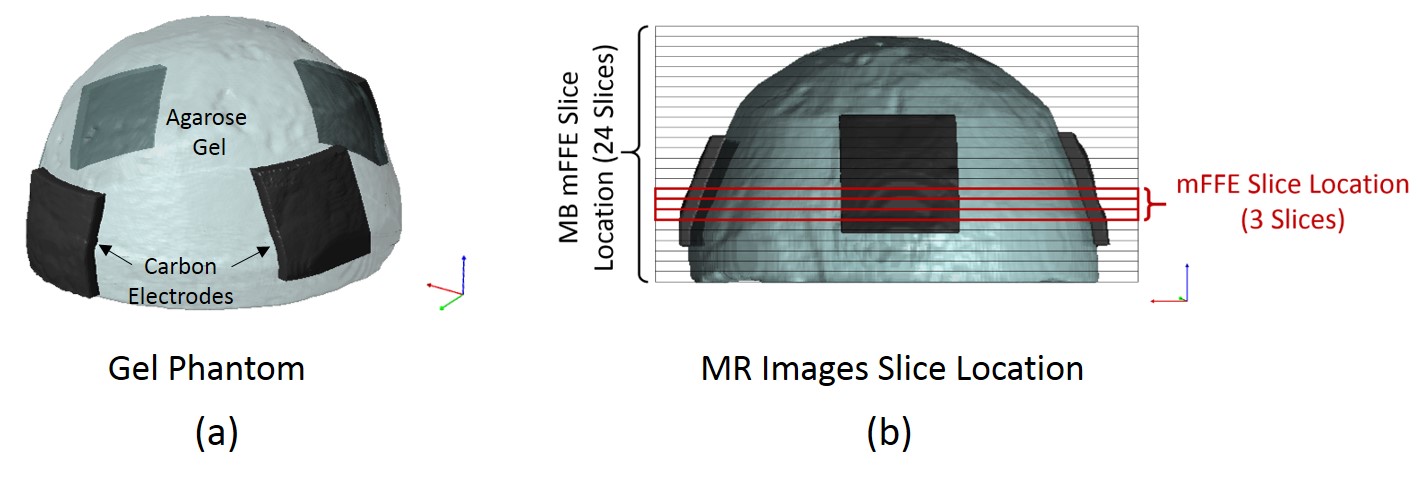
Imaging experiments were conducted using a hemispheric gel phantom that was 18-cm in diameter and 12-cm high. The phantom consisted of agarose gel material with an electrical conductivity of 1 S/m. Figure 1 shows schematic diagrams of phantoms and slice positions for both mFFE and MB-mFFE sequences. The gel phantom was imaged in a 3 T Philips Ingenia System (Barrow Neurological Institute, Phoenix Arizona, USA) during external tES-like current injections. A current intensity of 1.5 mA with a frequency of ~10 Hz was applied to the surface of gel via carbon electrodes (~36 cm2) in two independent and orthogonal directions (‘Horizontal’ and ‘Vertical’). The imaging parameters for phantoms were as follows. First, 3D FLASH T1-weighted structural images were collected with 224 mm (FH) x 224 mm (AP) x 140 mm (RL) field-of-view (FOV), and 1 mm isotropic resolution, centered on the mid-plane of the phantom. mFFE MREIT datasets were acquired with an in-plane FOV of 224 mm (RL) x 224 mm (AP) ), TR/TE= 50/7 ms, number of slices= 3, echoes= 10, echo spacing= 3 ms, acquisition matrix size=100 x 100, number of averages= 24 and total scan time= 6 min. MB-mFFE MREIT datasets were also acquired using the abovementioned parameters except, MB-factor= 8, SENSE-factor=1, number of slices= 24, number of averages= 6, total scan time= 6 min and the FOV in the foldover direction was increased to four times (896 mm (AP)) the original dimension to prevent aliasing. Other scans were obtained using: MB-factor=4, SENSE-factor=2 also produce overall eight-fold acceleration. Data was exported and processed offline with MATLAB 2018a (The MathWorks. Inc., Natick, MA, USA) to generate magnetic field maps. No-current MREIT datasets were also collected for mFFE and MB-mFFE to estimate T2* and optimize the current-induced magnetic field calculations2,3.Results and Discussion
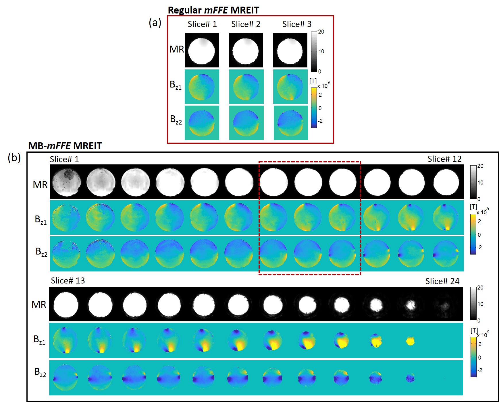
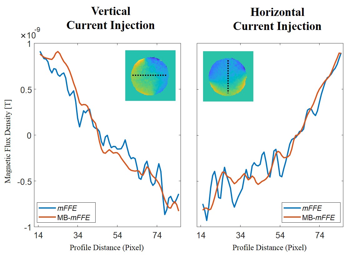
Figure 2 shows the MR magnitude and magnetic flux density images for mFFE and MB-mFFE data, with 8 simultaneous slices. The red box in Fig. 2(b) shows the slice locations of mFFE data. Figure 3 shows profiles of mFFE and MB-mFFE magnetic flux density data in a common slice through the phantom for vertical and horizontal current directions. Magnetic flux density profiles also showed good agreement between mFFE and MB-mFFE data. Images obtained with SENSE-factor=2 and MB-factor=4 acquisitions to achieve the same acceleration produced worse overall image quality.Conclusion
We computed magnetic flux density using regular mFFE and MB-mFFE MR sequences. Using MB-mFFE, we acquired 24 image slices within same scan time as the three slices in the original mFFE sequence, without compromising Bz quality. Our initial results show that MB-mFFE scans can be performed faster, which should improve MREIT techniques applied to the acquisition of full human head current density and conductivity images.Acknowledgements
We gratefully acknowledge the assistance of Dr Peter Boernert, Ulrich Katscher and Kay Nehrke (Philips Research, Hamburg) in providing access to sequences used in this study. Research reported in this abstract was supported by NIH award RF1MH114290 to RJS and Defense Advanced Research Projects Agency (DARPA) under the Targeted Neuroplasticity Training Program, award N66001-17-2-4018.References
1. Woo EJ, Seo JK. Magnetic resonance electrical impedance tomography (MREIT) for high-resolution conductivity imaging. Physiol Meas 2008;29; R1–R26.
2. Kasinadhuni AK, Indahlastari A, Chauhan M, et al. Imaging of current flow in the human head during transcranial electrical therapy. Brain Stimul 2017;10(4):764-772.
3. Chauhan M, Indahlastari A, Kasinadhuni AK, Schär M, Mareci TH, Sadleir RJ. Low-Frequency Conductivity Tensor Imaging of the Human Head in vivo using DT-MREIT: First Study. IEEE Transactions on Medical Imaging 2017;37(4):966-976.
4. Göksu C, Hanson LG, Siebner HR, Ehses P, Scheffler K, Thielscher A. Human in-vivo brain magnetic resonance current density imaging (MRCDI). NeuroImage2018;171: 26-39.
5. Jog M V, Smith R X, Jann K, Dunn W, Lafon B, Truong D, Wu A, Parra L C, Bikson M and Wang D J J 2016 In-vivo imaging of magnetic fields induced by transcranial direct current stimulation (tDCS) in human brain using MRI Scientific Reports 6 34385
6. Breuer FA, Blaimer M, Heidemann RM, Mueller MF. Griswold MA, Jakob PM Controlled aliasing in parallel imaging results in higher acceleration (CAIPIRINHA) for multi-slice imaging. Magn Reson Med 2005;53: 684-691
7. Larkman D J, Hajnal J V, Herlihy A H, Coutts G A and Young I R 2001 Use of multicoil arrays for separation of signal from multiple slices simultaneously excited. J. Magn. Res. Imag. 13 313–7
Figures