5045
Deep learning brain conductivity mapping using a patch-based 3D U-net1Philips Research Laboratories, Hamburg, Germany, 2University of Lubeck, Lubeck, Germany, 3University Medical Center Utrecht, Utrecht, Netherlands, 4Utrecht University, Utrecht, Netherlands, 5Department of Diagnostic and Interventional Radiology, Hokkaido University Hospital, Sapporo, Japan
Synopsis
Conventional Electrical Properties Tomography (EPT) suffers from reconstruction artifacts related to assumptions necessary for solving the equations analytically. To circumvent the necessity for these assumptions, in this study a deep learning approach is utilized to approximate the analytically unsolvable equations. For this purpose, a 3D convolutional neural network was trained on simulations and in-vivo data from healthy volunteers and cancer patients. Results demonstrate the potential of this method, as noise-free conductivity maps were obtained without anatomic apriori information in less than 1:30 min per reconstruction.
Introduction
Electrical Properties Tomography (EPT) derives conductivity and permittivity of tissue from complex B1 maps, as obtainable with standard MR systems and sequences [1]. In conventional (Helmholtz-based) EPT (HHEPT), the analytical formulation of the reconstruction equation gives rise to the transceive phase assumption, artifacts along tissue boundaries [2] and severe noise amplification in the reconstructed electrical properties maps. This work tries to overcome these challenges, as no satisfying solution for them has been found yet.
Following up on recent success of machine learning methods in medical imaging [3], first investigations have proven their potential for EPT [4,5]. In this study, the analytically unsolvable equations underlying EPT are approximated with a convolutional neural network (DLEPT). Following the work on simulations of full electrical properties maps by [4], this work is dedicated to challenges in reconstructing in-vivo data, with focus on phase-based conductivity reconstruction.
Methods
This work utilizes 3D patches as in [5] to maximize the local information. Empirical investigations led to the choice of a U-net with two downsampling steps and patch size of 24x24x24. To eliminate physically irrelevant phase offsets, the mean phase value was subtracted from each phase-patch. Data were augmented by mirroring patches at each axis. The first dataset consists of realistic EM-simulations with realistic Gaussian noise added. Transceive phase simulations were performed on a 2mm grid in Sim4life after applying geometrical deformations to virtual population models Duke and Ella [6]. The resulting 15 models were placed into a quadrature head coil model at 128 MHz. By training a network only on deformations of Duke and validating it on original Duke (excluded from training) and Ella, the impact of geometrical variance is investigated. For validation on in-vivo data, the full simulation dataset is used for training.
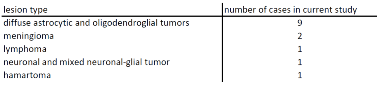
In-vivo datasets were acquired using commercial 3T scanner (Philips Healthcare, Netherlands, equipped with quadrature RF head coil). After obtaining informed written consent according to local Institutional Review Boards, 14 patients (mean age 42 +/- 17 yrs ) with various brain lesions (see Tab.1) and 18 healthy volunteers (mean age 44 +/- 7 yrs) were scanned with a bSSFP sequence (TR/TE=3.4/1.7ms, voxel size=1x1x1mm, flip angle=25°, 2 averages, scan duration 3:40 minutes). Two networks were trained on a GPU (Nvidia GeForce GTX 1080 Ti) using separate in-vivo datasets (volunteers/patients), and a third network was trained on the combined dataset. A three-fold cross-validation was performed using data from both in-vivo datasets. For quantitative evaluations, the correlation between DLEPT reconstruction and HHEPT used as ground truth reference was calculated. HHEPT reconstructions were performed according to [7]. Throughout this work, all validation data were excluded from training.
Results
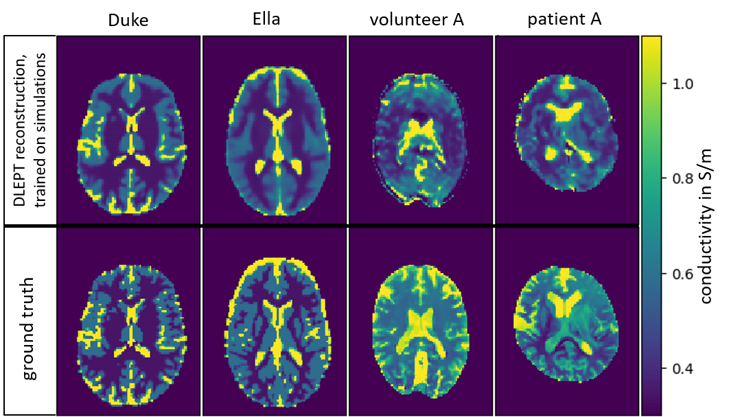
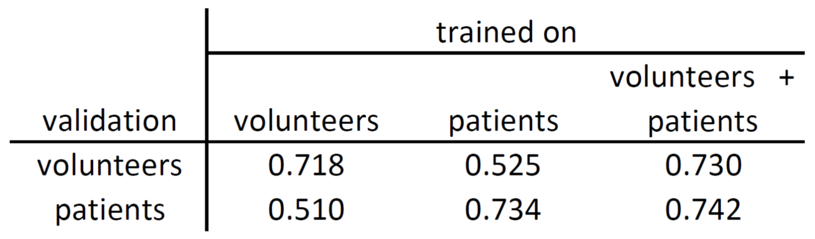
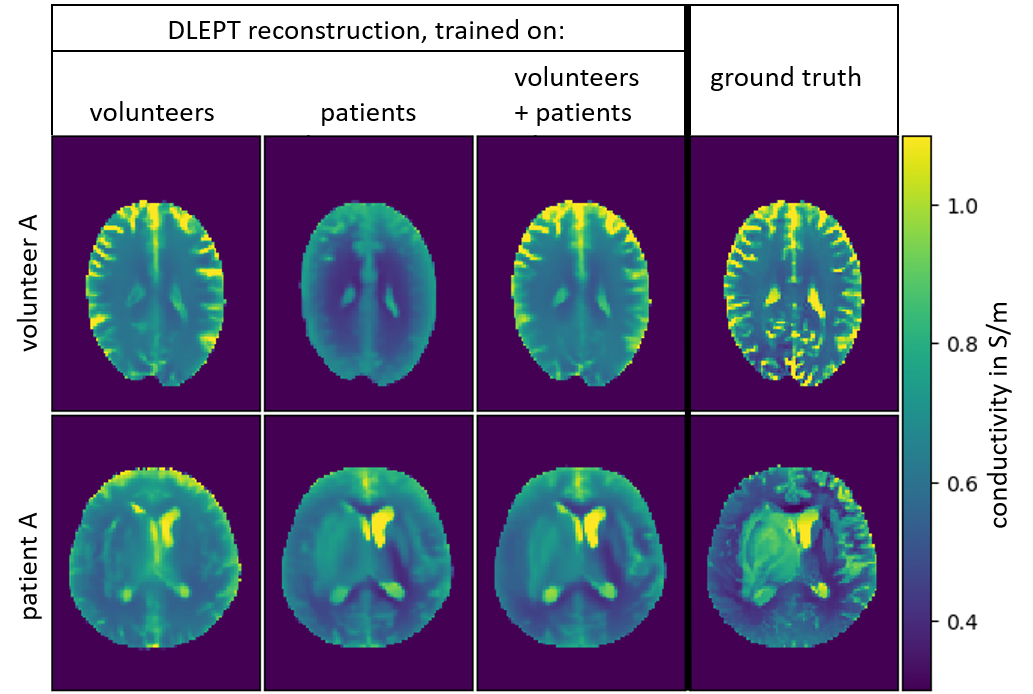
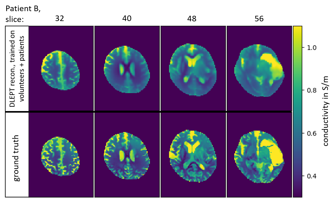
Fig.1 depicts example slices of reconstructions of simulations and in-vivo data by networks trained on simulations. The degradation of reconstruction quality from Duke to Ella illustrates the significance of geometrical variability in the training set. Additional artifacts occur in in-vivo reconstructions. Quantitative results of cross validations for networks trained on the in-vivo datasets increase in quality when combining volunteer and patient datasets for training (Tab.2). Exemplary reconstructions for these networks, depicted in Fig.2, do not show the in-vivo artifacts visible in Fig.1. However, the reconstruction of patients from the network trained on volunteers and vice versa yields only modest results. Training with the combined in-vivo datasets increases the reconstruction accuracy, in line with the correlations shown in Tab.2. In Fig.3, the reconstruction of patient B from the network trained on combined in-vivo datasets shows decent accuracy, also in the depicted lesion. All GPU times were under 1:30 min per reconstruction.Discussion
Networks trained on simulations show excellent results for reconstruction of simulations, which however degrade with increasing head geometry differences from training data. This, along with the performance improvement when combining both datasets, demonstrates that lacking geometric variability in training datasets of the given size is a major cause of reconstruction errors for DLEPT.
Promising reconstructions from networks trained on in-vivo data demonstrate the possibility of overcoming artifacts present in in-vivo reconstructions from networks trained on simulations, by including artifacts in training. Decreasing accuracy for the network trained on volunteers validated on patients and vice-versa (Tab.2, Fig.2) indicates dataset specificity of these in-vivo artifacts.
Conclusion & Outlook
Neural networks are proven to be a promising technique for EPT, allowing for fast, low-noise conductivity reconstructions without additional edge information. Simulations facilitate increasing geometrical variability for healthy tissue and lesions without HHEPT errors. Future investigations need to tackle the artifacts obtained when applying networks trained on simulations to in-vivo data, by including these artifacts in training.Acknowledgements
Cordial thanks to Mariya Doneva, Thomas Amthor, Christian Findeklee, Christoph Leussler, Jan Hendrik Wuelbern, Alfred Mertins and Philipp Koch for fruitful discussions.References
1. Katscher U, van den Berg CAT. Electric properties tomography: Biochemical, physical and technical background, evaluation and clinical applications. NMR Biomed. 2017;30. doi: 10.1002/nbm.3729.
2. Mandija S, Sbrizzi A, Katscher U, Luijten PR, van den Berg CAT. Error analysis of Helmholtz-based MR-electrical properties tomography. Magn Reson Med. 2018;80:90-100.
3. Zhu B1, Liu JZ, Cauley SF, Rosen BR, Rosen MS. Image reconstruction by domain-transform manifold learning. Nature 2018 21;555(7697):487-492.
4. Mandija S, Meliadò E, Huttinga N, Luijten P, van den Berg C. Opening a new window on MR-based Electrical Properties Tomography with deep learning. arXiv:1804.00016. 2018 Mar.
5. Hampe N, Herrmann M, Amthor T, Findeklee C, Doneva M, Katscher U. Dictionary-based Electric Properties Tomography. Magn Reson Med. 2018 Sep. doi:10.1002/mrm.27401.
6. Christ A, Kainz W, Hahn EG, Honegger K, Zefferer M, Neufeld E, Rascher W, Janka R, Bautz W, Chen J, Kiefer B, Schmitt P, Hollenbach HP, Shen J, Oberle M, Szczerba D, Kam A, Guag JW, Kuster N. The Virtual Family - Development of surface-based anatomical models of two adults and two children for dosimetric simulations. Phys Med Biol. 2010;55:23–38.
7. Tha KK, Katscher U, Yamaguchi S, Stehning C, Terasaka S, Fujima N, Kudo K, Kazumasa K, Yamamoto T, Van Cauteren M, Shirato H. Noninvasive electrical conductivity measurement by MRI: a test of its validity and the electrical conductivity characteristics of glioma. Eur Radiol. 2018;28:348-355.
Figures