5042
Measuring Regional Gas Transport in Injured Rabbit Lungs Using Hyperpolarized XenonYi Xin1, Kai Ruppert1, Maurizio Cereda2, Faraz Amzajerdian1, Hooman Hamedania1, Mehrdad Pourfathi1, Sarmad Siddiqui1, Ian Duncan1, Luis Loza1, Tahmina Achekzai1, Federico Sertic1, Ryan Baron1, Harrilla Profka1, Stephen Kadlecek1, and Rahim R. Rizi1
1Radiology, University of Pennsylvania, Philadelphia, PA, United States, 2Anesthesiology and Critical Care, University of Pennsylvania, Philadelphia, PA, United States
Synopsis
Hyperpolarized 129-Xenon MRI measures the regional content of tracer gas in the lungs; it can also differentiate between Xenon contained in the gas phase (GP) and in the dissolved phase (DP), allowing us to characterize regional gas diffusivity and uptake in the pulmonary capillary blood in addition to capturing parameters of alveolar aeration. By measuring absorbed Xenon signal in the left heart and aorta shortly after inhalation, it is theoretically possible to study the next step of gas transfer by measuring the gas that reaches the arterial blood. In this study, we explore the regional gas transport of injured rabbit lungs in two different states of recruitment.
Rationale
Clinical measurements of pulmonary function do not capture the contribution of each part of the lung to the overall amount of oxygen being transferred from the alveoli to the arterial blood, which depends on the distributions of ventilation and blood flow as well as on the local efficacy of alveolar-capillary diffusion. Hyperpolarized 129-Xenon MRI measures the regional content of tracer gas in the lungs; it can also differentiate between Xenon contained in the gas phase (GP) and in the dissolved phase (DP), allowing us to characterize regional gas diffusivity and uptake in the pulmonary capillary blood in addition to capturing parameters of alveolar aeration. By measuring absorbed Xenon signal in the left heart and aorta shortly after inhalation, it is theoretically possible to study the next step of gas transfer by measuring the gas that reaches the arterial blood. In this study, we explore the regional gas transport of injured rabbit lungs in two different states of recruitment.Methods
New Zealand rabbits (n=5) were anesthetized, tracheostomized and mechanically ventilated with frequency 40, FiO2 0.3 and VT 6ml/kg. Focal lung injury was generated by endobronchial instillation of hydrochloric acid (HCl 1.5ml/kg) in the lower lobes. MRI was performed at PEEP 0 and 9 cmH2O both before and after injury. 2D axial projection images of both gas-phase (GP) and dissolved-phase (DP) were acquired during end-expiratory breath holds following ventilation with 70% HP 129-xenon. A slab of saturation band was applied on the GP, and incrementally shifted in the ventro-dorsal direction. By selectively killing the GP signal, this technique allows us to assess the regional contribution of each examined region to the total transfer of dissolved xenon to the heart (Figure 1).Results
GP and DP defects due to regional HCl instillation were observed in the dorsal left lung at PEEP 0 cmH2O (Figure 2). GP signal was restored in this region at PEEP 9 cmH2O due to recruitment, while DP signal was also focally increased in the same area, suggesting locally higher Xenon uptake in injured tissue, possibly due to hyperemia. Post-injury, dorsal gas transport efficiency was lower at zero PEEP than at healthy baseline, and was more homogeneous during PEEP (Figure 3).Conclusion
Contributions of specific lung regions to overall gas uptake can be quantified using HP xenon-129 MRI. This technique could be used to evaluate the effects of lung disease and treatment on regional gas transport.Acknowledgements
Supported by NIH grants R01 EB015767, R01 HL129805, S10 OD018203 and R01 CA193050.References
No reference found.Figures
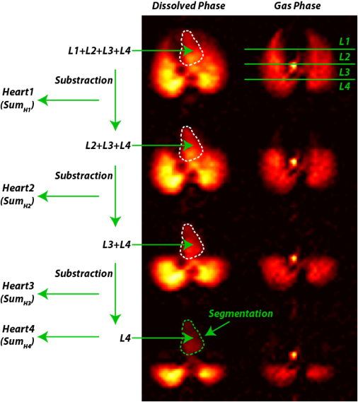
Figure 1. Schematic of the image analysis in a GP-saturation data set: a slab of GP
signal is abolished going from anterior to posterior lung regions, decreasing
the DP signal recovered in the heart and aorta; the value of DP signal after
each saturation is then subtracted from the total, yielding the contribution of
each individual slab of tissue to overall gas transfer.
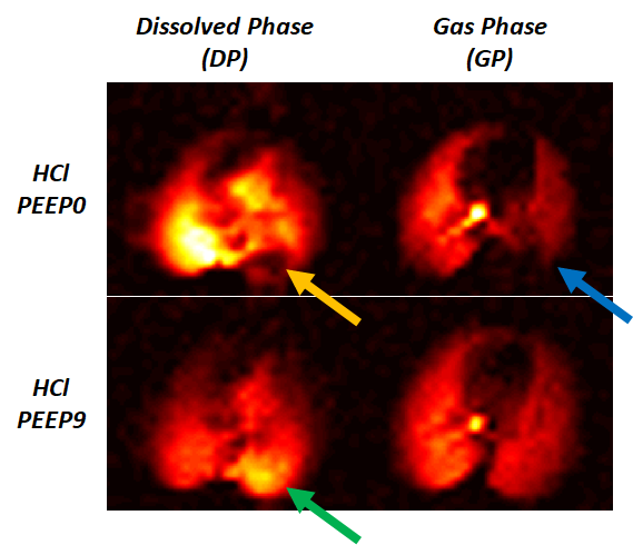
Figure 2. Axial DP-GP
projection maps for a breath hold at PEEP 0 and 9 cmH2O after HCl instillation:
a focus of decreased GP and DP signal was seen in the left dorsal lung (yellow
and blue arrows); GP signal was restored at PEEP, while DP signal was enhanced
in the same region (green arrow).
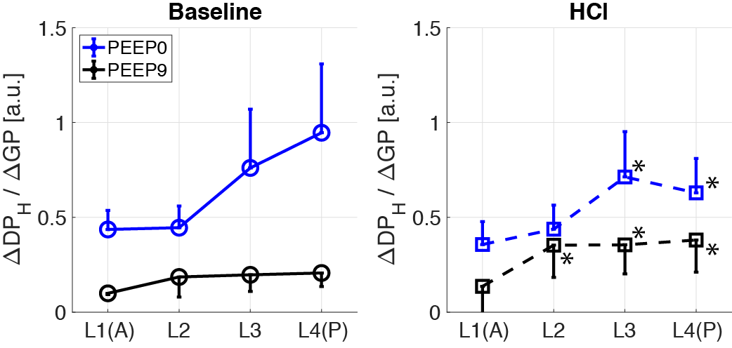
Figure 3. Changes in regional xenon transport
efficiency were measured by applying saturation bands before and after injury, with
and without PEEP. The change of GP signal was indexed by the change of GP for
each saturation step. After injury, gas transfer decreased in the posterior
lung (L4) at zero PEEP and was more homogeneous during PEEP. *:P<0.05 HCl
vs. baseline.