5038
A novel trimodality vascular contrast agent for “image-based systems biology” applications1Russell H. Morgan Department of Radiology and Radiological Science, Johns Hopkins University School of Medicine, Baltimore, MD, United States, 2Department of Orthopaedic Surgery, Johns Hopkins University School of Medicine, Baltimore, MD, United States
Synopsis
Preclinical vascular imaging has been instrumental in advancing our understanding of the role of blood vessels in health and disease. However,
Introduction
Preclinical vascular imaging techniques have advanced our understanding of the role of vasculature in health and disease1. More recently, preclinical microvascular images have been used in computational systems biology models of diseases such as cancer2,3. However, integrating multiscale and multimodality data for such applications remains challenging due to the unavailability of vascular contrast agents that are visible in MRI, CT, and optical imaging. Additionally, this makes “systems level” imaging of the vasculature and its interactions with the tissue microenvironment challenging4. Therefore, we developed a new vascular contrast agent that is water-soluble, radio-opaque, and fluorescent for imaging the vasculature within a single tissue with MRI, CT, and optical imaging, respectively. This is the first study to demonstrate the compatibility of a vascular contrast agent with different types of imaging contrast mechanisms such as tissue relaxation and diffusion-weighted (DW) contrast in MRI, bone-contrast in micro-CT (μCT), and with optical clearing methods for light-sheet microscopy5.Methods
The vascular contrast agent was prepared by combining 2ml of gadolinium-labeled albumin co-labeled with Rhodamine B (BioPAL Inc.) with 15 ml of water-soluble, radiodense BriteVu® (Scarlet Imaging). Healthy tissues (e.g. murine brain, hind leg and kidney) and pathological samples (e.g. breast tumor xenografts) were excised post perfusion, according to our protocol6. Ex-vivo MRI was performed on a 9.4T scanner (Bruker BioSpin) using a 10 mm volume RF coil. T1-weighted images were acquired using a 3D-FLASH sequence with the following parameters: flip angle = 30°, TE/TR = 4.2/40 ms, 4 averages, and isotropic resolution of 40 μm. Diffusion tensor imaging (DTI) data were acquired using a 3D diffusion-weighted GRASE sequence7 with TE/TR=32/800 ms, 12 echoes per excitation, 2 averages, diffusion gradient duration (d)/separation (D) = 2.8/10 ms, and 16 directions with b-value = 1700 s/mm2. Ex-vivo μCT was performed on agarose-embedded-samples on a high-resolution (7.5 μm isotropic resolution) scanner (SkyScan 1275, Bruker) with the following parameters: 55 kVp, 145 µA, 335ms exposure time, 0.2 rotation step and 3 averages. Thin tissue sections (~1mm) were optically cleared using CUBIC8 and imaged either using florescence microscopy or on a light-sheet microscope (LaVision BioTec GmbH) with the following parameters: 3.6 µm lateral resolution, 5 µm light-sheet thickness and 3 µm z-step-size. Fractional anisotropy (FA) and apparent density coefficient (ADC) maps were computed using DTIStudio9. Vessel segmentation was performed using a multiscale tubeness filter10. All data visualization was performed in ImageJ and Amira® (ThermoFisher Scientific).Results
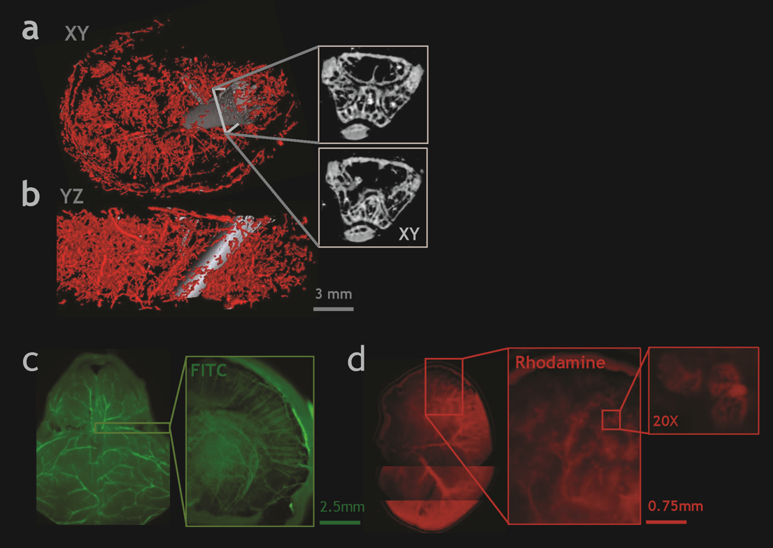
The trimodality contrast agent successfully enhanced the vasculature in each of ex-vivo MRI, μCT and light-sheet microscopy images (Fig. 1). We show the feasibility of performing DW-MRI on brain tissue to obtain directional diffusion tensor and anisotropy contrasts in the presence of a strong contrast-to-noise ratio (CNR) from cortical blood vessels (Fig. 2a-c). We observed excellent soft tissue contrast between the breast tumor and surrounding tissue on a T1-w MRI (Fig. 2d-f). Moreover, an ADC map acquired from the same tumor illustrated that necrotic areas were poorly vascularized (Fig. 2g-h). Fig. 3a-b show how vascular contrast from the murine hind leg vessels complements the µCT signal from the femur. Finally, we show that the fluorescence from the contrast agent can be adapted for unique applications in optical microscopy. For example, Fig. 3c-d show how fluorescent images of the optically-cleared murine brain (green channel) and kidney (red channel) vasculature can be obtained with fluorescence microscopy.Discussion
Integrating vascular data across MRI, CT, and optical imaging has remained challenging due to inherent differences in contrast mechanisms, spatial resolution, and sample preparation requirements. To the best of our knowledge, this is the first study to develop a trimodality imaging contrast agent and accompanying image processing pipeline to simultaneously visualize blood vessels in MRI, CT, and optical microscopy. This trimodality imaging platform has several advantages over conventional imaging methods. Firstly, since the vascular contrast agent did not interfere with conventional contrast mechanisms for each imaging modality, one could obtain complementary information on the vascular microenvironment. Secondly, it facilitated image registration and data integration across spatial scales via the presence of natural “vascular landmarks”. Finally, one can envision combining such multiscale/multimodality imaging data with computational modeling2,3,11 to develop functional atlases of both healthy and diseased vasculature with a range of preclinical applications.Conclusion
The results demonstrate the feasibility of obtaining vascular contrasts in MRI, CT and optical imaging using a single contrast agent. The ability to obtain multimodality and multi-scale information with complementary soft tissue contrasts has important implications for characterizing the healthy and diseased vascular microenvironment.Acknowledgements
Supported by NCI 1RO1CA196701-01References
1. McDonald DM, Choyke PL. Imaging of angiogenesis: from microscope to clinic. Nat Med. 2003;9 (6):713-25.
2. Stamatelos SK, Kim E, Pathak AP, Popel AS. A bioimage informatics based reconstruction of breast tumor microvasculature with computational blood flow predictions. Microvasc Res. 2014;91:8-21.
3. Kim E, Stamatelos S, Cebulla J, Bhujwalla ZM, Popel AS, Pathak AP. Multiscale imaging and computational modeling of blood flow in the tumor vasculature. Ann Biomed Eng. 2012;40(11):2425-41. 4. Megason SG, Fraser SE. Imaging in systems biology. Cell. 2007;130 (5):784-95.
5. Lagerweij T, Dusoswa SA, Negrean A, Hendrikx EML, de Vries HE, Kole J, et al. Optical clearing and fluorescence deep-tissue imaging for 3D quantitative analysis of the brain tumor microenvironment. Angiogenesis. 2017;20 (4):533-46.
6. Kim E, Zhang J, Hong K, Benoit NE, Pathak AP. Vascular phenotyping of brain tumors using magnetic resonance microscopy (muMRI). J Cereb Blood Flow Metab. 2011;31(7):1623-36
7. Aggarwal M, Mori S, Shimogori T, Blackshaw S, Zhang J. Three-dimensional diffusion tensor microimaging for anatomical characterization of the mouse brain. Magn Reson Med. 2010;64(1):249-61.
8. Susaki EA, Tainaka K, Perrin D, Yukinaga H, Kuno A, Ueda HR. Advanced CUBIC protocols for whole-brain and whole-body clearing and imaging. Nat Protoc. 2015;10(11):1709-27.
9. Jiang H, van Zijl PC, Kim J, Pearlson GD, Mori S. DtiStudio: resource program for diffusion tensor computation and fiber bundle tracking. Comput Methods Programs Biomed. 2006;81(2):106-16.
10. Cebulla J, Kim E, Rhie K, Zhang J, Pathak AP. Multiscale and multi-modality visualization of angiogenesis in a human breast cancer model. Angiogenesis. 2014;17(3):695-709.
11. Logsdon EA, Finley SD, Popel AS, Mac Gabhann F. A systems biology view of blood vessel growth and remodeling. J Cell Mol Med. 2014;18(8):1491-508.
Figures