5032
Dynamic Perfusion Tensor Imaging1Center for Medical Device Evaluation, NMPA, Beijing, China, 2MR Research China, GE Healthcare, Beijing, China
Synopsis
DCE-MRIprovides a method to continuously measure the spatial and temporal characteristics of local tissue perfusion. This study points out an interesting feature of DCE-MRI: the voxel wised correlation can be encoded in 26 directions, allowing for the measurement of perfusion tensor. We demonstrate this new method, dynamic perfusion tensor imaging (dPTI), facilitates the reconstruction of the local perfusion field, characterized by a perfusion tensor, from which can be derived quantities related to the structure of the local perfusion field, such as the mean perfusion and perfusion anisotropy.
INTRODUCTION
The ability to quantitative measurement of perfusion in the brain is important for many clinical applications. The traditional perfusion imaging methods cannot provide enough information on spatial and temporal heterogeneity. Velocity selective arterial spin labeling (VSASL) MRI has been adopted for perfusion tensor imaging (PTI) [1], but susceptible to low SNR, thereby limiting the clinical reproducibility. T1-weighted, dynamic contrast-enhanced (DCE) MRI is popular used for the estimating leakage of tissue with blood–brain barrier deficiency. DCE-MRI provides a method by which to continuously measure the spatial and temporal characteristics of local tissue perfusion [2], and employ multi-parts model to calculate the blood perfusion and permeability. We explored the DCE-MRI combining with a spatiotemporal correlation tensor model to determine the characteristics of time-dependent curve during the bolus passage of the brain capillaries[3]. The perfusion measurements based on DCE-MRI can then be characterized by a perfusion tensor, analogous to the diffusion tensor in DTI.METHODS
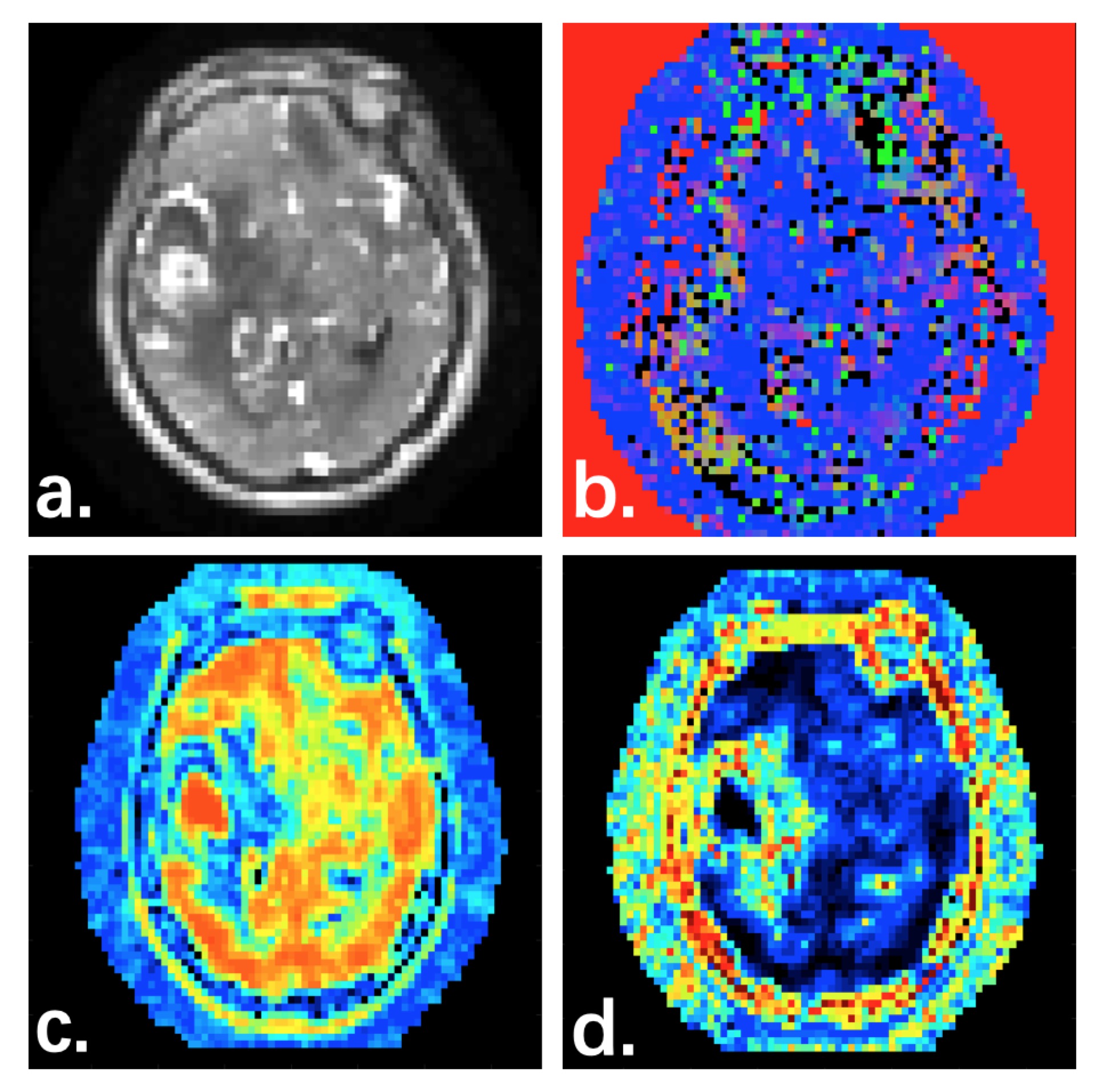
The voxel-wised DCE-MRI signal provides a time series that exhibits perfusion fluctuations. The temporal correlation between pairs of voxels indicates the degree of synchronous perfusion variation. The covariation of 26 neighboring voxels can be used to calculate the voxel-wised correlation tensors, which similar to the diffusion tensors of DTI [3], such as the observed temporal correlations along the 26 directions. A least square fitting method was using for the calculation of spatio-temporal correlation tensor. The eigenvector of perfusion tensor corresponding to the largest eigenvalue (the major eigenvector) represents the dominant direction of as well as the direction of regional blood feeding.This IRB-approved retrospective study included brain tumor patients who underwent DCE-MRI. The DCE-PTI maps were constructed using spatio-temporal correlation tensor evaluation of DCE-MRI time course (Figure 1). Total 10 patients with contrast-enhanced MR examination were included in this study (7 men and 3 women; mean age, 51 years; age range, 39–63 years). The T1-weighted DCE-MRI examination was performed on 3.0T scanner with an eight-channel phased array head coil. For dynamic MR imaging, a parallel 3D spoiled GRE sequence was performed: 2.9/1.3 ms TR/TE, a 15°flip angle, 7 mm slice thickness, 240 mm FOV, 128 ×128 matrix. Then a bolus of paramagnetic contrast agent (Omniscan) was injected with a dose of 0.20 mL/kg (equivalent to 0.10 mM/kg) and with the speed of 3 mL/s. A total of 60 phases were obtained at 1.3s intervals, with the injection occurring at the 10th phase, so that the bolus would typically arrive during the 5th to 20th images.The baseline T1 images were recorded before DCE-MRI. Eigenvalue decomposition of the perfusion tensor using first-level 26 encoding directions was performed using home-made software, including the Mean perfusion estimation, fractional anisotropy (FA).RESULTS AND CONCLUSION
DCE-MRI is widely explored in many clinical studies for noninvasive detection, characterization, and therapy monitoring of different diseases. In this study, we have demonstrated the use of dPTI based on DCE-MRI in the formation of perfusion tensor images from which can be derived the mean perfusion and the inflow anisotropy in each voxel (Fig.1). dPTI facilitates the reconstruction of the local perfusion field, which can be characterized by a perfusion tensor, including the mean perfusion, the perfusion anisotropy.The dPTI technique can be extended to combine the voxel distance and encoding directions to perform high resolution perfusion tensor imaging for the investigation of different level local inflow structure. This approach has the potential to improve image interpretation in a variety of applications for which the structure of the local inflow is desired, such as the investigation of the direction of blood supply and perfusion anisotropy to tumors.Furthermore, combined with the perfusion kinetic model, dPTI can be allow a full exploration of important perfusion properties.
Acknowledgements
References
1. Frank LR, Lu K, Wong EC. Perfusion tensor imaging.Magn Reson Med. 2008 Dec;60(6):1284-91.
2. Zhang Y, Wang J, Wang X, et al. Feasibility study of exploring a T₁-weighted dynamic contrast-enhanced MR approach for brain perfusion imaging. J Magn Reson Imaging. 2012 Jun;35(6):1322-31.
3. Ding Z , Newton A T , Xu R , et al. Spatio-Temporal Correlation Tensors Reveal Functional Structure in Human Brain. Plos One, 2013, 8(12):e82107.
Figures