5025
Imaging Blood Brain Barrier Permeability in a Human African Trypanosomiasis Mouse Model using Diffusion Weighted Multiple Boli Arterial Spin LabellingSamantha Paterson1, Antoine Vallatos2, Jean Rodgers3, and William Holmes1
1Neuroscience & Psychology, University of Glasgow, Glasgow, United Kingdom, 2University of Edinburgh, Edinburgh, United Kingdom, 3University of Glasgow, Glasgow, United Kingdom
Synopsis
Human African Trypanosomiasis is a parasitic disease that causes progressive blood brain barrier breakdown. We have developed a non-invasive high SNR ASL technique (mbASL) combined with bipolar diffusion gradients to determine the ratio of intravascular to extravascular signal from the brain. The ratio of this signal will change in a mouse brain infected with HAT due to the barrier breakdown. We have imaged this changing brain along with producing CBF maps, thus using a novel method in imaging a HAT infected mouse.
Introduction
Human African Trypanosomiasis (HAT) commonly known as Sleeping Sickness is a parasitic disease transmitted by the Tsetse fly. The parasite trypanosoma triggers a breakdown in the blood brain barrier (BBB) with the disease being fatal if left untreated1. Invasive treatments can cause more damage to patients with lumbar punctures used to examine the cerebral spinal fluid in humans to diagnose infection from the parasite2. MRI data is normally taken in later stages of the disease as the early stage is hard to diagnose. This means comparable data from before and after disease for MRI is scarce. Being able to image the BBB to look for permeability changes is crucial in HAT as well as multiple neurological diseases. The standard for imaging changes in the BBB is to use a contrast agent but these aren’t sensitive for imaging subtle changes in the barrier permeability and are invasive. We propose using the newly developed multiple boli arterial spin labelling3 (mbASL) accompanied with bipolar diffusion gradients to explore subtle changes in the BBB. mbASL is a high SNR sequence that can be used to non-invasively image perfusion changes in the brain along with acquiring cerebral blood flow (CBF) values. We present a novel method of imaging changes in the brain of mice infected with HAT. By using DW-mbASL to image the brain at multiple disease stages, we expect to see changes in the BBB permeability and CBF, which hasn’t been examined in the literature at this stage.Methods
Groups of female CD1 mice (n=6) were imaged at multiple disease points post infection: Day = 0,7,14,21,28. Experiments were performed on a Bruker PharmaScan 7T MRI system using a mouse surface head coil. DW-mbASL images were acquired at b = 0,25,50,75,100,200,300 s/mm^2, with CI = 5000ms, TI = 50ms. T1 maps and CE-MRI using a T1 weighted sequence were also acquired. After scanning, the mice were killed, and brains taken for histological purposes. Data was exported in DICOM format and analysed using in-house MATLAB code.Results & Discussion
Results and Discussion By using diffusion gradients after the ASL labelling, we can suppress the signal from the fast-flowing arterial blood in the intravascular compartment (capillaries) and compare this to the slower flowing arterial water that has diffused into the extravascular compartment (tissue). By fitting the data to a 2-compartmental model we can see the ratio of intravascular to extravascular signal (Figure 1). Early analysis suggests that this ratio of signal will change with the arterial water in the tissue compartment increasing due to the breakdown in the BBB. Figure 2 displays pilot data that shows the change in signal in the early stages of the disease. We expect to see a change in the ratio of intravascular to extravascular signal as the disease progresses to later stages where changes in the BBB can be seen using a contrast agent and confirmed using histology (experiment ongoing). Figure 3 demonstrates the high SNR DW-mbASL images in a non-infected mouse at multiple b values. As seen the signal drops when the gradients are applied, confirming the need for a high SNR sequence. This data can be fitted to the mbASL kinetic model and produce quantitative cerebral blood flow maps (analysis on going).Conclusion
We have produced a high SNR diffusion weighted ASL sequence that is being used to image subtle changes in blood brain barrier permeability. Using sleeping sickness to model BBB breakdown is a novel method, in which limited details about the brain evolution during the disease are known. By using DW-mbASL to image the brain during various stages of the disease, we have managed to explore new details about permeability and CBF that weren’t known previously in HAT.Acknowledgements
Many thanks to Jim Mullin and Lindsay Gallagher in their technical help and to the University of Glasgow and EPSRC for funding.References
1 Rodgers. J et al. (2017)
2http://www.who.int/trypanosomiasis_african/disease/diagnosis/en/
3Vallatos, A et al. (2017)
Figures
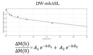
Fitting of DW-mbASL data to a bi-exponential model.
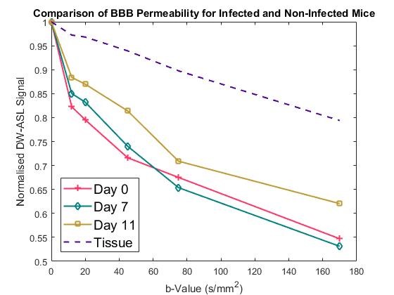
Comparison of DW-mbASL signal at multiple b-values for different post-infection days with comparison to a non-infected control group.
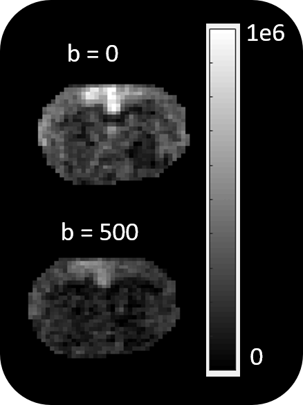
High SNR DW-mbASL images.