5018
Isotropic Whole-brain CEST Imaging with Fast SPACE Readout1Department of Biomedical Engineering, Zhejiang University, Hangzhou, China, 2Department of Radiology, Johns Hopkins University, Baltimore, MD, United States, 3MR Collaboration, Siemens Healthcare Ltd., Shanghai, China
Synopsis
For Chemical Exchange Saturation Transfer (CEST) imaging to be adopted as a routine clinical sequence, its spatial coverage and acquisition speed need ideally to be comparable to the current anatomical MRI sequences. To the best of our knowledge, whole-brain CEST imaging has only been demonstrated with 3D EPI readout, which, however, is vulnerable to susceptibility artifacts. Here, we propose a novel whole-brain isotropic-resolution CEST sequence utilizing the fast SPACE readout. The SPACE CEST sequence enables whole-brain 2.79mm isotropic CEST imaging in 5min without susceptibility artifacts, and should facilitate the translation of CEST MRI as a clinical routine sequence.
Introduction
Chemical Exchange Saturation Transfer (CEST) imaging1-3 is an emerging molecular MRI technique, which has been shown to be highly promising in multiple clinical applications4, especially neurological applications. For CEST to be adopted as a routine clinical sequence, its spatial coverage and acquisition speed need ideally to be comparable to the current anatomical MRI sequences, such as the 3D MPRAGE sequence5 which allows retrospective reconstruction along any desired plane. To the best of our knowledge, whole-brain CEST imaging has only been demonstrated with 3D echo planar imaging (EPI) readout6-8. However, it is well-known that the EPI sequence is vulnerable to susceptibility artifacts, and generates signal voids or distortion at the tissue-air interface, which might cause it to miss important pathological regions. Here, we propose a novel whole-brain isotropic-resolution CEST sequence utilizing the fast SPACE9 readout. The new SPACE CEST sequence is examined on human brains for APT-weighted (APTw)10 imaging.Methods
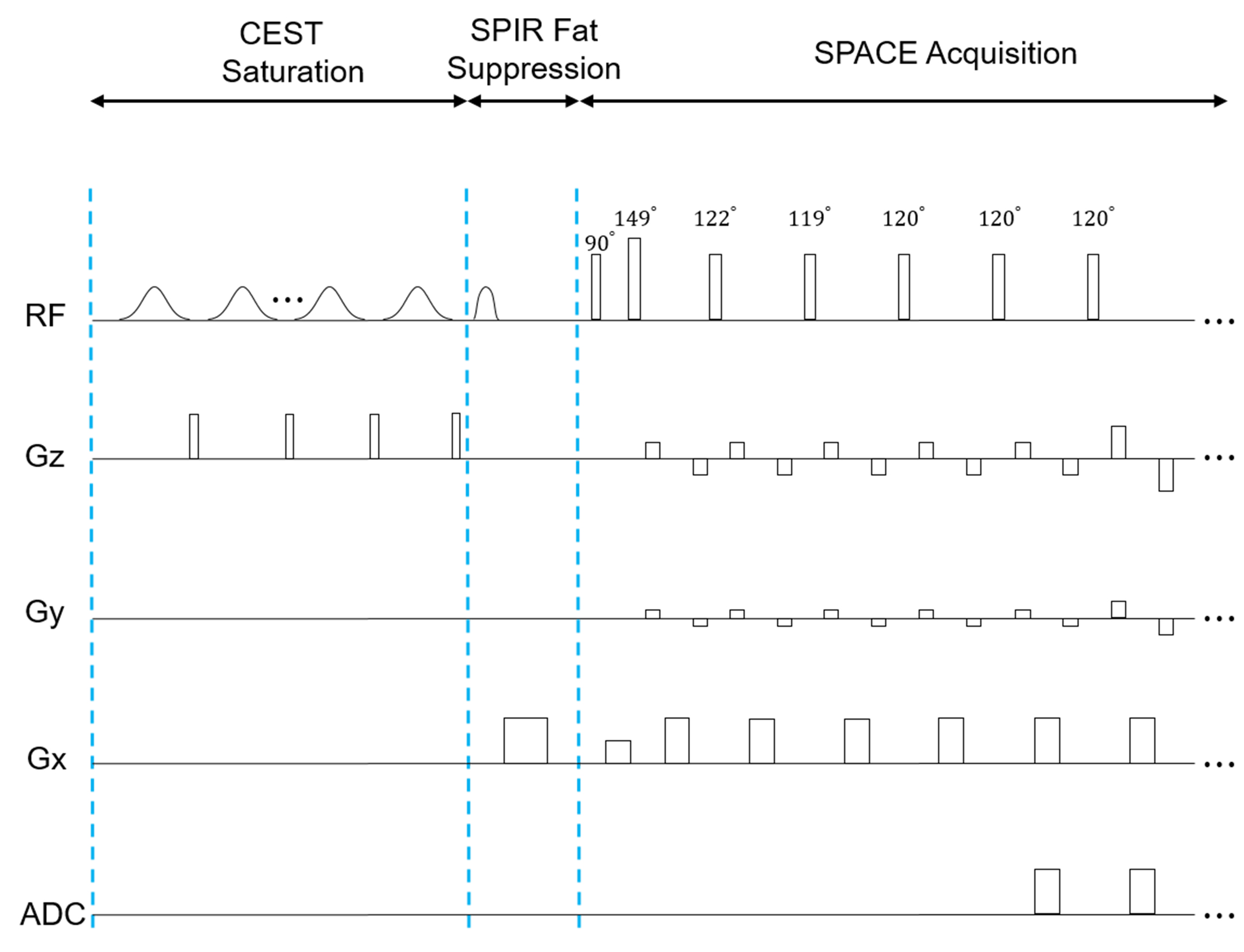
Five healthy volunteers were scanned on a 3T MAGNETOM Prisma (Siemens Healthcare, Erlangen, Germany) MRI system with a 64-channel head/neck coil. The study was approved by the local Institutional Review Board with consent forms obtained from each participant. The prototype SPACE CEST sequence as shown in Figure 1 consisted of three modules, i.e. CEST saturation, SPIR fat suppression and SPACE readout. The CEST saturation module had ten 100ms-long Gaussian pulses each with a root mean square power of 2.5uT. There was a 10ms gap between Gaussian saturation pulses during which period a 5ms-long, 15mT/m-strong crusher gradient was executed. As for SPACE readout, the following acquisition parameters were used: field of view (FOV) = 212x212x201mm3; matrix size = 76x76x72; resolution = 2.79x2.79x2.79mm3; repetition time (TR) = 3sec; echo time (TE) = 17ms; turbo factor = 140; GRAPPA11 factor = 2x2; sagittal orientation; 7 dynamics for APTw imaging with unsaturated (S0) and saturated frequency offsets of ±3ppm, ±3.5ppm, and ±4ppm; duration = 5min. The SPACE sequence had non-selective excitation and refocusing pulses, with four startup refocusing pulses before entering constant 1200 refocusing pulses. As for B0 field mapping, a gradient-echo sequence was utilized using the same FOV and resolution to the SPACE CEST sequence, with TR of 30ms, and dual TE of 4.92ms and 9.84ms. Then, both the SPACE and B0 images were interpolated to a matrix size of 152x152x152 and reformatted to generate images in different orientations. Finally, APTw maps were calculated using the SPACE CEST images after B0 correction12.Results and Discussion
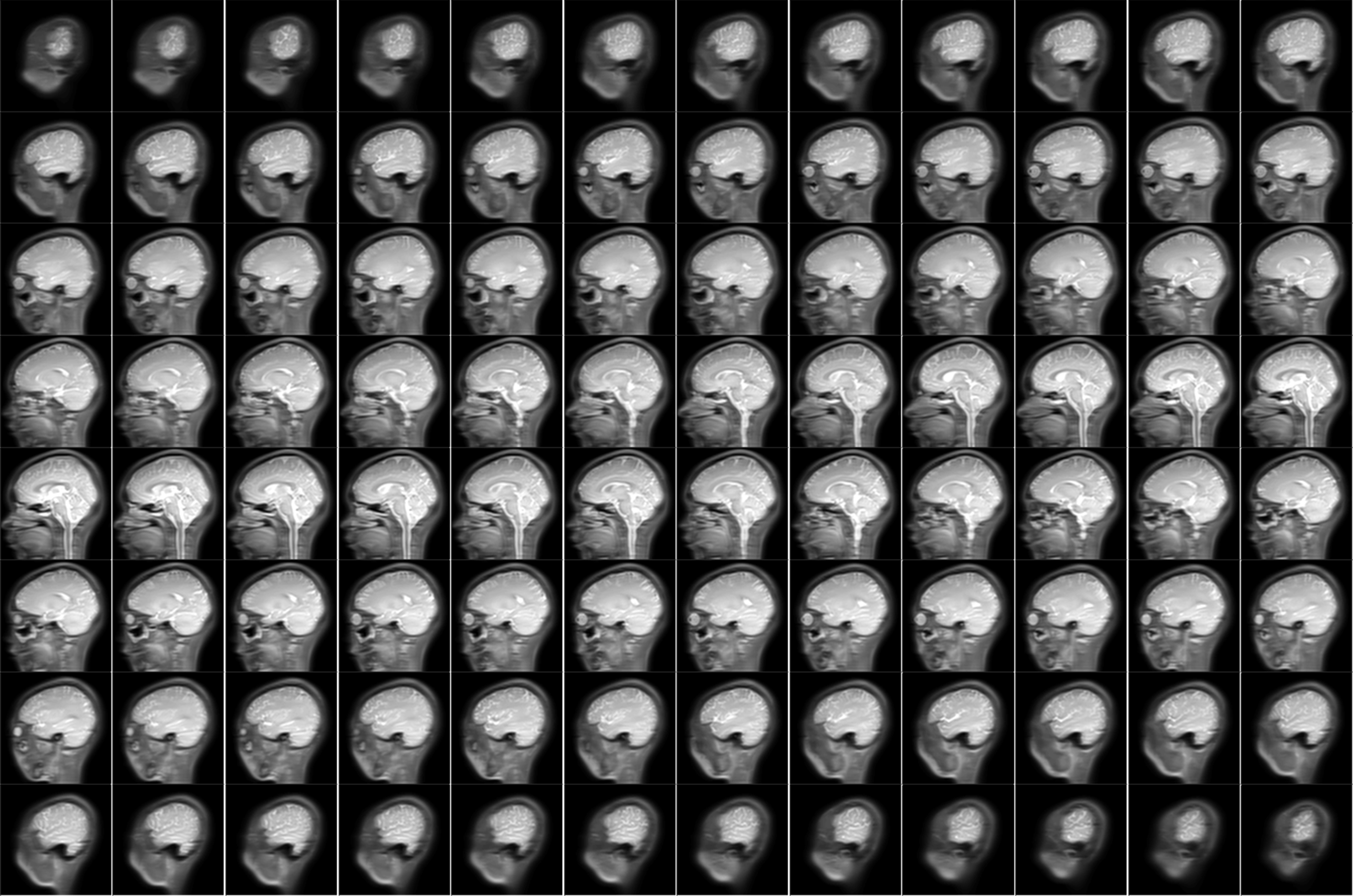
Figure 2 displays source S0 images obtained from the SPACE CEST sequence, covering the whole brain in a sagittal orientation. No discernible susceptibility artifact could be found on the SPACE CEST images, even in regions near the nasal cavity, which reflected the robustness of the new SPACE CEST sequence.
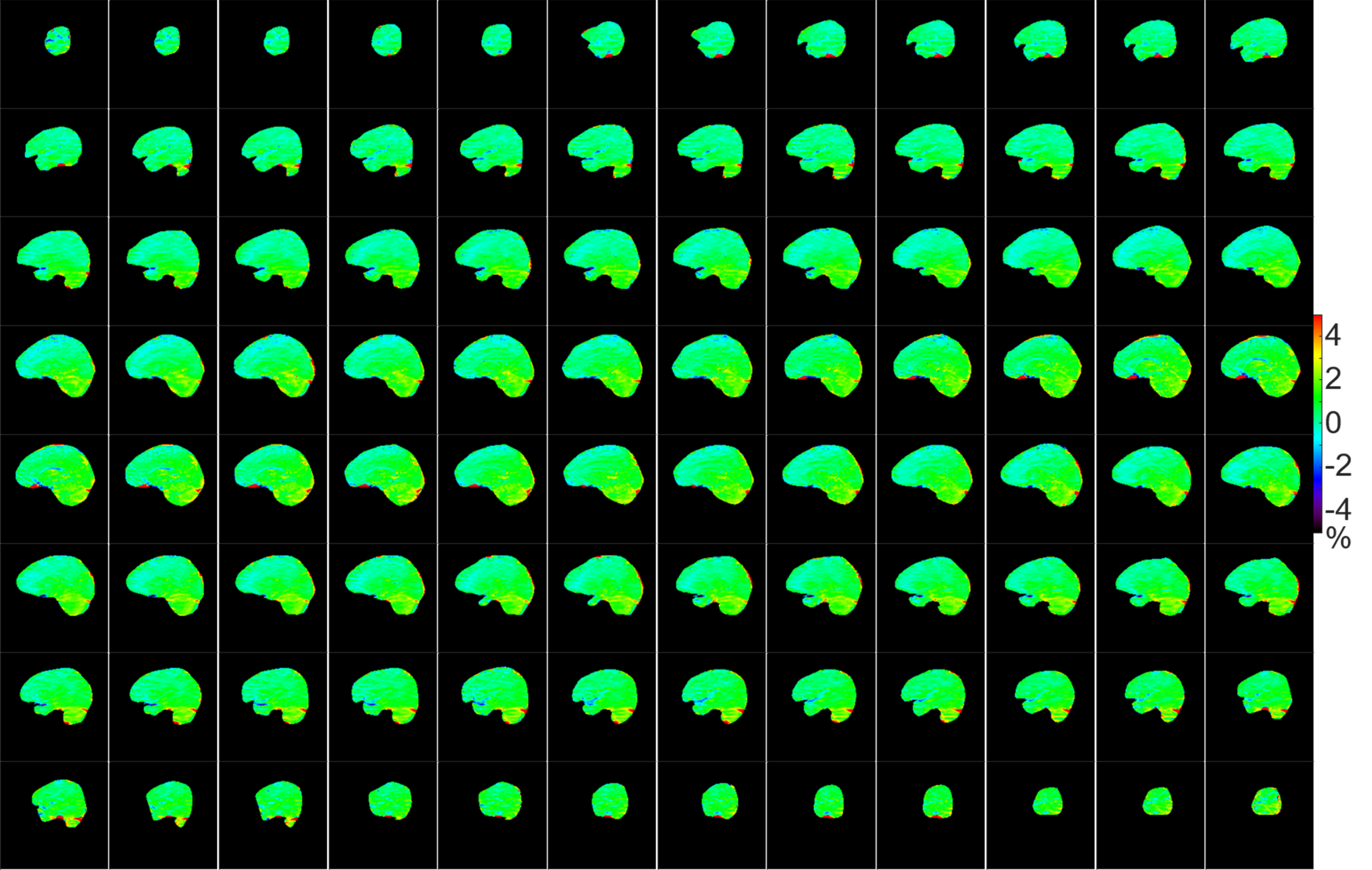
Figure 3 shows the skull-stripped APTw maps calculated from source images as shown in Figure 2. High-quality APTw images with good uniformity throughout the whole brain were obtained from the proposed SPACE CEST sequence. Minor artifacts exist in the cerebellum region, which is likely due to the suboptimal shimming in that region and can potentially be improved with region-adaptive shimming13.
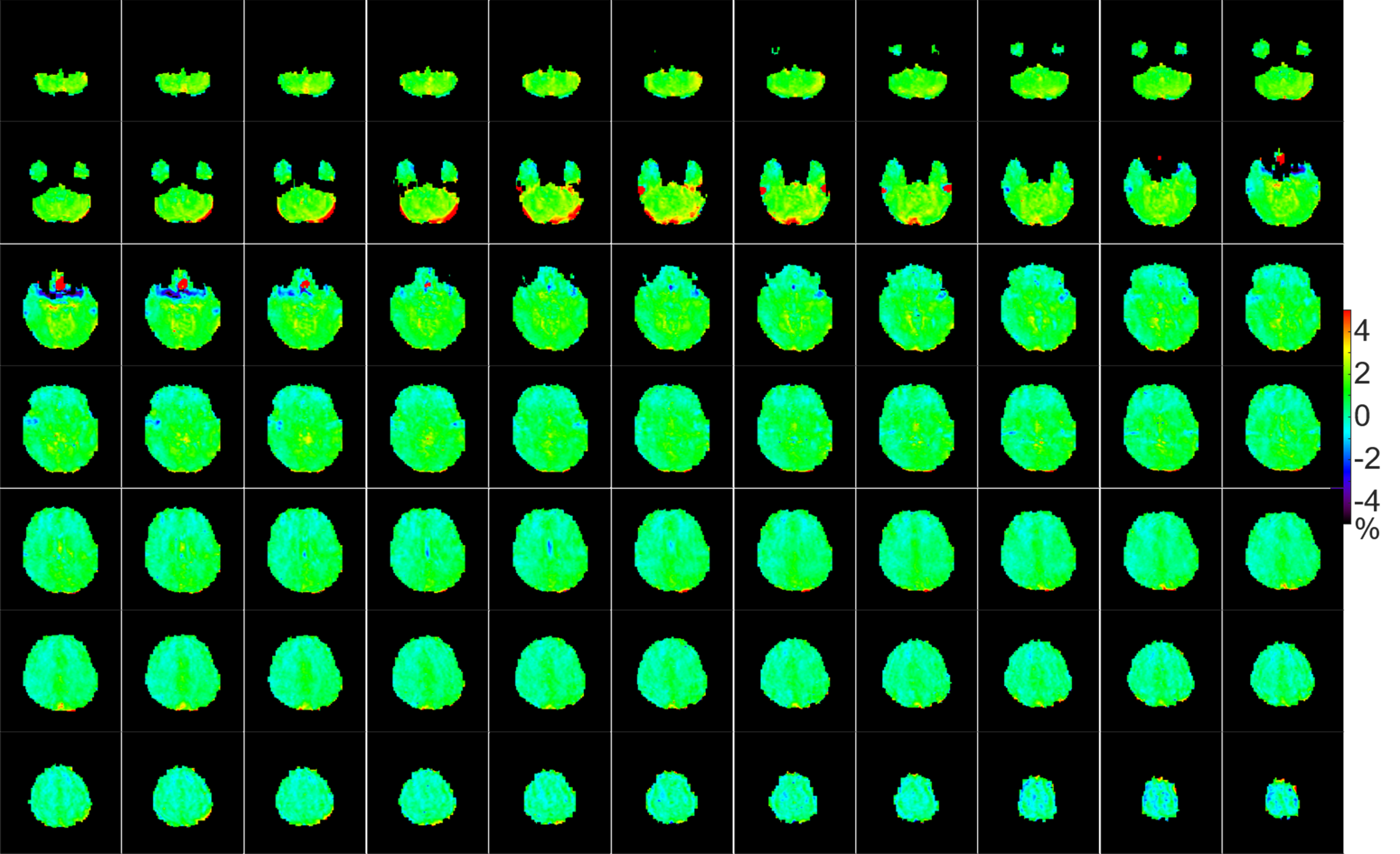
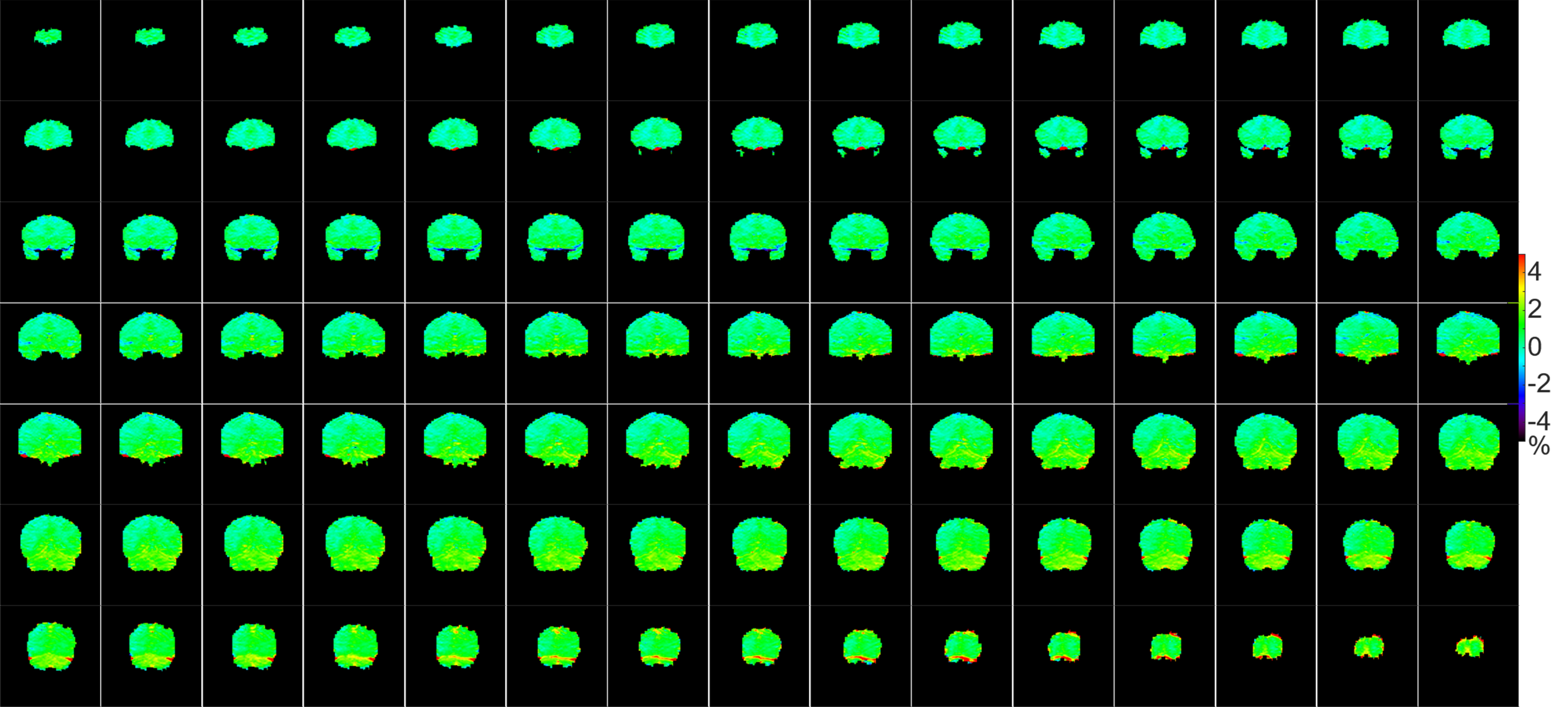
Figures 4 and 5 illustrate the APTw images in the transverse and coronal orientation, respectively, after retrospective multiplanar reconstruction. Again, these APTw maps are of high quality, and can potentially facilitate clinical diagnosis via revealing the target pathology from different orientations.
Lastly, the proposed SPACE CEST sequence achieved a higher duty cycle (91%) in the CEST saturation module than previous whole-brain EPI CEST sequences which had a duty cycle lower than 50%6-8. A higher saturation duty cycle will be beneficial for achieving the highest attainable CEST contrast within a limited saturation duration subject to scanner hardware14.
Conclusion
The proposed SPACE CEST sequence enabled whole-brain 2.79mm isotropic CEST imaging with no discernible susceptibility artifacts in 5min. This technical advance should facilitate the translation of CEST imaging into clinical routines.Acknowledgements
No acknowledgement found.References
1. Ward K, Aletras A, Balaban R. A new class of contrast agents for MRI based on proton chemical exchange dependent saturation transfer (CEST). J Magn Reson. 2000;143(1):79-87.
2. van Zijl P, Yadav NN. Chemical exchange saturation transfer (CEST): what is in a name and what isn't? Magn Reson Med. 2011;65(4):927-48.
3. van Zijl PC, Lam WW, Xu J, Knutsson L, Stanisz GJ. Magnetization Transfer Contrast and Chemical Exchange Saturation Transfer MRI. Features and analysis of the field-dependent saturation spectrum. Neuroimage. 2018;168:222-41.
4. Jones KM, Pollard AC, Pagel MD. Clinical applications of chemical exchange saturation transfer (CEST) MRI. J Magn Reson Imaging. 2018;47(1):11-27.
5. Mugler JP, Brookeman JR. Three-dimensional magnetization-prepared rapid gradient-echo imaging (3D MP RAGE). Magn Reson Med. 1990;15(1):152-7.
6. Jones CK, Polders D, Hua J, Zhu H, Hoogduin HJ, Zhou J, Luijten P, van Zijl P. In vivo three‐dimensional whole‐brain pulsed steady‐state chemical exchange saturation transfer at 7 T. Magn Reson Med. 2012;67(6):1579-89.
7. Heo HY, Jones CK, Hua J, Yadav N, Agarwal S, Zhou J, van Zijl PC, Pillai JJ. Whole‐brain amide proton transfer (APT) and nuclear overhauser enhancement (NOE) imaging in glioma patients using low‐power steady‐state pulsed chemical exchange saturation transfer (CEST) imaging at 7T. J Magn Reson Imaging. 2016;44(1):41-50.
8. Akbey S, Ehses P, Stirnberg R, Zaiss M, Stöcker T. Single-shot whole-brain CEST imaging using centric-reordered 3D-EPI. Proc Intl Soc Mag Reson Med. 2018(26):2231.
9. Mugler III JP. Optimized three‐dimensional fast‐spin‐echo MRI. J Magn Reson Imaging. 2014;39(4):745-67.
10. Zhou J, Lal B, Wilson DA, Laterra J, van Zijl P. Amide proton transfer (APT) contrast for imaging of brain tumors. Magn Reson Med. 2003;50(6):1120-6.
11. Griswold MA, Jakob PM, Heidemann RM, Nittka M, Jellus V, Wang J, Kiefer B, Haase A. Generalized autocalibrating partially parallel acquisitions (GRAPPA). Magn Reson Med. 2002;47(6):1202-10.
12. Zhou J, Zhu H, Lim M, Blair L, Quinones-Hinojosa A, Messina SA, Eberhart CG, Pomper MG, Laterra J, Barker PB. Three-dimensional amide proton transfer MR imaging of gliomas: Initial experience and comparison with gadolinium enhancement. J Magn Reson Imaging. 2013;38(5):1119-28.
13. Zhu H, Jones CK, van Zijl P, Barker PB, Zhou J. Fast 3D chemical exchange saturation transfer (CEST) imaging of the human brain. Magn Reson Med. 2010;64(3):638-44.
14. Schmitt B, Zaiß M, Zhou J, Bachert P. Optimization of pulse train presaturation for CEST imaging in clinical scanners. Magn Reson Med. 2011;65(6):1620-9.
Figures