5017
Fully Steady-State Chemical Exchange Saturation Transfer (CEST) imaging for Amide Proton Transfer (APT) at 3T MRI1Korea Advanced Institute of Science and Technology, Daejeon, Korea, Republic of
Synopsis
A steady-state chemical exchange saturation transfer (SS-CEST) MRI, which is based on the standard macromolecule transfer (MT) imaging method, has been developed for fast scanning 1,2. In this study, we proposed a SS-CEST imaging combined with steady-state free precession (SSFP) which is so-called fully SS-CEST imaging. The fully SS-CEST method was compared with
Introduction
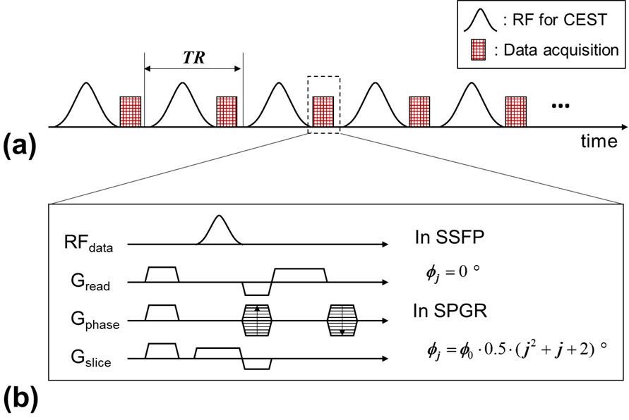
A steady-state chemical exchange saturation transfer (SS-CEST) MRI has been developed for fast scanning 1,2, which was adopted from the standard macromolecule transfer (MT) imaging method. As shown in Fig. 1, the irradiation scheme of RF pulse for CEST (RFCEST) is identical to that of pulsed-CEST but signal acquisitions are performed between two adjacent RFCEST. In this abstract, two types of acquisition sequences to be combined with the SS-CEST are compared, which are steady-state free precession (SSFP) and spoiled gradient echo (SPGR) schemes. Through the simulations and in-vivo experiments, we demonstrated that SS-CEST with SSFP that is so-called fully SS-CEST could provide higher CEST contrast than that with SPGR.Methods
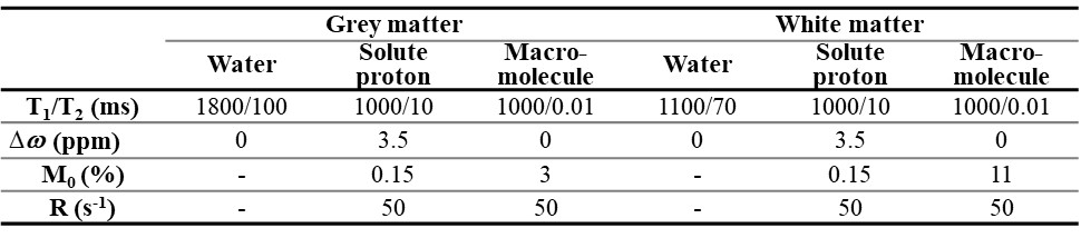
Numerical simulations for comparing two types of SS-CEST sequences were performed by using the modified Bloch-McConnell equations3. The Bloch-McConnell equations are composed of seven coupled equations for the three-pool model of bulk water, amide protons, and macromolecules. Each of bulk water pool and amide pool has three equations involving x, y and z components of magnetizations. The macromolecule pool has a single equation for the z component of magnetization, where the MT effect is modeled by a super-Lorentzian lineshape4. In SS-CEST with SPGR, the transverse magnetizations were set to zero immediately after data acquisition to simulate effects of dephasing gradients and RF spoiling. In SS-CEST with SSFP, we assume 16 spins in a voxel, influenced by different fields of the dephasing gradients, to simulate the non-zero steady-state magnetizations of SSFP. The simulations were conducted with the following sequence parameters: the flip angle of RFCEST is 200 °, repetition time of the sequence (TR) was 33 ms, the duty cycle of RFCEST was 0.75 and flip angle for imaging was 1~15 ° with a 2 ° increment. Table 1 presents the specific tissue parameters modeling amide proton transfer (APT) in the human brain.
In addition to the simulation, in-vivo experiments were performed using a 3.0T MRI system (Siemens MAGNETOM Verio, Erlangen, Germany). A 32-channel Rx head coil was used for human brain imaging. Two healthy subjects were scanned by SS-CEST with SSFP and SPGR with the following parameters: the flip angle of RFCEST is 180 °, repetition time of the sequence (TR) was 27 ms, the duty cycle of RFCEST was 0.5 and flip angle for imaging was 17 °. All in-vivo experiments were approved by the KAIST Institutional Review Board (IRB), and all participants signed the informed consent forms.
The CEST contrast is measured by the pool difference method2,5 as follow.
$$CEST_{cont} =S(\triangle\omega,0)-S(\triangle\omega,M_{c})$$
where $$$M_{c}$$$ is the concentration of the solute protons, $$$S(\triangle\omega,M_{c})$$$ is an acquired signal in a z-spectrum where RF saturation for CEST is applied to the chemical shift of exchangeable solute protons ($$$\triangle\omega$$$) and $$$S(\triangle\omega,0)$$$ is the corresponding z-spectrum signal where the concentration of the solute protons is zero. In in-vivo experiments, a Lorentzian curve was fitted to an acquired z-spectrum to quantify $$$S(\triangle\omega,0)$$$, where $$$S(\triangle\omega,0)$$$ has only macromolecule transfer (MT) and direct saturation (DS). All simulations and post-processing were performed using MATLAB (MathWorks, USA).
Results
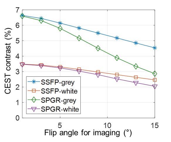
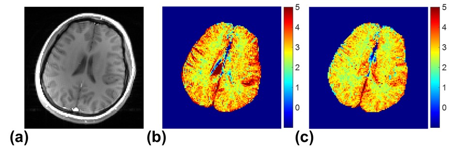
Fig. 2 shows the simulated results of APT contrast with respect to the flip angle for imaging in SS-CEST with SSFP and SPGR. The values of APT contrast generally decrease when the flip angle increases due to increased direct saturation (DS) of water by the RF pulse for imaging. In both of grey and white matters, the APT contrast from SS-CEST with SSFP is always higher than that from SS-CEST with SPGR. It shows that SS-CEST with SSFP is more robust to DS than SS-CEST with SPGR. Fig. 3 describes the resultant APT images of a human brain. The image from the SSFP approach also has higher APT contrast than that produced by the SPGR approach.Discussion & Conclusion
The different DS robustness in SSFP and SPGR is attributed to different signal models. In SPGR, once longitudinal magnetization of water is flipped to the transverse plane by RF excitation, the magnetization is fully dephased by the RF spoiling and dephasing gradient immediately after data acquisition. On the other hand, SSFP preserves the transverse magnetization so that some of them can be recovered back to the longitudinal axis by the next RF pulse. Therefore, the amount of longitudinal magnetization in SSFP is better maintained in comparison to those in SPGR. And, SS-CEST with SSFP could provide higher APT contrast than that achieved with SPGR.
Consequently, we proposed an SS-CEST with SSFP which were so-called fully SS-CEST. The simulations and MRI experiments demonstrated that the fully SS-CEST could provide higher APT contrast than SS-CEST with SPGR.
Acknowledgements
This research was supported by the Brain Research Program through the National Research Foundation of Korea (NRF) funded by the Ministry of Science, ICT & Future Planning (2014M3C7033999).
This research was supported by a grant of the Korea Health Technology R&D Project through the Korea Health Industry Development Institute (KHIDI), funded by the Ministry of Health & Welfare, Republic of Korea (grant number : HI14C1135)
References
1. Dixon WT, Hancu I, Ratnakar SJ, Sherry AD, Lenkinski RE, Alsop DC. A multislice gradient echo pulse sequence for CEST imaging. Magn Reson Med. 2010;63:253-256.
2. Jones CK, Polders D, Hua J, Zhu H, Hoogduin HJ, Zhou J, Luijten P, van Zijl PC. In vivo three-dimensional whole-brain pulsed steady-state chemical exchange saturation transfer at 7 T. Magn Reson Med. 2012;67:1579-1589.
3. Woessner DE, Zhang S, Merritt ME, Sherry AD. Numerical solution of the Bloch equations provides insights into the optimum design of PARACEST agents for MRI. Magn Reson Med. 2005;53:790-799.
4. Morrison C, Henkelman RM. A model for magnetization transfer in tissues. Magn Reson Med. 1995;33:475-482.
5. Khlebnikov V, Geades N, Klomp DW, Hoogduin H, Gowland P, Mougin O. Comparison of pulsed three-dimensional CEST acquisition schemes at 7 tesla: steady state versus pseudosteady state. Magn Reson Med. 2017;77:2280-2287.
Figures