4981
Physiologically Accurate Simulations of Endogenous Susceptibility-Based Contrast in Cancer Reveal the Importance of Intravascular Oxygen Variations on Transverse Relaxation1Neuroradiology Department, Heidelberg University Hospital, Heidelberg, Germany, 2Department of Radiology and Radiological Science, Johns Hopkins University School of Medicine, Baltimore, MD, United States
Synopsis
The heterogeneous nature of tumor vasculature and hemodynamics make it challenging to model transverse relaxation in such tissues. Here we imaged the intravascular oxygen saturation in healthy abdominal wall and breast tumor xenografts in mice using intrinsic optical signal (IOS) imaging, and used these quantitative data in realistic simulations of transverse relaxation. We found that the inclusion of de factooxygen distributions recapitulated the heterogeneity of tumor transverse relaxation rates in contrast to the traditional approach of assuming constant oxygen distribution for the tumor microvascular bed. These findings have important implications for BOLD MRI of tumors.
Introduction
The influence of micro- and mesoscopic blood vessel structures on transverse relaxation during MR experiments has been studied extensively with analytical and numerical approaches]1,2,3. A common simplification in such studies is the modeling of the magnetic susceptibility of blood with a constant value throughout the microvasculature, implying homogeneous intravascular oxygen saturation and hematocrit. However, this assumption is not valid for most tissues, especially the tumor4. In this study, we independently assessed intravascular oxygen saturation within breast tumor xenografts and healthy abdominal walls of mice using intrinsic optical signal (IOS) imaging. We then incorporated these quantitative oxygen saturation data in models of extravascular spin dephasing and compared transverse relaxation rates to those obtained using the homogeneous oxygenation simplification. Our motivation was to assess the implications of varying intravascular oxygenation on BOLD MRI of tumors.Methods
We interrogated the healthy abdominal wall and breast tumor xenografts in mice with widefield IOS imaging at 570 nm and 600 nm with 5 μm spatial resolution. The modified Beer-Lambert equation was applied to these images to compute intravascular oxygen saturation5. A finite perturber method3 was implemented in two dimensions to calculate microscopic magnetic field distortions based on the local susceptibility Δχ=Δχ0(1-Y)Hct of blood with Δχ0=3.39 ppm4, hematocrit Hct=0.4, and measured intravascular oxygen saturation Y using Matlab R2018a (Mathworks, Natick, MA, USA). Parallelized random walks, written in C++11, were conducted to simulate extravascular spin dephasing during free induction decay (FID) with physiological water diffusion set to D=1 µm2/ms within virtual MRI voxels of 1 mm3. We selected 5 different regions of interest (ROI) within each tissue type. Analogous simulations were conducted with homogeneous oxygenation Y, set to the mean of the measured values for each dataset. Experimental scenarios with different external field strengths B0 between 1.5 and 10 Tesla, and Hct-values in the physiological range were included in the simulations.Results
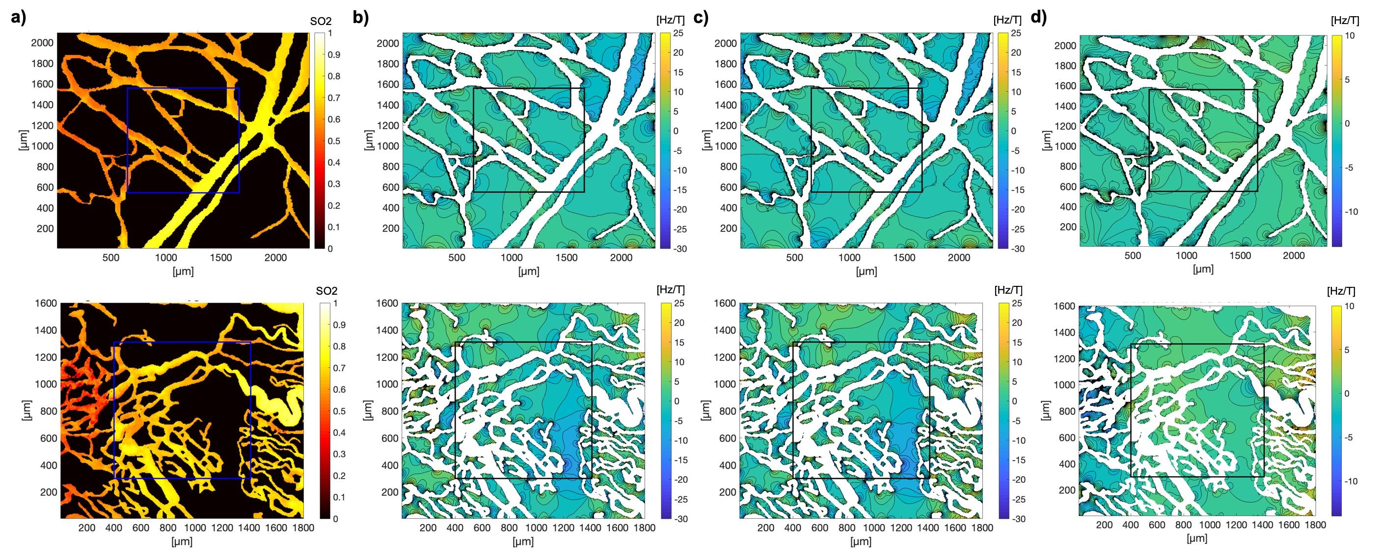
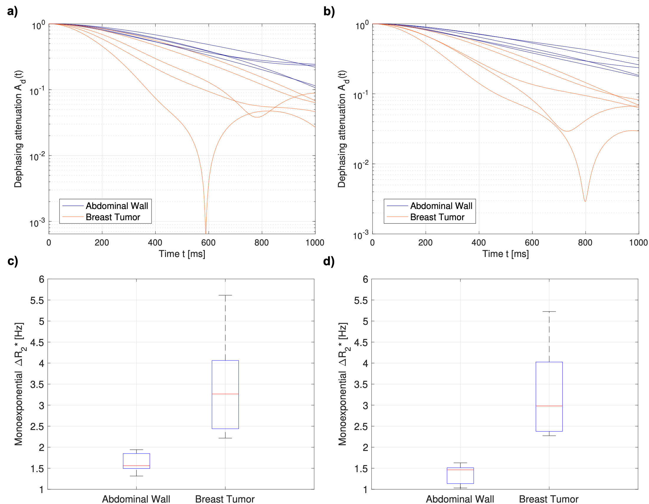
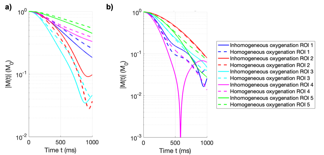
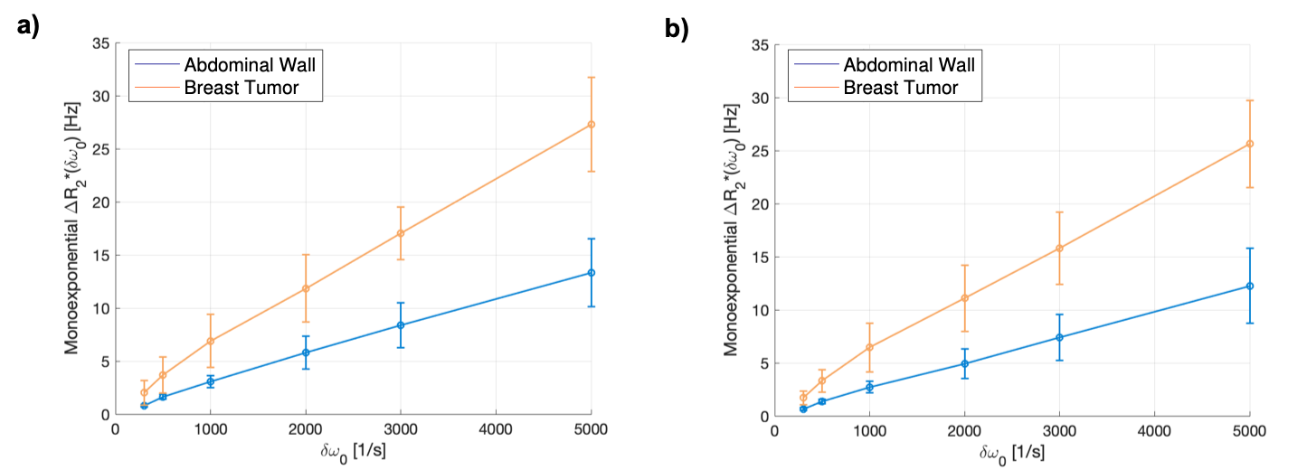
The inclusion of blood oxygen saturation data from IOS imaging in the calculations of vessel-induced magnetic field inhomogeneities resulted in more heterogeneous field distortions with a broader range of off-resonance Larmor frequencies (Fig. 1). In most ROI, this led to faster spin dephasing, and more heterogeneous inter-voxel decay characteristics compared to those obtained for the constant intravascular oxygenation scenario (Fig. 2). A trend towards enhanced transverse relaxation was observed in 4 of 5 healthy ROI and 3 of 5 tumor ROI (Fig. 3). With increasing external field strength (B0) or hematocrit (Hct), summarized in the off-resonance factor δω0, the fundamental trends were maintained with tissue types being differentiated by transverse relaxation rates more clearly (Fig. 4).Discussion
Our results show that the effects of inhomogeneous blood oxygenation distributions on spin dephasing varied strongly with location and deoxyhemoglobin content of proximal vessels in healthy and tumor tissues. We were able to successfully recapitulate well-known susceptibility-based phenomena such as stronger dephasing arising from the abnormal and heterogeneous tumor microvasculature relative to that in healthy tissue.Conclusion
Physiological changes in blood flow, oxygenation, and microvessel architecture all affect transverse relaxation and are relevant when imaging with endogenous susceptibility contrast mechanisms, such as BOLD. It is important to be able to disentangle the effects of different physiological variables on the MR-signal in order to develop robust biomarkers, sensitive to aspects of the underlying pathological vasculature. An omission of blood oxygenation information may lead to biased results when using BOLD-derived biomarkers for tumor studies such as vessel size index (VSI) or cerebral blood volume (CBV). We are currently working on quantifying the intravascular hematocrit distribution for developing a physiologically accurate model of spin dephasing in tumor microvascular networks.Acknowledgements
A. Hahn and F. T. Kurz received funding from grant DFG KU 3555/1-1. F. T. Kurz was also supported by the Hoffmann-Klose foundation of Heidelberg University Hospital.References
1. Troprès I, Pannetier N, Grand S, et al. Imaging the Microvessel Caliber and Density: Principles and Applications of Microvascular MRI. Magn Reson Med. 2015 Jan; 73:325-41.
2. Dickson JD, Ash TWJ, Williams GB, et al. Quantitative phenomenological Model of the BOLD Contrast Mechanism. J Magn Reson. 2011; 212:17-25.
3. Pathak AP, Ward BD, Schmainda KM. A Novel Technique for Modeling Susceptibility-Based Contrast Mechanisms for Arbitrary Microvascular Geometries: The Finite Perturber Method.NeuroImage. 2008; 40:1130-1143.
4. Döme B, Hendrix M, Paku S, Tóvari K. Alternative Vascularization Mechanism in Cancer. Am J Pathol. 2007; 170: 1-15.
5. Hillman EM. Optical brain imaging in vivo: techniques and applications from animal to man. J Biomed Opt. 2007; 12: 051402.
Figures