4972
The influence of fat on T1 mapping of the liver: a comparison of Look-Locker and variable-flip-angle techniques1Institute of Radiology, University Hospital Regensburg, Regensburg, Germany, 2MR Applications Predevelopment, Siemens Healthcare GmbH, Erlangen, Germany
Synopsis
In 383 patients two methods for T1 mapping – 2D Look-Locker (LL), and 3D variable-flip-angle (VFA) combined with a 2-point-Dixon technique – were compared and their correlation with the intrahepatic proton density fat fraction (PDFF) was evaluated. T1_LL showed a moderate positive correlation with PDFF, while there was an intermediate negative correlation between T1_VFA_in (T1 calculated from water and fat in-phase signal) and PDFF; T1_VFA_W (T1 calculated from water only signal) was nearly independent of PDFF. In patients with PDFF above 5%, LL, VFA_in, and VFA_W yielded significantly different results for T1.
Introduction
T1 mapping has been proposed as a novel, non-invasive tool for detecting or staging of liver disease. Up to now, there is only sparse information about the influence of intrahepatic fat on the measured T1 relaxation time: depending on the T1 mapping technique and the sequence parameters the presence of fat might increase or decrease T1 1, 2. This study aimed to compare the influence of intrahepatic fat on T1 measured with a Look-Locker (LL), and a variable-flip-angle (VFA) combined with a 2-point-Dixon technique in a large patient collective.Methods
423 patients underwent MRI to assess suspected liver lesions, or as follow-up examination in case of known liver disease on a clinical 3T scanner (MAGNETOM Skyra, Siemens Healthcare, Erlangen, Germany). In addition to our routine MRI protocol, T1 mapping with LL and VFA as well as mapping of proton density fat fraction (PDFF) were performed in all patients: A prototype 2D LL technique based on snapshot-FLASH imaging 3 was applied for 3 transverse slices through the porta hepatis with inline calculation of T1 maps (T1_LL). Sequence parameters were chosen as TR 3ms, TE 1.32ms, flip angle 8°, 16 contrasts acquired following the inversion pulse, measured voxel size 3.0mm x 2.1mm x 8.0mm, and acquisition time (TA) 16s. T1 mapping of the whole liver was performed with a VFA prototype 3D gradient echo sequence using a protocol with TR 5.79ms, TE 2.46, 3.69ms, flip angle 1°, 7°, 14°, measured voxel size 3.6mm x 2.5mm x 4.8mm, and TA 17s. The VFA sequence was combined with a preceding B1-map for an inline correction of B1 inhomogeneities. Using a 2-point Dixon method, T1 maps from in-phase (T1_VFA_in) and from water (T1_VFA_W) signals were calculated inline. PDFF mapping of the whole liver was performed with a 6 echo prototype 3D VIBE sequence (TR 9.2ms, TE 1.23 to 7.38ms, flip angle 4°, measured voxel size 2.9mm x 2.6mm x 6.4mm, and TA 15s). T1 relaxation times and PDFF were evaluated in a single coaligned ROI.
A t-test was applied to compare T1 relaxation times calculated from different methods, Pearson correlation coefficient was used to quantify the influence of PDFF on T1. Results with p<0.05 were considered to be statistically significant.
Results
21 of 423 patients had to be excluded for further evaluation due to global fat-water swaps in T1 and/or fat mapping, 19 patients were excluded due to severe motion artifacts. T1_LL and PDFF were only moderately correlated (R = 0.37), while there was an intermediate negative correlation between T1_VFA_in and PDFF (R = -0.46); T1_VFA_W was not correlated with PDFF (R = -0.14) (Fig. 1). In patients with low intrahepatic fat fraction (PDFF < 5%, n = 289), no significant difference was found between T1_LL and T1_VFA_in (p = 0.231), T1_VFA_W was higher than T1_LL (p = 0.015), and T1_VFA_in and T1_VFA_W were significantly different (p < 0.001); in patients with increased fat fraction (PDFF ≥ 5%, n = 94) T1 results were significantly different for all T1 techniques (Figs. 2-5).Discussion
T1 of the liver varied in a broad range in our patient collective – even in patients with low intrahepatic fat. This can be partially attributed to the heterogeneity of patients concerning liver function / chronic liver disease. Nevertheless, there was also a clear trend showing the influence of PDFF on T1 and this trend was different for the T1 mapping techniques used: due to the short T1 of fat there was a decrease of T1 with increasing PDFF when using the water and fat in-phase signal for T1 calculation (VFA_in). The minimal effect of fat on T1_VFA_W might be explained by a potentially incomplete separation of water and fat using a 2-point Dixon method. The correlation between PDFF and T1_LL was only moderate, but the T1-prolonging effect of fat might be caused by using an opposed-phase echo time in the FLASH readout of the LL sequence.Conclusion
Intrahepatic fat has a significant influence on the measured T1 relaxation time of the liver and the kind of influence – increasing or decreasing T1 – depends on the technique used. This result might be important when using quantitative results of T1 mapping in patients with increased intrahepatic fat. Calculating T1 from a water-only signal will minimize the influence of fat.Acknowledgements
No acknowledgement found.References
1. Le Y, Dale B, Akisik F, Koons K, Lin C. Improved T1, contrast concentration, and pharmacokinetic parameter quantification in the presence of fat with two-point Dixon for dynamic contrast-enhanced magnetic resonance imaging. Magn Reson Med. 2016;75:1677-1684.
2. Mozes FE, Tunnicliffe EM, Pavlides M, Robson MD. Influence of fat on liver T1 measurements using modified Look-Locker inversion recovery (MOLLI) methods at 3T. J Magn Reson Imaging. 2016;44:105-111.
3. Deichmann R and Haase A. Quantification of T1 values by SNAPSHOT-FLASH NMR imaging. J Magn Reson Imaging.1992;96:608-612.
Figures
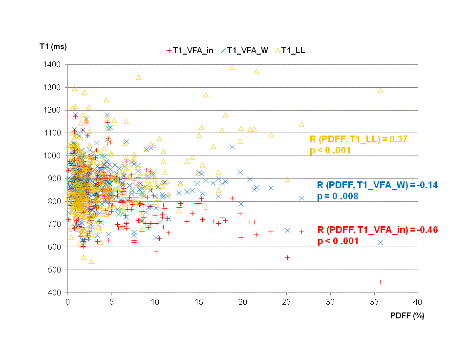
Fig. 1:
Scatterplot of T1 mapping results from three different techniques versus PDFF and correlation coefficient R:
Moderate (positive) correlation between T1_LL and PDFF, nearly no correlation between T1_VFA_W and PDFF, intermediate negative correlation between T1_VFA_in and PDFF.
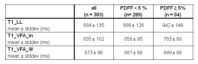
Fig. 2:
Descriptive statistics of T1 in 383 patients and in two subgroups (PDFF < 5% and PDFF ≥ 5%) using LL and VFA technique.
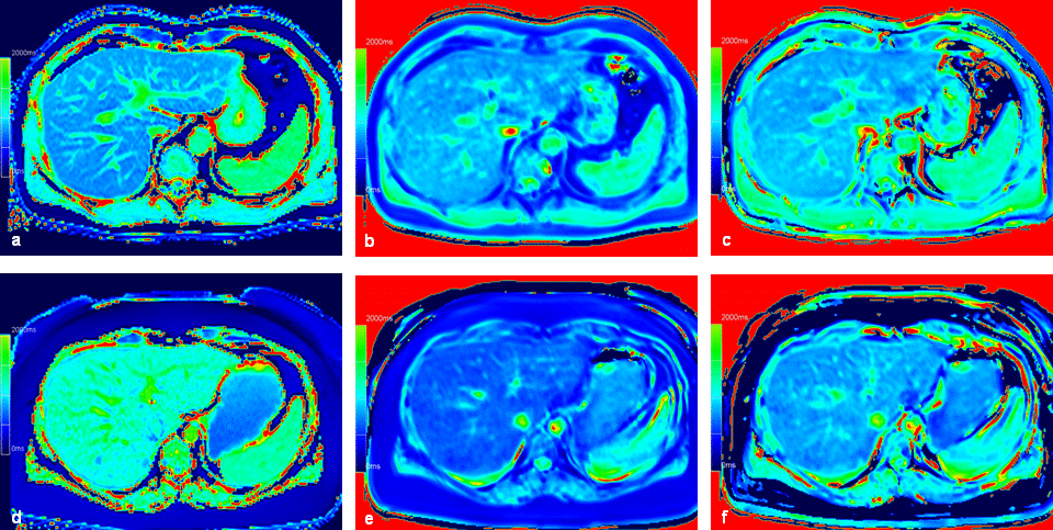
Fig. 3:
Similar T1 results using different T1 mapping techniques in a patient with normal intrahepatic fat, but clearly different results in a patient with increased intrahepatic fat. Identical window and center settings in all figures.
a-c: T1 mapping in a 43-year-old man with PDFF = 2.3%: T1_LL = 820 ms (a), T1_VFA_in = 819 ms (b), T1_VFA_W _ 835 ms (c).
d-e: T1 mapping in a 38-year-old woman with PDFF = 23.2%: T1_LL = 1093 ms (d), T1_VFA_in = 667 ms (e), T1_VFA_W = 860 ms (f).
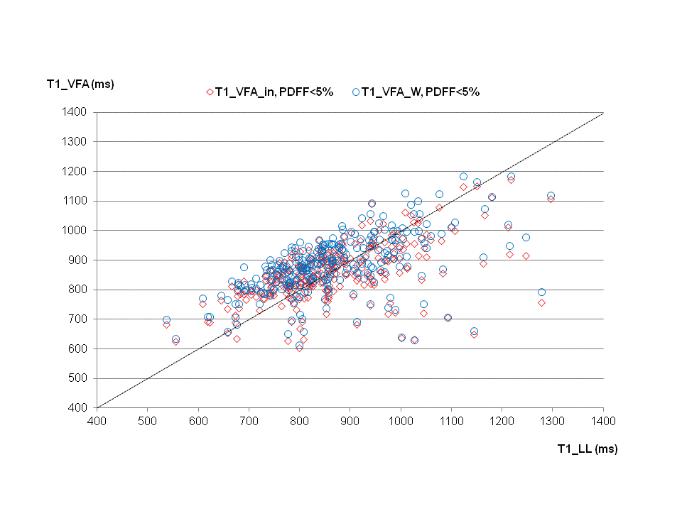
Fig. 4:
Scatterplot of T1 results using a VFA technique combined with 2-point Dixon (VFA_in, VFA_W) versus T1 using a LL technique in patients with low intrahepatic fat (PDFF < 5%):
Similar results in the majority of patients (black dotted line: line of identity), but larger deviations in some patients.
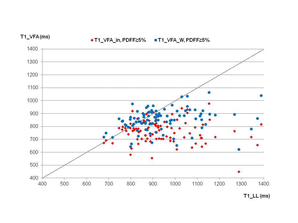
Fig. 5:
Scatterplot of T1 results using a VFA technique combined with 2-point Dixon (VFA_in, VFA_W) versus T1 using a LL technique in patients with increased intrahepatic fat (PDFF ≥ 5%):
Relevant differences between T1 mapping techniques (black dotted line: line of identity).