4963
Robust and SAR-efficient whole-brain pseudo-continuous ASL at 7T1German Center for Neurodegenerative Diseases (DZNE), Bonn, Germany, 2Department of Physics and Astronomy, University of Bonn, Bonn, Germany
Synopsis
In this work, a modified pseudo-continuous ASL sequence is presented, which reduces the SAR deposition by ~50% and provides robust labeling efficiency in the presence of off-resonances between -300Hz and 300Hz. The sequence was successfully tested on two coils with different coverage of the neck region at two labeling positions. The method allows PCASL experiments at UHF without a pre-scan in significantly reduced scan time and, therefore, exploits the advantage of UHF for perfusion imaging.
Purpose
Pseudo-continuous ASL (PCASL)(1) has many advantages at ultra-high field (UHF >3T)(2). Most prominent are the longer T1,blood, leading to slower decay of the label, and the intrinsically higher SNR. But the rapid application of thousands of RF pulses causes significant energy deposition (SAR) forcing the user to use longer TRs, which decreases the sensitivity per unit time. A second problem is the loss of labeling efficiency in the presence of B0 inhomogeneities, which can lead to a nonuniform labeling across the brain feeding arteries(3),(2).
In this work, modifications to the PCASL pulse train are presented, which reduce the SAR deposition and increase the robustness against off-resonances by means of shorter pulse spacing and variable rate RF pulses. This enables a prescan-free acquisition of background-suppressed whole brain PCASL at 7T within a TR of 8s.
Methods
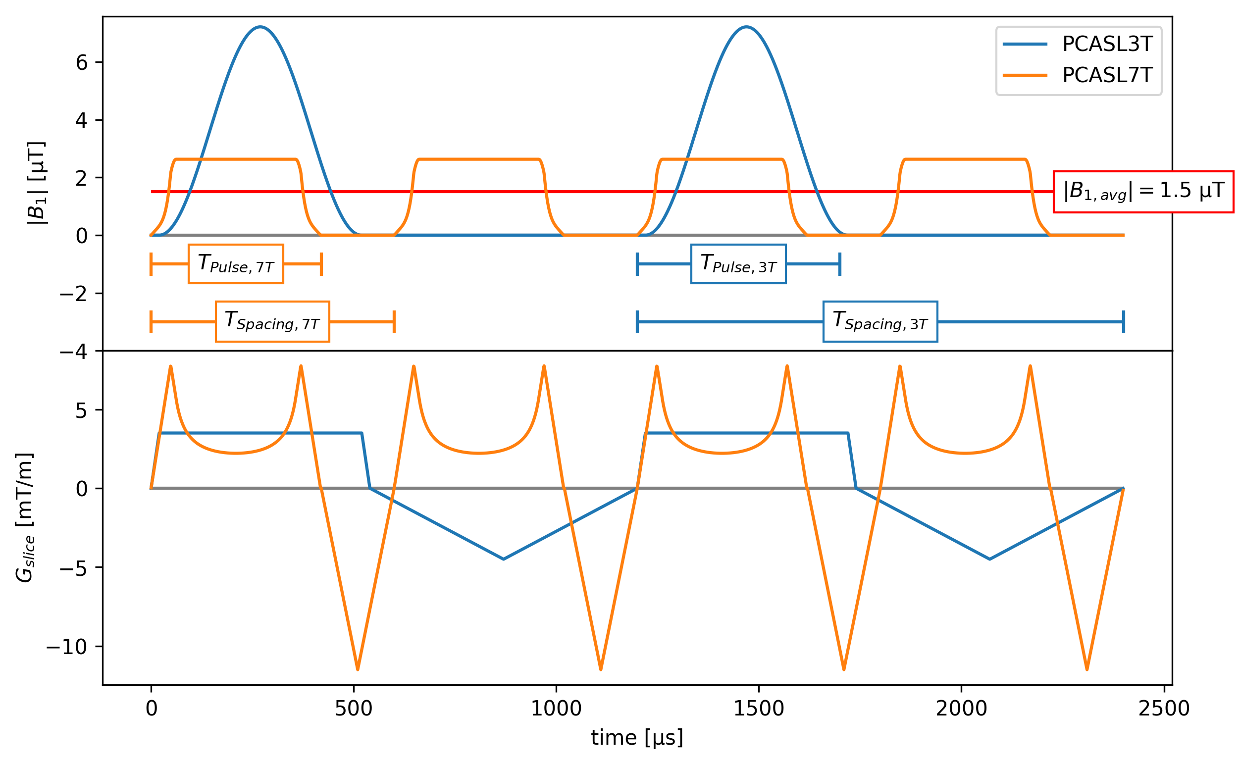
PCASL labeling was simulated as described in (3) to find optimal pulse spacing and duration. The parameter set presented in the aforementioned publication (unbalanced PCASL at 3T, TPulse=500 µs, TSpacing=1200 µs, B1,avg=1.5 µT, gmax=3.5 mT/m, gaverage=0.5 mT/m) served as a starting point for further optimization. Since the off-resonance effects of PCASL are a direct result of the introduced gaps between RF pulses, the gaps were minimized. The pulse duration maximized to distribute the RF energy deposition over as much time as possible to reduce SAR. Along with hardware restrictions due to acoustic resonance frequencies of the gradient system, gradient limits and RF duty cycles, this leads to a pulse spacing of 600 µs and a pulse duration of 420 µs. To further reduce SAR, the VERSE scheme presented in (4) was applied to the Hann-shaped RF pulses and corresponding gradients, significantly reducing RF amplitude while timing is maintained. The initial PCASL sequence (PCASL3T) and the optimized one (PCASL7T) are shown in Fig. 1.
This PCASL scheme was implemented on a Siemens Magnetom 7T (Siemens Healthineers, Erlangen) equipped with an additional parallel transmit (pTX) system (8-channel, version 2.3). A centric-reordered gradient echo 3D-EPI readout was used, which helps to minimize SAR due to its small excitation flip angles. For background suppression a WET(5) pre-saturation with 4 pulses and two GOIA-WURST-16-4(6) pulses (6.7 ms duration, 16.8 kHz bandwidth, 14 µT B1,max) for inversion were used.
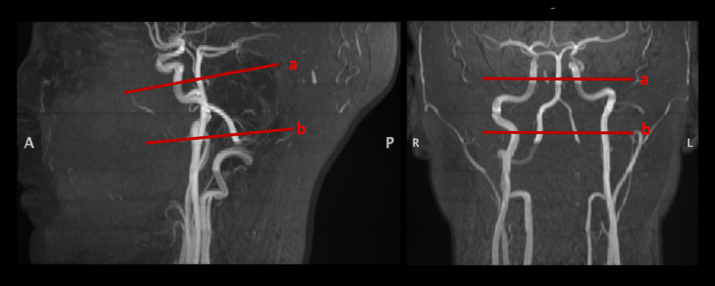
Two subjects (2 female, 34 +/- 6 years) were scanned at 7T. The imaging protocol included a flip angle map (3D-DREAM)(7), a B0 shimming sequence, an anatomical MPRAGE scan (1mm3) and a PCASL scan (Resolution=2.5x2.5x2.5 mm3, TE=6.8 ms, TR=8 s, GRAPPAPE1=2x, Partial-fourier-factorPE1=6/8, repetitions=22, acquisition time=02m:56s, labeling duration=1.5s, PLD=1.5s). Reference voltages were derived from the flip angle map and the PCASL pulse voltage was then increased by 25% to mitigate low transmission efficiency in the neck region. The first subject was scanned in single channel transmit mode at labeling position (a) as shown in Fig. 3. The second subject was scanned with the 8-channel pTX system in so called protected mode (conservative RF power limits given by global worst-case estimates). Here, labeling position (b) as in Fig. 3 was possible, since the flip angle map indicated a more efficient transmission in the lower regions for the pTX coil (32RX/8TX Nova Medical) compared to the single channel transmit coil (32RX/1TX Nova Medical).
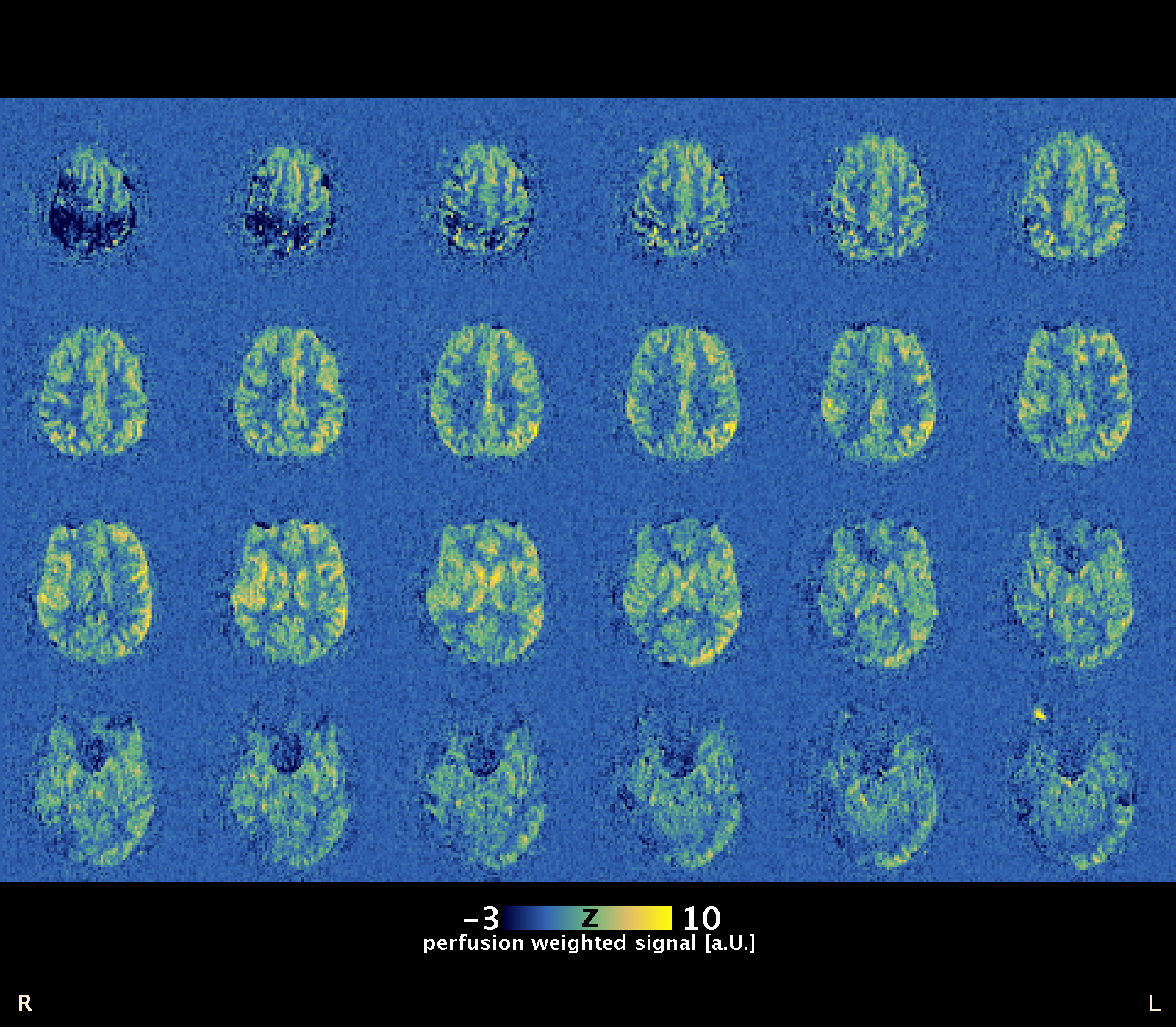
Results
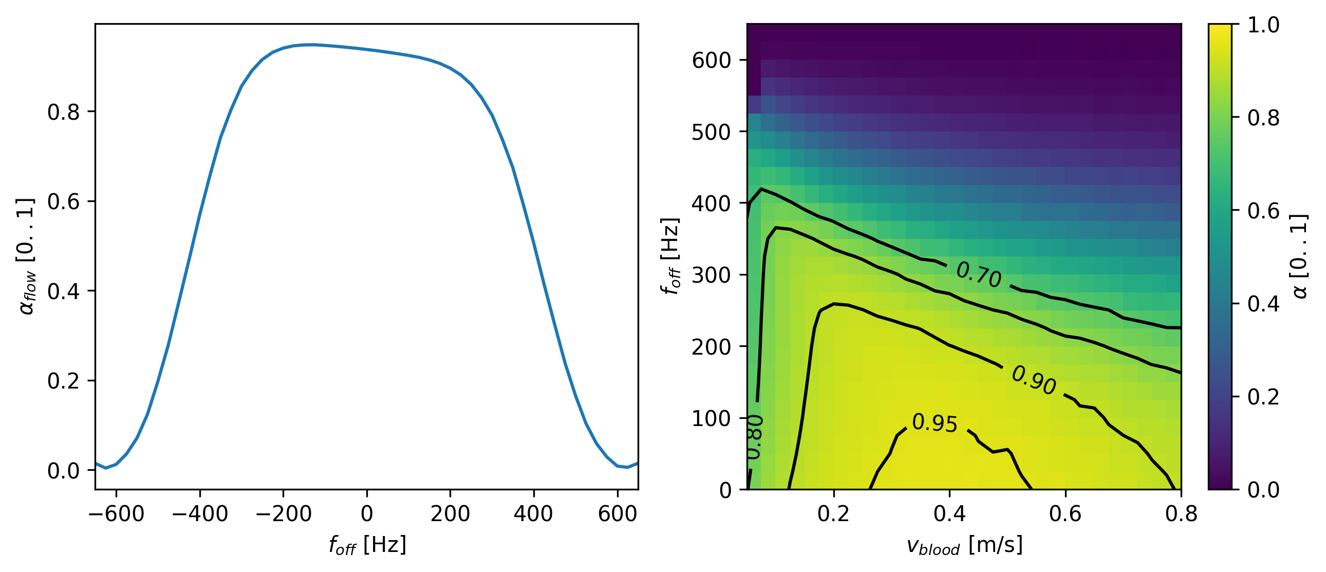
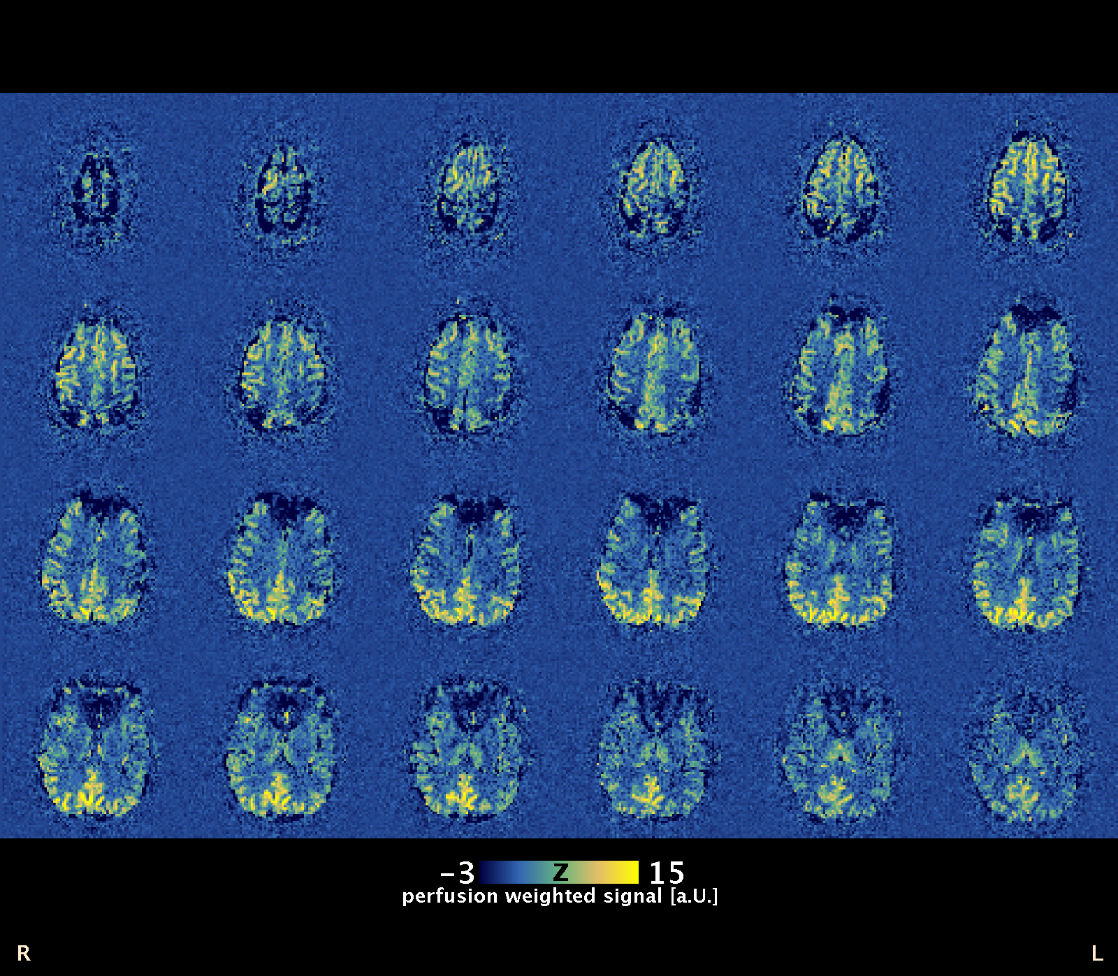
Figure 2 depicts the simulation results, showing that the proposed labeling scheme has a high inversion efficiency (see (3)) over a wide range of velocities (5cm/s to 80 cm/s) and off-resonances (+/- 300 Hz). This is reflected in both in vivo measurements (see Fig. 4, Fig. 5). They show a uniform perfusion weighting from all arteries. Signal dropouts in Fig. 4 are not related to the labeling scheme but to pronounced susceptibility artifacts above the sinus (GRE 3D-EPI) in this particular scan.Discussion
An improved PCASL labeling pulse train was designed and simulated. It reduces the energy deposition by ~ 50% and is insensitive to off-resonances from -300Hz - 300Hz. In two subjects a successful PCASL experiment without a pre-scan was conducted with a single channel and an 8 channel transmission coil. The longer pTX coil yielded better coverage of the neck region and enabled labeling at lower positions. Currently, pTX local SAR supervision is not possible due to the fast application of RF pulses in the PCASL labeling scheme. Once this technical limitation is solved, the sequence will provide even more efficient labeling at UHF. Nevertheless, our preliminary results already indicate that the theoretical advantages of UHF for perfusion imaging can be realized in routine applications with the proposed optimized PCASL sequence, providing 2.5 mm isotropic whole-brain perfusion-weighted images in only 3 min scan time.Acknowledgements
No acknowledgement found.References
1. Dai W, Garcia D, De Bazelaire C, Alsop DC. Continuous flow-driven inversion for arterial spin labeling using pulsed radio frequency and gradient fields. Magn. Reson. Med. 2008;60:1488–1497. doi: 10.1002/mrm.21790.
2. Ghariq E, Teeuwisse WM, Webb AG, Osch MJP van. Feasibility of pseudocontinuous arterial spin labeling at 7 T with whole-brain coverage. MAGMA 2012;25:83–93. doi: 10.1007/s10334-011-0297-0.
3. Zhao L, Vidorreta M, Soman S, Detre JA, Alsop DC. Improving the robustness of pseudo-continuous arterial spin labeling to off-resonance and pulsatile flow velocity. Magn. Reson. Med. 2016;00. doi: 10.1002/mrm.26513.
4. Hargreaves BA, Cunningham CH, Nishimura DG, Conolly SM. Variable-rate selective excitation for rapid MRI sequences. Magn. Reson. Med. 2004;52:590–7. doi: 10.1002/mrm.20168.
5. Ogg RJ, Kingsley PB, Taylor JS. WET, a T1- and B1-insensitive water-suppression method for in vivo localized 1H NMR spectroscopy. J. Magn. Reson. B 1994;104:1–10. doi: 10.1006/jmrb.1994.1048.
6. Andronesi OC, Ramadan S, Ratai EM, Jennings D, Mountford CE, Sorensen AG. Spectroscopic imaging with improved gradient modulated constant adiabaticity pulses on high-field clinical scanners. J. Magn. Reson. 2010;203:283–293. doi: 10.1016/j.jmr.2010.01.010.
7. Brenner D, Stirnberg R, Pracht ED, Stöcker T. Rapid MRI System Calibration using 3DREAM. Proceedings of the International Society for Magnetic Resonance in Medicine 2015;23.
Figures